
Don't wanna be here? Send us removal request.
Statistics
We looked inside some of the posts by uberstrainerusa and here's what we found interesting.
Average Info
Notes Per Post
2
Likes Per Post
2
Reblog Per Post
0
Reply Per Post
0
Time Between Posts
21 days
Number of Posts By Type
Text
14
Last Seen Tumblr Blogs


Fun Fact
1,644 Tumblr posts in 1 second.
Text
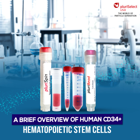
A Brief Overview of Human CD34+ Hematopoietic Stem Cells
CD34 expression in hematopoietic stem cells is significant for cell isolation techniques. CD34+ cells have diverse functions and potential applications in medical research. However, isolating and preserving these cells pose challenges.
The human body consists of several interconnected systems that rely on one another to preserve equilibrium. Within these systems, there are specialized cells that carry out specific functions essential for various processes. One such example is the immune system, which safeguards the body against foreign invaders using specialized blood cells. Among these blood cells, hematopoietic stem cells found in the bone marrow play a crucial role in the development of different types of blood cells.
This blog delves into the significance of CD34 expression and highlights the techniques used for cell enrichment in the study of hematopoietic stem cells.
What Are Hematopoietic Stem Cells?
Hematopoietic stem cells (HSCs), characterized by the presence of the CD34+ marker, possess a unique characteristic known as multipotency. This means that they have the ability to give rise to a diverse range of mature blood cells, including leukocytes, erythrocytes, and platelets. HSCs exhibit self-renewal capabilities, enabling them to regenerate and sustain themselves. They have the remarkable capacity to differentiate into almost any type of blood cell required, making them an integral component of the hematopoietic system.
Understanding CD34: A Cell Marker for Hematopoietic Stem Cells
Cell markers, also known as surface antigens, are molecules present on the cell membrane that serve specific functions related to the cell's role in the body. These proteins are crucial for identifying and characterizing different cell types. CD34, also known as sialomucin, is a transmembrane protein that spans the cell membrane once. It is primarily expressed in certain vascular and hematopoietic tissues.
The Significance of CD34 Expression
The presence of CD34 markers and the utilization of effective cell enrichment techniques are essential in the identification and isolation of hematopoietic stem cells. Scientists employ various techniques for cell separation, including those based on the physical properties of cells and those that utilize antibodies to target specific surface antigens. CD34 expression on the cell membrane enables the binding of CD34-specific antibodies to hematopoietic stem cells, facilitating their identification and isolation.
Using Pluribead to Enrich CD34+ Cell Samples
Pluribead is a technique where a particular antibody directly binds to the target cells, enabling their separation from other undesirable cells during subsequent enrichment steps. The unbound cells are removed, leaving only the desired cells. To immobilize the labeled cells, a cell strainer or magnets can be used when coupled with a solid phase. This approach can be used with various sample materials, such as PBMC, secretion or excretion material, buffy coat, whole blood, brain homogenate, spleen, liver, and so on.
In conclusion, hematopoietic stem cells (HSCs) with CD34 expression play a critical role in the development and regeneration of different types of blood cells. Understanding the importance of CD34 markers and employing effective cell separation techniques are key to studying and harnessing the potential of these stem cells for medical research and treatment. Explore the possibilities of cell enrichment techniques and uncover the potential of hematopoietic stem cells in advancing medical science. Start your journey today!
0 notes
Text
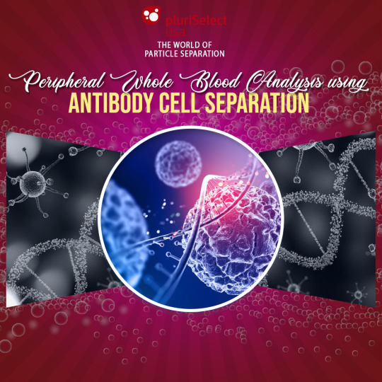
Peripheral Whole Blood Analysis using Antibody Cell Separation
This blog emphasizes antibody cell separation in peripheral whole blood analysis, utilizing PluriBead and PluriSpin systems for high-quality cell isolation. Read on to discover more about these innovative products.
Peripheral whole blood (PWB) is a vital biological sample utilized in numerous medical research investigations, such as identifying diseases, developing drugs, and creating treatments. The blood consists of different types of cells, including red blood cells, white blood cells, and platelets.
Among the white blood cells, peripheral blood mononuclear cells (PBMCs) are especially significant since they have a critical role in the immune system. Nevertheless, isolating these cells from the intricate mixture of blood components requires an effective method of cell separation like antibody cell separation.
A Brief on Peripheral Whole Blood
Peripheral whole blood is a blood sample collected from a peripheral vein, usually from the arm. It is a complex mixture of various components, including red blood cells, white blood cells, and platelets, suspended in a liquid called plasma. The blood is collected in tubes containing anticoagulants, which prevent the blood from clotting.
What are the Components of PBMCs
Peripheral blood mononuclear cells (PBMCs) are a group of white blood cells, including lymphocytes, monocytes, and dendritic cells. They play a crucial role in the immune system and are involved in the recognition and elimination of pathogens and infected cells. PBMCs are essential for medical research, as they can be used to investigate immune responses to various diseases and treatments.
Antibody Cell Separation for Peripheral Whole Blood Analysis
Isolating PBMCs from whole blood is essential in medical research, as it allows for the investigation of immune responses to diseases and treatments. Antibody cell separation is a technique used to separate specific cell types from the complex mixture of blood components. This technique uses antibodies that specifically bind to the surface markers of the target cells, allowing for their separation from the rest of the blood components.
Using the Right Antibody Cell Separation Method
Several methods are available for antibody cell separation, including positive selection and negative selection. Positive selection involves using antibodies that specifically bind to the target cells, allowing for their isolation, while negative selection involves using antibodies that bind to non-target cells, allowing for the isolation of the target cells. The choice of antibody cell separation method depends on the research question, the target cell type, and the downstream analysis.
PluriBead
Pluribead is a technique where a particular antibody directly binds to the target cells, enabling their separation from other undesirable cells during subsequent enrichment steps. The unbound cells are removed, leaving only the desired cells. To immobilize the labeled cells, a cell strainer or magnets can be used when coupled with a solid phase. This approach can be used with various sample materials, such as PBMC, secretion or excretion material, buffy coat, whole blood, brain homogenate, spleen, liver, and so on.
Plurispin
Plurispin involves binding all cells to specific antibodies and separating them, except for the cells of interest. Unlike positive cell enrichment, this approach eliminates all unwanted cells. During this method, the desired cells remain unbound and "untouched" by the antibodies or beads, resulting in highly purified and viable cells. This approach reduces the likelihood of damage to the activated cells.
Conclusion
For efficient and reliable antibody cell separation tools, choose Uberstrainer, a leading provider of high-quality cell separation products and services.
0 notes
Text
What is the Function of TwinSpin in Buffy Coat Preparation?
You can go to our website for more details. Use our products to experience the distinction for yourself. Start using our density gradient centrifugation products for cell separation right away!
A buffy coat is an accumulation of platelets and white blood cells (WBCs). We'll discuss how TwinSpin helps in the preparation of a buffy coat.
A sample of peripheral whole blood contains less than 1% white blood cells (WBCs) and platelets. When scientists spin the blood sample in a centrifuge, these WBCs and platelets combine to form their own layer that is suspended between the red blood cells (RBCs) and supernatant plasma. This thin layer is referred to as a buffy coat because of its color (yellowish to brownish).
We will also find out how TwinSpin density gradient centrifugation tubes are used in buffy coat extraction.
A Brief On TwinSpin Tubes
Spin Medium can be used to separate peripheral blood mononuclear cells (PBMC) from bone marrow and whole blood into TwinSpin centrifuge tubes. A standard 15-inch tire and an inner tube make up the TwinSpin. The open bottom of the inner tube is submerged in the density gradient medium (DGM).
Instructions for Using the TwinSpin Tube
1. Ensure that the (diluted blood) sample, the TwinSpin, and the centrifuge are all at room temperature.
1.1 Check that the inner tube is partially filled and immersed in the DGM. If not, maintain the vertical position while rotating the TwinSpin device.
1.2 Take out and throw away the transport stopper.
2. Include Sample Material
2.1 Pipette the sample material on top of the DGM in the inner tube by tilting the TwinSpin 45° and allowing the sample to run down the inside of the dropper.
2.2 Push the elastic cap firmly into the opening to close the TwinSpin. PluriSpin PLT Depletion can be used to reduce platelet contamination.
3. Spin
3.1 Centrifuge at 800 x g for 20 minutes at room temperature in a swing bucket rotor, brake off. If the blood is more than 4 hours old, centrifuge it for 30 minutes at 1000g.
4. Collect
4.1 Remove the inner cell separation tube by unscrewing it.
4.2 Push the elastic cup down to collect cells in the opaque layer in a new tube.
5 Wash (if necessary)
5.1 Add wash buffer to the reaction tube.
5.2 Cells are spun down for 10 minutes with 300 x g at 4 °C (no or small break).
5.3 Remove the supernatant and set the reaction tube on the table for 20 seconds.
Excess wash buffer will flow down the tube wall and accumulate at the bottom. Aspirate the majority of the liquid above the pellet. The liquid will appear foggy because it contains mostly platelets; aspiration will improve the washing result.
5.5 Carefully pipette the pellet with 1 ml of wash buffer.
5.6 Re-do steps 5.1–5.4.
5.7 Refill the pellet to the desired volume.
Preparation of buffy coat from scratch
Buffy coats can be prepared in two different ways. To separate the whole blood into red cells, plasma, and buffy coat, the whole blood donation is centrifuged on the one hand. The second option is to filter the blood and keep the leukocytes from circulating.
0 notes
Text
What is TCR Therapy, CAR T Cell, and What are They Used For?
The antigen-binding theory is the foundation of CAR-T and TCR-T. Let's examine how their recognition and action characteristics vary.
Medical research and cancer treatment strategies are constantly evolving to meet new challenges. When more traditional cancer treatments fail, patients may resort to adoptive cell therapy as a last resort.
Adoptive cell therapy combines human T cells with scientific enhancements to treat diseases that the body is ill-equipped to handle.
CAR T-cell therapy and engineered TCR therapy are two T-cell therapies that involve the modification of receptors for the purpose of cancer treatment.
We will discuss the Pluribead cascade straining and how it helps in the gentle separation of T cells.

Chimeric Antigen Receptors (CAR)
CAR T-cell therapy involves binding proteins called chimeric antigen receptors to patient-derived activated T cells. These receptors have been engineered to target naturally occurring antigens on the surface of cancer cells. This allows patients to receive treatment for cancers that the body cannot fight on its own.
CAR T Cell Activity
Currently, three types of FDA-approved CAR T-cell therapies are available for clinical use. These techniques have enabled the treatment of certain types of lymphoma and leukemia.
Other receptor-antigen combinations are undergoing clinical trials. As research advances and more cell therapies are approved by the FDA, the number of cancers that can be treated grows.
TCR Cell Therapy Function
The MHC, or major histocompatibility complex, is a group of genes that code for antigens that the immune system can recognize. The MHC identifies foreign pathogens by attaching proteins to their surfaces that T lymphocytes can bind to.
Engineered TCR therapy imbues activated T cells with specific receptors that target cancer antigens. This greatly improves treatment personalization and increases the likelihood of positive outcomes for patients.
TCR vs. CAR T
CAR T-cell therapy is a type of TCR therapy that has been engineered. While these two strategies have many similarities, their differences determine what purpose they serve. The receptors programmed into the cell are the main difference between CAR T-cell therapy and engineered TCR therapy.
CAR T cells, as previously stated, have receptors that target naturally occurring antigens. The immune system plays no role in CAR T-cell therapy other than to lend T lymphocytes. TCR therapy, on the other hand, uses MHC to label cancer cells with identifiable antigens.
Pluribead For T Cells Separation
This research can be simplified by streamlining various stages of the manufacturing process. T-cell separation takes a long time and requires expensive equipment.
Previously, researchers used complex machines to perform the time-consuming task of sorting and purifying large volumes of lymphocytes. Innovative cell separation strategies, such as Pluribeads, are more efficient and produce better results at a lower cost.
Pluribead cascade straining, which operates without the use of any magnetic components, aids in the gentle and safe isolation of Dendritic Cells. The procedure is straightforward: your pluriBeads (which contain bound target cells) are sieved through a strainer, with unwanted cells passing through and your target cells remaining on top.
After detaching, you are now ready to proceed with your target cells. Our team of scientific experts is committed to the success of the researchers with whom we collaborate, and we look forward to providing you with our novel pluriBeads technology as yet another tool in your arsenal as you work toward the next big scientific breakthrough.
1 note
·
View note
Text
What Are Effector T Cells And The Different Cell Types?
Pluribead, a leading cell enrichment technology, is specifically designed for fast and gentle sample preparation, resulting in a highly-enriched population of healthy, viable cells. Learn more about our T cell isolation products, which can help you overcome long-standing sample preparation challenges.
The term "effector T cell" refers to a type of T cell that actively responds to a stimulus, such as co-stimulation. Learn more about our unique Effector T cell separation technologies.

Effector cells are immune system cells that have gone through the process of differentiation and maturation. In the event of a stimulus, these are the cells that mount specific responses. As part of the immune response against a pathogen or a self cell, the body's immune system generates effector cells (in case of autoimmune disorders).
We'll look at how Pluribead cell enrichment aids in Effector T Cells isolation.
T Cell Activation
When a T cell encounters a recognized APC, it receives a signal to mature. If a cell gets all three signals, it will develop into an effector cell. If a cell only receives one of the signals (TCR or BCR), it becomes ineffective.
Effector Cells
Depending on the APC encountered, a naive cell can develop into an effector T cell. Effector T cells have relatively short lifespans and perform immune response functions. T cells can be a cytotoxic, helper, or regulatory.
Cytotoxic T Cells
The primary function of cytotoxic T cells, also known as CD8+ cells, is to kill toxic/target cells. When they are recognized, their purpose changes to the removal of virally infected cells, bacteria, and tumor fragments (such as cancer cells) via a process known as apoptosis. Apoptosis occurs when the internal organelles of a cell are destroyed, causing the cell to die from the inside out.
Helper T Cells
T helper cells, also known as CD4+ cells, are similar to cytotoxic cells but perform a broader range of functions. These cells are critical to cell immunity because they are required for the majority of adaptive immune responses. When exposed to antigens, T helper cells become activated and have the ability to differentiate into cell subtypes.
Regulatory T Cells
The regulatory T cell is the final type of effector cell. Once the threat has been eliminated, regulatory T cells are tasked with suppressing the autoimmune response. After helper T cells and cytotoxic T cells bind to a pathogen and work together to eliminate it from the body, they are no longer useful. Regulatory T cells prevent them from taking up space or attacking healthy cells until they die of apoptosis.
While these three types of effector cells handle the majority of the immune response, they are not the only T cell variations. Even after a pathogen has been removed, some types of T lymphocytes remain. These long-living lymphocytes are memory T cells that are highly capable of responding to antigens when reintroduced—which aids the immune system.
These cells are formed following infection and are extremely important because they have the ability to multiply into a large number of effector T cells when exposed to familiar antigens. Memory cells are distinct in that they remember pathogens and infectious cells faster than other cells, allowing them to fight bacteria and viruses more effectively.
Try Our Cell Separation Products Right Now!
0 notes
Text
Protocol For Buffy Coat Preparation From Whole Blood
In this blog, we’ll explore what is buffy coats and how it is prepared from whole blood. We will also learn how Pluribead and Plurispin help in this process.
Millions of cells are suspended in a liquid called plasma to form blood. Red blood cells (which carry oxygen), white blood cells (which fight infection), and platelets are examples of these cells (which help with clotting). Examining red blood cells is simple due to their abundance; thousands of red blood cells can be seen in a single drop of blood. White blood cells, on the other hand, are present in very small numbers and are more difficult to examine.
Examining a buffy coat smear is the quickest way to examine large numbers of white blood cells. The buffy coat is simply a collection of all of the white blood cells and platelets in a blood sample.
Let’s find out how Pluribead Antibody Cell Separation technology helps in buffy coat extraction.
Preparation Of Buffy Coat From Scratch
Making your own buffy coat is an alternative. Therefore, simply adhere to the following succinct protocol:
Mix one part washing buffer with one part whole blood.
Centrifuge the diluted whole blood for 10 minutes at 200 x g while the brake is turned off.
Remove the interphase leukocytes (buffy coat)
It's important to remember that a whole blood sample could contain pathogens, so these samples must be handled with the same care as if they were capable of transmitting infectious diseases. The time has come to separate the buffy coat from a whole blood sample after all the necessary preparations have been made.
Buffy Coat Extraction
The buffy coat is separated from the plasma and RBC by centrifuging the prepared whole blood sample. After centrifugation, there will be a thin layer between the RBC and plasma that accounts for about 1% of the sorted sample – the buffy coat. The experimenter uses a tiny pipette to gather and transfer the buffy coat to a different container.
The buffy coat is in direct contact with isolated RBCs, while PBMCs are isolated from the other particles in the sorted sample by density gradient centrifugation. Using a manual extraction technique like pipetting increases the possibility that contaminating RBCs will be present in the buffy coat sample because of the size and abundance of RBCs (Buffy coats are referred to as "pink" before further processing because of this). Researchers frequently combine centrifugation with another cell separation technique, like BACS, to get rid of any remaining RBC contamination in order to enrich the buffy coat even more.
How can Plurispin be useful?
A brand-new technique for isolating negative cells from whole blood, buffy coat, or cord blood is called the PluriSpin system. This new technique isolates live, unaltered, and highly purified cells in a single step without the use of magnets or a column. As a result, there is less chance of activating or harming the target cells.
How PluriBead can help?
PluriBead is a one-of-a-kind Antibody Cell Separation technology that doesn't rely on magnetic components. The process is simple: Your pluriBeads are sieved through a strainer with your bound target cells on top, while the unwanted cells pass through. After detaching, you have your target cells ready.
For more information, you can visit our website. Experience the difference for yourself by using our products. Get started with our cell separation products today!
0 notes
Text
How do Regulatory T Cells Work?
Regulatory T cells (Tregs) are a type of T cell that suppresses immune responses by secreting negative regulatory (anti-inflammatory) cytokines. Let’s find out about the Regulatory T-cells separation technologies.
T cells that regulate or suppress other immune system cells are known as regulatory T cells (also known as Tregs). They regulate the immune response to self and foreign particles (antigens), which aids in the prevention of autoimmune disease. Tregs are produced naturally by a healthy thymus. 'Adaptive' Treg are formed by the differentiation of naive T cells outside the thymus, i.e. the periphery, or in cell culture.
CD4+ T cells are commonly classified as regulatory T (Treg) cells or T helper (Th) cells. By activating other effector immune cells, Th cells regulate adaptive immunity against pathogens and cancer. Treg cells are CD4+ T cells that are in charge of suppressing potentially harmful Th cell activities.
We'll look at how Pluribead cascade straining aids in Regulatory T Cells isolation.
Identification of Treg cells remains difficult because evidence suggests that all of the currently used Treg markers (CD25, CTLA-4, GITR, LAG-3, CD127, and Foxp3) are general T-cell activation markers rather than Treg-specific.
What Are The Functions Of Treg Cells?
Treg cells' primary function was originally defined as the prevention of autoimmune diseases through self-tolerance. Several additional functions have been proposed over the years, and it will be critical to determine what Treg cells actually do in the immune system.
Prevention of autoimmune diseases through the development and maintenance of immunologic self-tolerance.
Allergic and asthmatic symptoms are reduced.
Oral tolerance is the induction of tolerance against dietary antigens.
Induction of maternal-fetal tolerance.
Pathogen-induced immunopathology is suppressed.
Regulation of the immune response's effector class.
T-cell activation is suppressed in response to weak stimuli.
Effector T cells regulate the magnitude of the immune response.
How To Identify Treg Cells?
Molecular markers are critical tools for defining and analyzing immune cell subpopulations. The failure to identify specific markers for suppressor T cells contributed significantly to the decline of suppressor T cells at the end of the 1980s.
The most commonly used Treg cell markers are:
CD25 is an abbreviation for cytotoxic T lymphocyte-associated antigen 4. (CTLA-4).
Gene related to the glucocorticoid-induced tumor necrosis factor receptor family (GITR).
Lymphocyte activation gene-3 (LAG-3) (LAG-3).
P3 transcription factor box forkhead/winged-helix CD127 (Foxp3).
All T cells express CD25, the -chain of the interleukin-2 (IL-2) receptor, which is a T-cell growth factor important for T-cell clonal expansion. CTLA-4 is a T-cell activation negative regulator that is upregulated on all CD4+ and CD8+ T cells 2-3 days after activation.
Pluribeads For Regulatory T Cells Separation
Pluribead cascade straining helps in the gentle and safe isolation of Dendritic Cells and operates without the use of any magnetic components.
The method is simple: your pluriBeads (which contain bound target cells) are sieved through a strainer, with your target cells remaining on top and unwanted cells passing through. You are now ready to proceed with your target cells after detaching.
You can see the difference for yourself by using our products. Order our cell separation products right away, and don't forget to look over our Programs and Special Offers!
0 notes
Text
How Do Human Macrophages and Monocytes Differ?
Monocytes and macrophages are cells that are very similar with a few key differences and different applications. This blog will go over the distinctions between Macrophages and Monocytes.
The immune system includes lymphocytes, macrophages, monocytes, neutrophils, and other cells such as basophils, eosinophils, and natural killer cells. Macrophages and monocytes are large, irregularly shaped white blood cells that stimulate the body's antibody production.
We'll look at how Pluribead cascade straining aids in monocyte isolation, as well as the differences between human macrophages and monocytes.
What is a Monocyte?
Monocytes are immune cells found in the blood that can migrate to tissues by differentiating into macrophages. They have the ability to differentiate into dendritic cells as well.
Monocytes play an important role in innate immunity, serving as the host's first line of defense. They also activate the adaptive immune system by inducing an inflammatory response. Monocytes produce cytokines such as IL-1, IL-2, and TNF, as well as chemokines such as monocyte chemotactic protein-1 and -3. Monocytes migrate into tissue in response to inflammation within 8-12 hours.
Monocytes are classified into three types in blood-based on the receptors found on their surfaces. CD14 is a surface receptor found on classic monocytes. Non-classical monocytes have CD16 as well as CD14. On the cell surface, intermediate monocytes have CD14 and low levels of CD16 receptors.
What exactly is a macrophage?
Macrophages are immune cells that are found in extracellular fluid. They are distinct from monocytes. Macrophages are large cells that can engulf dead cells and ingest foreign material such as bacteria and viruses by forming pseudopodia around them.
Enzymes for the digestion of engulfed material are found in macrophage cytoplasm granules. Macrophages are phagocytes that work professionally. Macrophages are found in Langerhans cells in the skin, Kupffer cells in the liver, the pigmented epithelium of the eye, and microglia in the brain. Macrophages in the spleen remove old and defective RBCs from circulation.
The main distinction between monocytes and macrophages
The primary distinction between monocytes and macrophages is that monocytes are precursors to some macrophages, whereas macrophages are professional phagocytes that engulf pathogens that infiltrate the body.
Monocytes and macrophages are two types of cells found in organisms' immune systems. They are regarded as the host's first line of defense. Monocytes are small bean-shaped cells, whereas macrophages are large irregular-shaped cells. Monocytes and macrophages can both secrete cytokines and chemokines.
Pluribead for Monocyte Enrichment
Many conventional monocyte enrichment techniques have been reported to cause harm to delicate cell populations and to be expensive to execute. The magnetic field in magnetic-based cell separation techniques can cause cells to lyse. The gentle Pluribead cascade straining preserves cell viability throughout the isolation procedure.
Pluribead
Because all undesirable cells are unbound when the specific antibody binds directly to the cells using positive selection, all undesirable cells are separated from the labeled and desired cells during the subsequent enrichment steps. A cell strainer, when coupled to a solid phase, is the simplest way to keep labeled cells in place.
Conclusion
Monocytes and macrophages are immune system cells that participate in both innate and adaptive immunity. Blood contains monocytes. Monocytes migrate to the extracellular fluid that surrounds the inflammatory tissue and differentiate into macrophages in response to inflammation.
0 notes
Text
TwinSpin and centrifugation: How it works?
TwinSpin centrifuge tubes support density gradient centrifugation. Let’s find out how and we will also discuss the protocol for the use of TwinSpin in combination with density gradient media.
Scientists divide materials using the centrifugation technique based on their size and shape. The two types of centrifugal methods used to separate particles are differential centrifugation and density gradient centrifugation. This blog will discuss TwinSpin's functionality as well as how it supports density gradient centrifugation.
Peripheral blood mononuclear cells (PBMC) can be separated from whole blood and bone marrow using TwinSpin centrifuge tubes that have already been pre-filled with PBMC Spin Medium. The TwinSpin is made up of an inner tube and a standard 15. The inner tube's open bottom is buried in the density gradient medium (DGM). The blood sampling tube can be directly pipetted into the TwinSpin with anticoagulated blood or bone marrow.
The sample is placed on top of the DGM inside the inner tube. Depending on the density gradient used (Leuko Spin, PBMC Spin, PBMC 24+, or PLT Spin Medium), leukocytes, lymphocytes, and PBMCs are separated from unwanted erythrocytes and granulocytes during Density Gradient Centrifugation.
Target cells above the DGM are therefore enriched in the interphase. Through the DGM, the erythrocytes will exit the inner tube and fall to the bottom of the outer tube. Once the inner tube has completely separated, just take it out.
As a valve, the elastic cap is used. The collection tube can be converted into a pipette by removing the cap. Drop by drop, the contents can be collected.
Instructions for Using the TwinSpin Tube
1. Ensure that the (diluted blood) sample, the TwinSpin, and the centrifuge are all at room temperature.
1.1 Check that the inner tube is partially filled and immersed in the DGM. If not, maintain the vertical position while rotating the TwinSpin device.
1.2 Take out and throw away the transport stopper
2. Include Sample Material
2.1 Pipette the sample material on top of the DGM in the inner tube by tilting the TwinSpin 45° and allowing the sample to run down the inside of the dropper.
2.2 Push the elastic cap firmly into the opening to close the TwinSpin. PluriSpin PLT Depletion can be used to reduce platelet contamination.
3. Spin
3.1 Centrifuge at 800 x g for 20 minutes at room temperature in a swing bucket rotor, brake off. If the blood is more than 4 hours old, centrifuge it for 30 minutes at 1000g.
4. Collect
4.1 Remove the inner separation tube by unscrewing it.
4.2 Push the elastic cup down to collect cells in the opaque layer in a new tube.
5 Wash (if necessary)
5.1 Add wash buffer to the reaction tube.
5.2 Spin down cells for 10 minutes at 4°C with 300 x g (no or small brake).
5.3 Remove the supernatant and set the reaction tube on the table for 20 seconds.
Excess wash buffer will flow down the tube wall and accumulate at the bottom. Aspirate the majority of the liquid above the pellet. The liquid will appear foggy because it contains mostly platelets; aspiration will improve the washing result.
5.5 Carefully pipette the pellet with 1 ml of wash buffer.
5.6 Re-do steps 5.1–5.4.
5.7 Refill the pellet to the desired volume.
Want to see TwinSpin in action? Check out our line of products that supports density gradient centrifugation, and let us know if you have any questions.
0 notes
Text
How to Determine Cell Viability in a Sample
The ratio of live to dead cells is used to perform a cell viability assay. This blog discusses Cell Viability and how to pluribead helps assessing cell viability.
The percentage of alive, healthy cells in a sample is known as cell viability. When choosing the best conditions for cell growth in culture or in response to extracellular stimuli, chemicals, or therapeutic treatments, cell viability assays are used to assess the physical and physiological health of cells. Researchers must optimize their methods for differentiating healthy cells from dead or damaged cells in order to perform accurate cell viability tests.
Our cell separation technologies identify and remove unwanted dead or degraded cells from a sample in a gentle and efficient manner, leaving healthy cells for further study or downstream research applications.
Cell Viability
The number of living cells in a population is referred to as cell viability. Understanding cell behavior and assessing efficacy require determining the viability of cells in a sample throughout research and treatment development. Cell viability assays enable researchers to infer the overall health of cells in samples, improve experimental procedures, and determine whether cells remain healthy after therapeutic testing.
Pluribead Enhances Cell Viability
Cell degradation in a population can occur as a result of natural cell death, therapeutic testing, or experimental procedure. When cells die or are damaged, they leave behind cellular debris. Both unhealthy cells and the debris they produce can impair the accuracy of cell viability assays if they are counted as whole cells or are unable to be differentiated.
The sample population being tested must be made up of viable cells in order to determine how a particular process or treatment will affect healthy cells in the body. As a result, the ability to distinguish healthy cells from cellular debris or dead cells within a sample is critical for accurately determining cell viability and the efficacy of therapeutic testing.
Pluriselect has created a breakthrough technology for healthy cell separation from dead or damaged cells that is inexpensive, simple to use, and produces viable cell populations of high purity.
Pluribead
When the specific antibody binds directly to the cells using positive selection, all undesirable cells are separated from the labeled and desired cells during the subsequent enrichment steps because they are all unbound. When coupled to a solid phase, a cell strainer is the simplest way to keep labeled cells in place. Any type of sample material, such as PBMC, secretion or excretion material, whole blood, buffy coat, spleen, liver, and so on, may be used.
Key features of Pluribead
No Sample Preparation: Use a sample volume ranging from 200l to 45 ml with no erythrolysis, gradient centrifugation, or other techniques.
Any type of sample material, such as PBMC, secretion/excretion material, brain homogenate, spleen, liver, buffy coat, whole blood, and so on, can be used.
Suitable for Isolation from a Wide Range of Species: Isolate from sheep, mice, rats, cows, dogs, and other animals.
Cell division can begin within five minutes of isolation.
PluriBead Cascade Simultaneous Cell Isolation: Separate two different cell types from the same sample material at the same time.
Using sequential cell isolation, you can isolate up to six different targets from a single sample.
To learn more about the importance of cell viability and how Pluribead is revolutionizing cell enrichment and cell viability assays, you can visit our website or talk to our support team.
0 notes
Text
How To Improve Sample Purity Using Unique Cell Separation Technologies?
When working with cells, scientists must examine sizable, healthy populations of particular cell types. Here is everything you need to know about improving the purity of your sample.
Given the vast array of factors that researchers must be aware of and take into account in order to produce useful results, cell biology is complex. It is common practice to reduce experimental complexity by using isolated populations of cells rather than heterogeneous mixtures of cells.
Cell biologists can thus with certainty attribute observed effects and responses to a specific cell type. For this reason, any cell biologist would benefit from becoming proficient in the fundamental methods of cell enrichment.
What does Cell Purification actually mean?
To remove outside influences on the results, unwanted cells are taken out of a biological sample through the process of cell purification.
What Circumstances Require Cell Culture Purification?
When getting ready to conduct an experiment that requires a single isolated cell population with high cell viability and health, cell purification is an essential step. This procedure can be done either before or after the main cell separation method to remove contaminants.
The Most Common Contaminants
The two types of cells that are most frequently known to cause issues in purified cell samples are red blood cells (RBCs) and dead cells. If allowed to metabolize, RBCs create waste and change the pH of a solution, which can affect target cells' behavior.
When isolating cells from a blood sample, such as T cells or B cells, a lot of RBCs are likely to be present. These RBCs must be eliminated in order to conduct an experiment with reliable, significant results.
Other elements you might want to remove from a sample, depending on the cells you're trying to isolate, include:
Macrophages
Dendritic cells
Circulating Tumor Cells
Platelets
T cell and B cell variants
Residual Tissue
You can more precisely purify your sample before or after cell enrichment if you are aware of the components you want to eliminate.
Purification of Cell Samples
Numerous techniques can be used to purify cell culture. The gold standard for isolating heterogeneous mixtures with high purity in cell purification is currently affinity molecules like antibodies. Antigen-specific antibodies are used in antibody binding methods to mark target cells and make them more visible or accessible later in the process.
Try Pluriselect’s Cell Separation Products Today
If you're looking for the most efficient way to separate desired cells from unwanted cell populations, pluriBead and pluriSpin technology provides an exceptionally gentle method for cell enrichment that preserves the physiology and health of delicate immune cells.
Pluribead and Plurispin can be used as the primary method or to further purify samples that have already been isolated using different techniques. Our distinctive products eliminate the need for additional purification steps by combining a high level of purity, sample viability, and throughput. They don't have abrasive magnetic fields or swiftly moving liquids that could endanger cell physiology and health.
If you’re still curious about what Pluribead and Plurispin are and how they work, visit our website. Don’t forget to check out our Programs and special offers section.
0 notes
Text
Achieve Gentle White Blood Cells Using Pluribead And Plurispin
White blood cells, also known as leukocytes, are critical components of the immune system. This blog discusses the functions and how advanced cell separation technologies can be used to gently separate them.
White blood cells, also known as leukocytes or white corpuscles, lack hemoglobin and thus protect the body from infection and disease. In this blog, we will discuss how Pluribead and Plurispin, the best cell enrichment technologies, aid in the rapid isolation of white blood cells.
Major classes of white blood cells
Lymphocytes, which are further subdivided into B cells and T cells, are in charge of recognizing and removing foreign agents from the host. Antibodies, which are proteins that bind to and destroy foreign microorganisms in body tissues, are secreted by B lymphocytes.
T cells typically recognize and destroy virally infected or cancerous cells, or they act as helper cells to aid B cell antibody production. Because of their inherent ability to kill a wide range of target cells, natural killer (NK) cells are also included in this category. In a healthy person, lymphocytes account for 25 to 33 percent of white blood cells.
Granulocytes, the most numerous white cells, are responsible for ridding the body of large pathogenic organisms such as protozoans and helminths, and they also play important roles in allergy and other forms of inflammation. Neutrophils, eosinophils, and basophils are the three types of granulocytes based on how their granules take up dye in the laboratory. Neutrophils are the most abundant granulocytes, accounting for 50 to 80% of all white cells.
Eosinophils, basophils, and mast cells are usually the last to show up. Basophil and mast cell granules contain a variety of chemicals that are important in inducing allergic inflammatory responses, such as histamine and leukotrienes. Eosinophils are parasite killers that also modulate inflammatory responses.
Monocytes, which account for 4 to 8% of all white blood cells in the blood, migrate from the blood to infection sites, where they differentiate further into macrophages. The main phagocytic cells in the body are neutrophils and macrophages, but macrophages are much larger and live much longer than neutrophils. Some macrophages play an important role as antigen-presenting cells, which phagocytose and degrade microbes before presenting fragments of these organisms to T lymphocytes, activating the specific acquired immune response.
Our Unique Cell Separation Products- Pluribead and Plurispin
PluriBead technology, if you're looking for the most efficient way for cell enrichment, provides an exceptionally gentle method that preserves the physiology and health of delicate immune cells.
PluriBead
Using positive selection, when the specific antibody will bind directly to the cells, all undesirable cells will be separated from the labeled and desired cells during the subsequent enrichment steps because they are all unbound. A cell strainer is the simplest way to keep labeled cells in place when coupled to a solid phase.
Plurispin
Negative cell selection eliminates all unwanted cells. Unlike positive cell enrichment, all cells will be bound to specific antibodies and separated, with the exception of the cells of interest. While the undesirable cells are extracted from the sample, the desired cells remain unbound and "untouched" by antibodies or beads.
Traditional cell enrichment methods subject samples to intense mechanical or magnetic forces and require the use of expensive equipment, whereas our Cell Separation products are gentle, cost-effective, and easy to use.
0 notes
Text
What are Neutrophile Granulocytes and how do they function?
Granulocyte neutrophils are the most highly abundant myeloid cell type, comprising 40% to 70% of total white blood cells.
Granulocyte neutrophils are the most common myeloid cell type, accounting for 40% to 70% of total white blood cells. They are the first cells to be recruited to acute inflammatory sites and are motile phagocytic leukocytes. Neutrophils are short-lived cells that are produced in the bone marrow in response to granulocyte colony-stimulating factor stimulation.
They ingest, kill, and digest microbial pathogens, and their functions are dependent on special proteins found in primary and secondary granules (containing cationic defensins and myeloperoxidase) (iron chelators, lactoferrin, and digestive enzymes).
Multiple antimicrobial agents are found in neutrophilic granules, including oxygen-independent lysozyme (peptidoglycan degradation) and lactoferrin (iron chelator). In this blog, we will discuss the innovative Antibody Cell Separation technologies that will help in Granulocyte neutrophils separation.
Function of neutrophils
Neutrophils are extremely mobile cells. They move towards, phagocytose, and degrade a variety of particulate material, including bacteria and damaged tissue cells. Neutrophils are drawn to sites of infection or inflammation by chemotactic gradients that form around such sites. Chemotactic factors include activated complement components (C3a, C5a, C567), membrane phospholipids and other factors released by tissue cells, lymphokines released by activated lymphocytes, products of mononuclear phagocytes (e.g., tumor necrosis factor, IL-8), platelet-derived factors (platelet factor 4, the -thromboglobulin neutrophil-activating peptide 2 (NAP-2), platelet-derived growth factor), IL-8, platelet factor 4, and NAP-2 bind to CXC chemokine receptors on neutrophil surfaces and activate them.
Activated neutrophils bind to endothelial cells via cell membrane adhesion molecules. The arrival of neutrophils at sites of inflammation is most likely aided by increased permeability of adjacent blood vessels caused by activated complement components like C3a and C5a. The adherence of the neutrophil to the particle is the first stage in the phagocytosis of a particle such as a bacterium.
Adhesion is mediated by specific receptors on the neutrophil cell membrane, such as Fc (IgG1, IgG3) and C3 receptors. The interaction of such particles with opsonizing factors such as C3 generated via the classical or alternative complement activation pathway, antibody, and mannose-binding lectin improves both adherence and subsequent ingestion.
Pluribeads For Neutrophile Granulocytes Separation
Pluribead is one such cell separation technology that helps in the gentle and safe isolation of Memory T Cells. Unusual cell separation technology called PluriBead operates devoid of any magnetic components.
The process is easy: Your pluriBeads (which contain bound target cells) are sieved through a strainer; the pluriBeads containing your target cells remain on top while the unwanted cells pass through. You are prepared with your target cells after detaching.
Plurispin For Neutrophile Granulocytes Separation
Negative cell selection eliminates all unwanted cells. Unlike positive cell enrichment, all cells will be bound to specific antibodies and separated, with the exception of the cells of interest. While the undesirable cells are extracted from the sample, the desired cells remain unbound and "untouched" by antibodies or beads.
PluriSpin, a PluriSelect system, isolates viable, untouched, and highly purified cells in a single step without using magnets or columns. As a result, the activated cells are less likely to be damaged.
You can find out more about us by going to our website. By using our products, you can see the difference for yourself. Order our cell separation products right away and don’t forget to check our Programs and Special Offers section!
0 notes
Text
What Are Schwann Cells And How To Isolate Them Efficiently?
Schwann cells surround neurons, keeping them alive and occasionally encasing them in a myelin sheath. To learn more about them, keep reading.
The neural crest is the embryological source of Schwann cells. They act as the primary glial cells of the peripheral nervous system (PNS), myelinating peripheral nerves and supplying axons with nutrients and insulation.
Conduction velocity along the axon is accelerated by myelination, enabling saltatory conduction of impulses. Nonmyelinating Schwann cells still offer trophic support and cushioning to the unmyelinated axons even though they do not wrap axons to improve conduction.
When it comes to cell separation, stop losing your precious samples to outdated processes! Let’s find out about the innovative cell separation technology.
Structure of Schwann Cells
Numerous Schwann cells are required to myelinate the length of an axon because each Schwann cell contributes to one myelin sheath on a peripheral axon, and a different Schwann cell creates each subsequent myelin sheath. The central nervous system's (CNS) myelinating cell, the oligodendrocyte, forms myelin sheaths for numerous surrounding axons, in contrast to this arrangement.
Unlike oligodendrocytes, Schwann cells are surrounded by a basal lamina. Nodes of Ranvier, or gaps of about 1 micrometer, exist between adjacent myelin sheaths. The node, the site of saltatory conduction, contains a concentration of voltage-gated sodium channels. In densely myelinated neurons, Schmidt-Lanterman incisures are cytoplasmic outpouchings that disrupt compact myelin.
The function of Schwann Cells
The PNS's myelinating cell and the peripheral neurons' support cells are both Schwann cells. By concentrically wrapping its plasma membrane around the inner axon, a Schwann cell creates a myelin sheath. The inner turn of the glial cell membrane spirals around the axon to add membrane layers, or lamellae, to the myelin sheath while the nucleus stays fixed.
Extremely high levels of lipids can be found in the plasma membrane of Schwann cells, and cholesterol plays a crucial role in the construction of the myelin sheath. The axon segment is protected by the compact myelin sheath, which also increases conduction velocity and significantly lowers membrane capacitance. The survival and maturation of Schwann cell precursors depend on neuregulin-type III expression on axons, and the amount of neuregulin present on the axon surface determines the degree of myelination.
Axons are also given energy metabolites by Schwann cells, which shuttle them through monocarboxylate transports that are located on the axon's surface and inside the Schwann cell. In response to PNS axon damage and axon regeneration, Schwann cells are essential.
Wallerian degeneration will happen away from the area of injury. When the distal axon segment dies, Schwann cells and then macrophages clear the contents of the dead cells and encourage axon regeneration.
Pluribeads For Schwann Cell Separation
Pluribead is one such cell separation technology that helps in the gentle and safe isolation of Schwann cells. Unusual cell separation technology called PluriBead operates devoid of any magnetic components. The process is easy: Your pluriBeads (which contain bound target cells) are sieved through a strainer; the pluriBeads containing your target cells remain on top while the unwanted cells pass through. You are prepared with your target cells after detaching.
You can find out more about us by going to our website. By using our products, you can see the difference for yourself. Order our cell separation products right away and don’t forget to check our Programs and Special Offers section!
1 note
·
View note