#MicroGraphic Image
Explore tagged Tumblr posts
Visit Tumblr Blog
Explore Tumblr blogs with no restrictions, modern design and the best experience.
Last Seen Tumblr Blogs


Fun Fact
69% of Tumblr users are millennials.
Text
OCR04811: Spelunker: Cave Diving - TSori, Dewey Newt, GuitarSVD, Kenny Jr., MarimbaMatt, myuik, Reuben Spiers, Sean R. Hanson, Siolfor the Jackal, Xander
[Remix primarily of Stage 1, with all other songs and jingles on the soundtrack being referenced]
from OverClocked ReMix; follow the links below to hear more by each of the artists.
song credits:
Arrangement & Trumpets: TSori
Classical guitar: GuitarSVD
Acoustic guitar: Reuben Spiers
Electric guitar: Siolfor the Jackal
Hand percussion: Sean R. Hanson
Steel drum: MarimbaMatt
Drum set: Kenny Jr.
Piano: Xander (ZeldaRocks)
Trombone: Dewey Newt
Bass, mixing: myuik
#TSori#spelunker#spelunker nes#Tim Martin#Dewey Newt#GuitarSVD#Kenny Jr.#MarimbaMatt#myuik#Reuben Spiers#Sean R. Hanson#Siolfor the Jackal#Xander#Tamtex#Tose#MicroGraphic Image#broderbund#overclocked remix#ocremix#ocr#video game remix#video game music#vgm
0 notes
Text

A close up micrograph of the infestations on your skin.
#infestation#micrograph#ai artwork#AI#AI art#AI art generation#AI artist#AI artwork#AI generated#AI image#AI model#AI woman#computer art#computer generated image#digital#digital art#digital drawing#digital illustration#digital painting
5 notes
·
View notes
Text
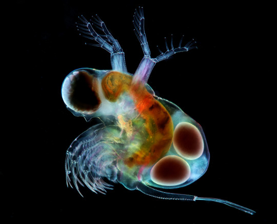
The image presents Polyphemus, a predatory cladoceran with eggs. Micrograph taken in combined technique of the illumination, dark field and polarized light.
201 notes
·
View notes
Photo

🌴 On the germination, development, and fructification of the higher Cryptogamia London: Published for the Ray Society, by Robert Hardwicke ..., 1862. Original source Image description: Historical scientific illustration plate showing detailed cellular and structural drawings of Pteridophyta reproductive organs and development stages. Nine primary diagrams depict various elongated and rounded plant structures with distinct cell patterns and internal compartments, likely representing sporangia and gametophyte development phases. Smaller insets highlight early cell division stages and spores. The intricate cellular outlines emphasize germination and fructification processes in higher Cryptogamia, focusing on tissue differentiation and reproductive cell arrangement. The plate is labeled "PLATE XII" and features fine line work, typical of 19th-century botanical micrographic studies.
20 notes
·
View notes
Text
The Real Life Biology of the Three Body Problem Series
In the first book of Liu Ci Xin's Three Body Problem series, we are introduced to our main antagonists, the Trisolarans. Whilst we never get to see them directly, we are shown some of their biology via the game that our protagonist plays.

ID: A grand domed palace in a chinese style sits in the background of the image. The foreground has hundreds of ancient Chinese soldiers holding white placards on sticks. Two people dressed in Chinese armour can be seen riding horses towards the palace.
In the game it is revealed that Trisolaris, the planet in the Alpha Centauri system on which the aliens reside, revolves around not one, but three suns. As such, the system is subject to the classic physics conundrum of the three body problem (after which the first book in the series is named), which states that for most initial conditions the trajectories of three celestial bodies is chaotic and difficult to predict.
This means that Trisolaris experiences very extreme, unpredictable conditions, divided into "stable eras" and "chaotic eras". Stable eras come about when Trisolaris settles into orbit around one of its three suns, bringing relative prosperity to the planet. However, chaotic eras result in disasters, such as extreme droughts, seemingly endless nights, and even changes in gravity. The first novel partially revolves around the Trisolarans attempting to see if humans could collectively solve the three body problem and bring some level of predictability to their planet.
During the course of the game, it is revealed to the protagonist (and us, the readers), that in order to cope with the devastation and unpredictability of chaotic eras, the Trisolarans can dehydrate themselves and enter a spore-like state, hibernating until the next stable era comes. This allows them to bypass some of the extreme conditions and ensures the survival of the species as a whole.
Believe it or not, we have our very own Trisolarans here on Earth. In fact, there's loads of examples, from bacteria to triops, to my favourite of the bunch, Bdelloid Rotifers.
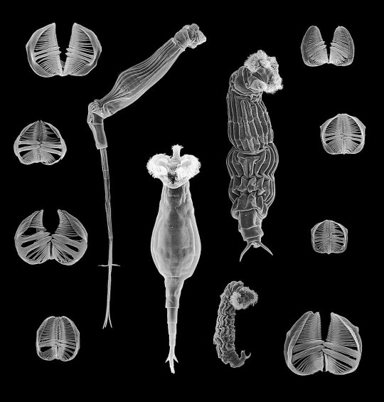
ID: An electron micrograph of some Bdelloid Rotifers and their mouthparts. They are long and slender, with a distinct mouth and tail section. Their mouthparts look like two semicircles lined with a comb-like structure.
These microscopic animals look freaky, because they are. If you've got any media literacy you've probably picked up by now that I am segueing here because they are somewhat similar to the aliens in the Three Body Problem, except this time they are very much real. Like the Trisolarans, Bdelloids live in very ephemeral environments: their usual haunts are the very thin film of water on moss and lichen. As you can imagine, these do not last all that long, and thus when they dry up, so do the Bdelloid Rotifers; in biology, we call this process anhydrobiosis.
"Ok, that's all well and good Ocean Sunfish Hater, but why do you like these guys more than the other anhydrobiotic creatures that roam our good, green Earth?" I hear you ask.
So you know how things that reproduce asexually don't have all that much genetic variation, and how sexual reproduction gives you an edge over asexual populations since you can keep that genetic variation fun and funky fresh, and how that has been the cornerstone for eukaryotic reproduction? Well. Well. Just like me, Bdelloid Rotifers have been completely celibate for 35-40 million years, with some people even bringing that number up to 100 million years, when they diverged from their sister clade. So how do these turbo-virgins not go extinct, racking up tonnes of deleterious mutations, not having any advantageous innovations, and eventually exploding into a genetic soup?
The secret lies in their ability to dehydrate. Not only is it a really handy dandy way to stay alive when your only source of water is gone, it literally rips apart their cells and genes! And why! Why the fuck does that help? It sounds like the opposite of helping!
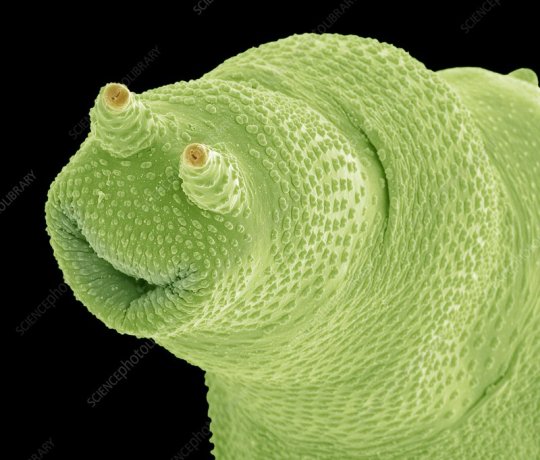
ID: An electron micrograph of the foot of a Bdelloid Rotifer. It has been shaded a light green. The structure looks almost like a face, with a smile and two stalk-like structures that could be mistaken for eyes. But this is not a face.
Having this mild-to-moderate level of cell membrane and chromosomal damage enables the Bdelloids to take up genetic material from their environment, mostly via their digestive systems, where their last meals are slowly being broken down to reveal that juicy DNA inside. When the water returns and the Bdelloids rehydrate, this genetic material gets incorporated into their chromosomes as their cells get back to work repairing themselves. And they sure ain't picky. In fact, it has been shown that in some species of Bdelloids, up to 8% of their genetic material has non-animal origins. How cool is that?
This is probably what has allowed them to continue adapting and evolving, even when they have been reproducing asexually for so long. This strategy has been so successful that the Bdelloids have managed to diversify into over 450 species. Pretty impressive for a class of animals that haven't had sex in over 40 million years.
Perhaps the Trisolarans might have a similar mechanism as part of their biology (even if they do reproduce sexually as stated in the book). Maybe they've managed to survive for this long because they have been able to absorb useful genes from their home planet, just like Bdelloids have been doing here on Earth. I don't know if these are what Liu Ci Xin had in mind when he wrote the Three Body Problem, but they sure were what I was thinking of when I read the book.
If you're still here, thanks for reading! I know this was a bit of a longer post, but I just wanted to use the new Netflix show to talk about one of my favourite books and one of the weirdest, most underappreciated animals.
#I would like trisolaris because the ocean sunfish would not survive there#bdelloid rotifers are so fucking cool and i think more people need to know about them#biology#ecology#Trisolaris#three body problem
106 notes
·
View notes
Text


Electron Micrographs
(Part 2 of 4)
Fugitive Glue, Lollipop, and Strawberry DNA
Fugitive Glue


I received a junk mail credit card offer, and I decided to take the piece of rubbery stuff that held the fake card to the paper to look at under the microscope. I originally referred to it as rubber cement, but I think it's actually called fugitive glue, among other names. Even exposed to air for a few days, it was still rubbery and malleable.
Unfortunately, being an organic compound, it did start to melt from the current of electricity that the microscope runs through it, which you can see happening in these images.
Lollipop Fragment


We had a bunch of leftover lollipops from Halloween at my house, so I took one of the flavors my brother didn't like to smash up and look at under the microscope.
In the first image, don't get confused by the background. The big angular piece is the lollipop, and the round things in the back are from the special tape used for keeping things on the stage (probably some bubbles trapped under it). I forget what kind of tape he said it was, but it was something able to conduct electricity.
The images of the sugar crystals are unclear and blurry because they, too, were actively melting while we were looking at them. Organic compounds just tend to do that. That said, you can still see the shapes of some of the crystals that hadn't yet melted.
Strawberry DNA


Having extracted some of my own DNA before, which I keep in an Eppendorf tube on my desk, I thought maybe it would be interesting to look at under the microscope.
However, I didn't want to potentially ruin the sample I had, so I followed NileRed's video to refresh myself on the process and used strawberries like he did. I also did it at 1/4 scale of what he did, because I really didn't need as much as he got. (By the way, DNA feels disgusting to touch. So much so that I used tweezers and forceps to handle it instead.)
Unfortunately, as you can see, the images aren't super clear or detailed, possibly for a few reasons.
The sample may still have had some moisture in it, which does interfere with how the machine sees it, I think.
It was also very clumped, because when you extract DNA, it all clumps together and I couldn't really separate the piece I was using into a thin layer.
It also may just be the size of DNA in general, which is molecular (just a very long one). I was hoping we'd see at least something, but I guess the indications of lines is all we could get.
I'm keeping the rest of the DNA we didn't use in some more Eppendorf tubes on my desk, because why not.
In the BDS Full images, the white parts are the hotter parts, where more electrons are being conducted through.
The scale in the bottom left corner of the images is in micrometers. 1 millimeter = 1000 micrometers
#microbes in hats#microbe posting#non-microbe post#electron microscopy#scanning electron microscopy#long post
20 notes
·
View notes
Text
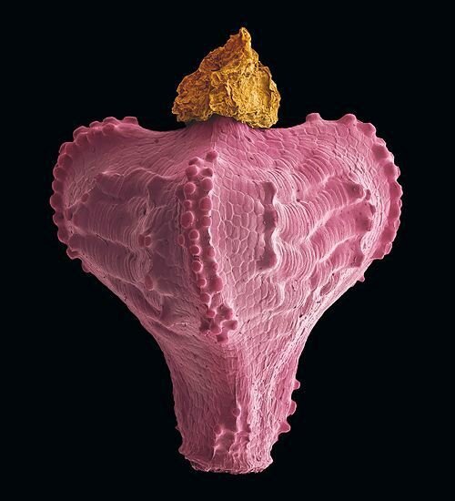
Lily Orchard recently started using the term tardigrade as an insult because she would really like to call her detractors a different word and tardigrade sort of sounds similar, I guess. This has inspired me put together a little educational aid:

And just in case you need a little more help spotting the difference, here's a higher res image:

The original tardigrade scanning electron micrograph is from Steve Gschmeissner/Science Photo Library
#lily orchard#lily orchard critical#lily posting#Lily's not cool enough to be a tardigrade#stockholm#retardigrade
36 notes
·
View notes
Text

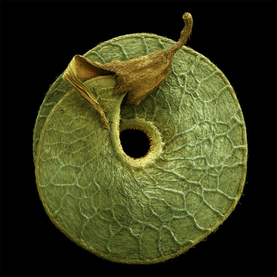
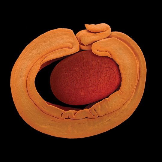
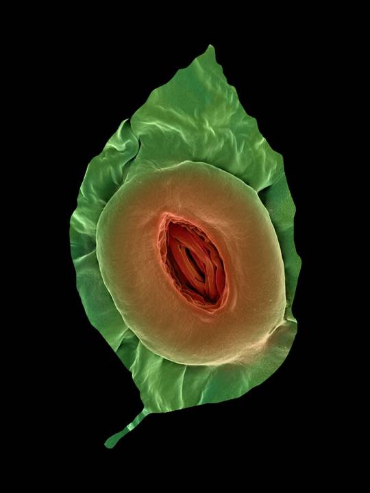
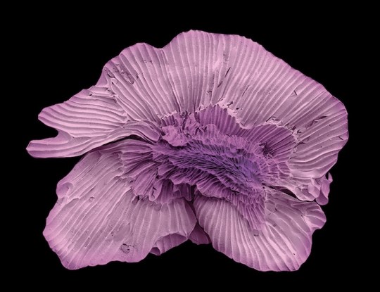
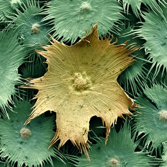
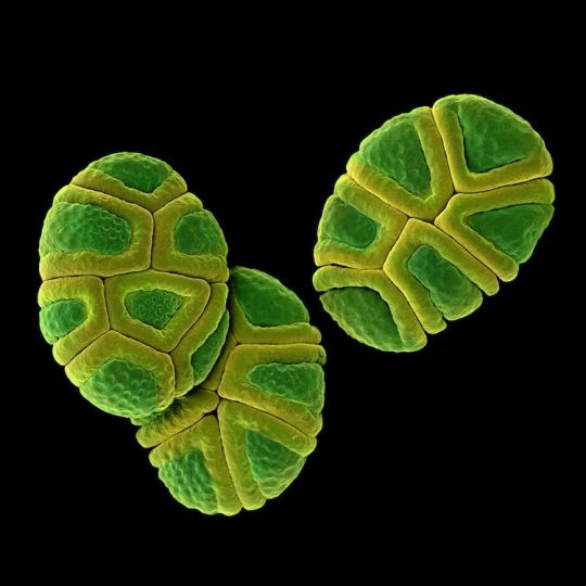
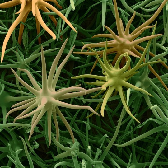
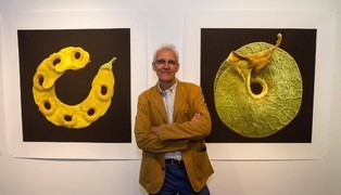
Rob Kesseler is a visual artist and Emeritus Professor of Arts, Design & Science at Central Saint Martins, London. For the past twenty years he has worked with botanical scientists and molecular biologists around the world to explore the living world at a microscopic level. Using a range of complex microscopy processes he creates multi-frame composite images of plant organs.
Using scanning electron microscopy and a mix of microscopic, scientific, digital, and manual processes, artist Rob Kesseler develops coloured micrographs of the intricate patterns within pollen and seed grains, plant cells, and leaf structures. The photographs feature specifics of cellular composition that are undetectable without magnification.
Kesseler tells that as a child, his father gifted him a microscope, marking a pivotal moment in his creative career. “What the microscope gave me was an unprecedented view of nature, a second vision,” he writes, “and awareness that there existed another world of forms, colours and patterns beyond what I could normally see.” The artist says his use of color is inspired by the time he spends researching and observing, and that just like nature, he employs it to attract attention.
36 notes
·
View notes
Text

Gut check: You hear it all the time: Keep your gut healthy! Why? Because it can impact your digestion, immune system, and even your mood. But what exactly is the gut microbiome—and how can you keep it healthy? We let you know. (Above, a bacteria in the gut dividing into daughter cells.)
MICROGRAPH BY MARTIN OEGGERLI, NAT GEO IMAGE COLLECTION
#martin oeggerli#photographer#national geographic#digestion#immune system#gut microbiome#micrography#micro photography#nature#bacteria#daughter cells
11 notes
·
View notes
Text
youtube
The confocal microscope at Imperial College's Sir Alexander Fleming Building lab is used for imaging the interior of living plant and animal cells.
During my PhD project, I used the confocal microscope to view the interior of Nicotiana benthamiana plant cells which were expressing Green Fluorescent Protein (GFP) tagged genes of interest. I aimed to find out where the proteins encoded by the genes of interest were localised in the plant cell, which turned out to be in the cytoplasm.
From Wikipedia's entry on Confocal Microscopy: "Confocal microscopy, most frequently confocal laser scanning microscopy (CLSM) or laser scanning confocal microscopy (LSCM), is an optical imaging technique for increasing optical resolution and contrast of a micrograph by means of using a spatial pinhole to block out-of-focus light in image formation. Capturing multiple two-dimensional images at different depths in a sample enables the reconstruction of three-dimensional structures (a process known as optical sectioning) within an object. This technique is used extensively in the scientific and industrial communities and typical applications are in life sciences, semiconductor inspection and materials science. Light travels through the sample under a conventional microscope as far into the specimen as it can penetrate, while a confocal microscope only focuses a smaller beam of light at one narrow depth level at a time. The CLSM achieves a controlled and highly limited depth of field."
Music by the Fiechter Brothers
Images by Katia Hougaard & the Facility for Imaging by Light Microscopy at Imperial College London
#katia plant scientist#botany#plant biology#plants#plant science#biology#science#science and technology#confocal microscopy#microscopy#microscope#scientific instruments#laboratory#research#phdblr#phd#phd life#molecular biology#cell biology#green fluorescent protein#plant scientist#Youtube
4 notes
·
View notes
Text
This volume, the third in a series describing the history, characteristics, and scientific analysis of artists’ pigments, covers 10 pigments, from some of the earliest man-made colorants, such as Egyptian blue and gamboge, to the 20th century’s titanium dioxide whites. Also included are orpiment and realgar; indigo and woad; madder and alizarin; Vandyke brown; Prussian blue; emerald green and Scheele’s green; and chromium oxide greens. More than 200 images depict artworks created with the pigments described, as well as x-ray diffraction patterns, scanning electron micrographs, and thin-layer chromatographs. The chapters provide details on nomenclature and general composition; history of use; color, permanence, compatibility, and painting and handling qualities; composition; characterization and identification through technical methods and procedures; and notable instances of use by a particular artist or in a specific work.
now i will have actual samples of scheele's green and paris green to colour match at the paint store!
3 notes
·
View notes
Text
Pathogen assigned: Vittaforma corneae
Cause of keratitis, type of microsporidian fungus

(Image from: https://www.researchgate.net/figure/Transmission-electron-micrograph-of-a-human-lung-fibroblast-cell-depicting-a_fig1_15363825)
Spread via: Traumatic insertian into stroma


Rodent marriage insult! Single power!
18K notes
·
View notes
Text

Microbug
Midjourney prompt: <https://s.mj.run/ixe3-38G-xI> <https://s.mj.run/_iVlS8QLF48> <https://s.mj.run/mmpIrW3eX1c> a full color electron micrograph of a transculect lady microbe monster --chaos 25 --weird 25 --s 0 --style raw
#micrograph#midjourney#AI#AI art#AI art generation#AI artist#AI artwork#AI generated#AI image#computer art#computer generated
0 notes
Photo

🌿 Etudes sur la fronde des Zygoptéridées. Lille, Impr. L. Danel, 1909. Original source Image description: Historical illustration titled "Etudes sur la fronde des Zygoptéridées" showing seven detailed botanical micrographs of frond cross-sections. Each image highlights cellular structures, vascular bundles, and tissue organization, labeled with red alphanumeric notations. The samples include varieties of frond rachises from species such as A. B. VAR. WESTPHALIENSIS, A. GRAYI, ASTEROCHLÆNA, and CALAMOPITYS. The layout displays diverse internal anatomy of fronds, emphasizing vascular tissue and cellular arrangement under magnification. The images are black and white, printed with fine texture and clarity to support botanical study from 1909.
11 notes
·
View notes
Text

An unexpected mineral in a Ryugu grain
Serendipitous discovery of djerfisherite in Ryugu grain challenges current paradigm of the nature of primitive asteroids.
The pristine samples from asteroid Ryugu returned by the Hayabusa2 mission on December 6, 2020, have been vital to improving our understanding of primitive asteroids and the formation of the Solar System. The C-type asteroid Ryugu is composed of rocks similar to meteorites called CI chondrites, which contain relatively high amounts of carbon, and have undergone extensive aqueous alteration in their past.
A research team at Hiroshima University discovered the presence of the mineral djerfisherite, a potassium-containing iron-nickel sulfide, in a Ryugu grain. The presence of this mineral is wholly unexpected, as djerfisherite does not form under the conditions Ryugu is believed to have been exposed to over its existence. The findings were published on May 28, 2025, in the journal Meteoritics & Planetary Science.
“Djerfisherite is a mineral that typically forms in very reduced environments, like those found in enstatite chondrites, and has never been reported in CI chondrites or other Ryugu grains,” says first and corresponding author Masaaki Miyahara, associate professor at the Graduate School of Advanced Science and Engineering, Hiroshima University. “Its occurrence is like finding a tropical seed in Arctic ice—indicating either an unexpected local environment or long-distance transport in the early solar system.”
Miyahara’s team had been carrying out experiments to understand the effects of terrestrial weathering on Ryugu grains. While observing the grains by field-emission transmission electron microscopy (FE-TEM) for effects of weathering, they found djerfisherite in the number 15 grain of sample plate C0105-042.
“The discovery of djerfisherite in a Ryugu grain suggests that materials with very different formation histories may have mixed early in the solar system’s evolution, or that Ryugu experienced localized, chemically heterogeneous conditions not previously recognized. This finding challenges the notion that Ryugu is compositionally uniform and opens new questions about the complexity of primitive asteroids,” Miyahara elaborates.
Ryugu is a part of a larger parent body that formed between 1.8 to 2.9 million years after the beginning of the Solar System. This parent body is thought to have originated in the outer region of the solar system, where water and carbon dioxide existed in the form of ice. Inside the parent body, heat generated by the decay of radioactive elements caused the ice to melt around 3 million years after its formation. The temperature during this process is estimated to have remained below approximately 50℃.
In contrast, the parent bodies of enstatite chondrites, which are known to contain djerfisherite, are believed to have formed in the inner region of the solar system. Thermodynamic calculations indicate that djerfisherite in enstatite chondrites formed directly from high-temperature gas. In addition, hydrothermal synthesis experiments have shown that djerfisherite can also form through reactions between potassium-bearing fluids and Fe-Ni sulfides at temperatures above 350 ℃.
This led the team to propose two hypotheses for its presence in the Ryugu grain: either it arrived from another source during the formation of Ryugu’s parent body; or, it was formed intrinsically when the temperature of Ryugu was raised to above 350 ℃.
Preliminary evidence indicates that the intrinsic formation hypothesis is more likely to be true. The next steps will be to conduct isotopic studies of this and other Ryugu grains, to determine their origins. “Ultimately, our goal is to reconstruct the early mixing processes and thermal histories that shaped small bodies like Ryugu, thereby improving our understanding of planetary formation and material transport in the early solar system,” Miyahara concludes.
TOP IMAGE: Scanning electron micrograph of the number 15 grain of sample plate C0105-042 from Ryugu, in which djerfisherite was discovered. (Hiroshima University/Masaaki Miyahara). Credit Hiroshima University/Masaaki Miyahara
LOWER IMAGE: Bright-field transmission electron micrograph of the djerfisherite inclusion in the number 15 grain of sample plate C0105-042 from Ryugu. (Hiroshima University/Masaaki Miyahara). Credit (Hiroshima University/Masaaki Miyahara)

1 note
·
View note
Text

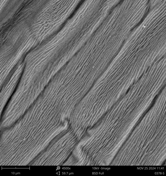
Electron Micrographs
(Part 1 of 4)
Flower Petal & Stamen
The images in this post are all of a flower petal, except the last one, which is the stalk of a stamen. (I don't know what kind of flower it was. Sorry to all the botanists out there.)

Here on we can see the plant cells on the petal, with some wrinkles along them. The wrinkling may be a normal part of the petal, or it could be due to the vacuum that the microscope pulls (which would therefore be pulling moisture out of the cells and causing them to wrinkle).


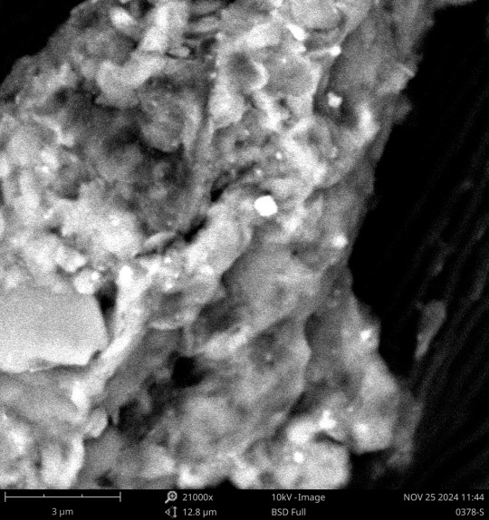
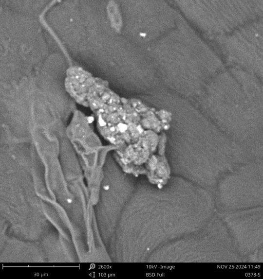
We also kept finding these little granules all over the petal, though we couldn't figure out what they were. Referencing images of pollen under SEM, it doesn't seem to match up.
I messaged my former biology professor about these images, and she suggested that one possibility is that they could be the conidia (reproductive spores) of a plant fungus called Alternaria alternata. She noted it looks similar and is approximately the right size.

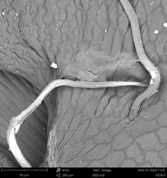


We also found this odd part with little stalks sticking out of it. There also seems to be some sort of layer on top, which got a bit torn. I messaged my biology professor about this as well and she suggested that they look like plant hairs, which are similar to root hairs.

This part here is the stalk of a stamen. He (the lab manager) was hoping to get the pollen part but it seems he accidentally messed it up when trying to place it on the stage and it got destroyed/removed. Oh well.
The scale in the bottom left corner of the images is in micrometers. 1 millimeter = 1000 micrometers
#microbes in hats#microbe posting#non-microbe post#electron microscopy#scanning electron microscopy#long post
25 notes
·
View notes