anatomyhelp-blog
Anatomy
Outlines and help for things to do about anatomy.
4 posts
Don't wanna be here? Send us removal request.
Last Seen Blogs

irrlicht-writes
irrlicht-writes

vobat
vobat

ownsdoggies
ARMY OF ME.

pinkyshiv
newgenroys

realcozy
THE COZIEST
Text
Sarcoidosis
Sarcoidosis is a disease in which inflammation occurs in the lymph nodes, lungs, liver, eyes, skin or other tissues.
I. Causes, Incidence and Risk Factors
Generally the cause of the disease is unknown. In sarcoidosis, tiny clumps of abnormal tissue (granulomas) form in certain organs of the body. Granulomas are clusters of immune cells.
The disease can affect almost any organ of the body but it most commonly affects the lungs.
Possible Causes:
extreme immune response to infection
high sensitivity to environmental factors
genetic factors.
The condition is more common in African Americans than Caucasions, especially in Caucasions of Scandinavian heritage. Females are usually affected more often than males.
Disease typically begins between ages 20 and 40. It's very rare in young children.
A person with a close blood relative who has sarcoidosis is nearly five times as likely to develop the condition.
II. Symptoms
There may be no symptoms. When symptoms do occur, they can involve almost any body part or organ system in your body. Almost all patients have lung or chest symptoms:
Chest pain (most often behind the breast bone)
dry cough
shortness of breath.
Symptoms of general discomfort or uneasiness often occur:
fatigue (most common symptoms of chidlren)
fever
joint achiness or pain (arthralgia)
overall feeling of discomfort, illness or lack of well-being
weight loss (common symptom in children)
2. Skin Symptoms
hair loss
raised, red, firm skin sores (erythema nodosum) almost always on the front part of the lower legs
rash
scars that have become raised or inflamed
3. Nervous System Symptoms may include:
headache
seizures
weakness on one side of the face
4. Eye Symptoms:
burning
discharge from the eye
dry eyes
itching
pain
vision loss
5. Other Symptoms
Dry mouth
fainting spells if the heart is involved
nosebleeds
swelling in the upper part of the abdomen
III. Signs and Tests
A physical exam may show the following:
abnormal breath sounds (such as rales)
enlarged liver
enlarged lymph glands
enlarged spleen
rash
Often, the disease is found in patients with visible physical signs who have an abnormal chest x-ray.
Different imaging tests may help diagnose sarcoidosis:
chest x-ray: check to see if the lungs are involved or lymph nodes are enlarged.
CT scan of the chest
Lung gallium scan
To diagnose this condition, a biopsy is required. Biopsy of the lung using bronchoscopy is usually done. Biopsies of other body tissues may be needed.
The disease may affect the results of the following lab tests:
Calcium levels (urine, ionized, serum)
CBC
immunoelectrophoresis - serum
liver function tests
quantitative immunoglobulin (nephelometry)
serum phosphorus
IV. Treatment
Sarcoidosis symptoms will often get better on their own slowly without treatment.
Patients whose eyes, heart, nervous system or lungs are involved may need to be treated with corticosteroids (prednisone or methylprednisolone). Therapy may continue for 1 to 2 years. The mostseverely affected patients may need lifelong therapy.
Drugs that suppress the immune system (immunosuppressive medicines) are sometimes also needed:
Drug used most often is methotrexate, but azathioprine and cyclophosphamide are also sometimes recommended.
Hydroxychloroquine is useful for skin sarcoidosis.
Rarely, some people with end stage heart or lung damage may need an organ transplant.
V. Expectations (prognosis)
Many people with sarcoidosis are not seriously ill, and the disease may get better without treatment. About 30-50% of cases get better without treatment in 3 years. About 20% of people whose lungs are involved will develop lung damage.
The overall death rate from sarcoidosis is less than 5%. Causes of death include:
bleeding from lung tissue
heart damage, leading to heart failure and abnormal heart rhythms
lung scarring (pulmonary fibrosis)
VI. Complications
fungal lung infections (aspergilloma)
Glaucoma and blindness from uveitis (rare)
Kidney stones from high calcium levels in blood or urine
Osteoporosis and other complications of taking corticosteroids for long periods of time
Pulmonary hypertension
VII. Calling a Health Care Provider
Notify your health care provider if:
difficulty breathing
palpitations
vision changes
other symptoms of this disorder
22 notes
·
View notes
Text
The Cellular Level of Organization
I. Parts of a Cell
plasma membrane: cell's flexible outer surface, separating the cell's internal environment from external. regulates flow of material in and out with selectivity.
cytoplasm: all cellular contents between membrane and the nucleus. two compartments:
- cytosol: fluid portion containing H2O, dissolved solutes and suspended particles.
- organelles: little organs; each has a characteristic shape/function. ie cytoskeleton, Golgi complex, lysosomes.
II. The Plasma Membrane
flexible yet sturdy barrier that surrounds and contains the cytoplasm of the cell; fluid mosaic model
A. The Lipid Bilayer
Two back-to-back layers made up of phospholipids, cholesterol and glycolipids
Phospholipids: 75% of the bilayer. Contain phosphate groups.
cholesterol: 20% of the bilayer. Steroid with a -OH group.
glycolipids: 5% of the bilayer. lipids with attached carbohydrate groups.
amphipathic: ie lipids. Have polar and nonpolar parts. In phospholipids, head is polar (hydrophilic), tail is nonpolar (-phobic). orient themselves so heads face interstitial liquid and cytosol.
cholesterol is weakly amphipathic. -OH group forms H bonds with the polar parts of phosphilipids and glycolipids.
glycolipids appear in only the layer facing the extracellular fluid.
B. Arrangement of Membrane Proteins
Integral Proteins: extend into or through the lipid bilayer among fatty acid tails and firmly embedded in it
transmembrane proteins: span entire lipid bilayer and protrude in both cytosol and extracellular; attached by covalent bonds
amphipathic
2. peripheral proteins: associate with polar heads of membrane lipids or with integral proteins at inner or outer surface of the membrane
3. glycoproteins: proteins with carbohydrate groups attached to ends protruding into the extracellular fluid.
oligosaccharides: chains of 2-60 monosaccharides; straight or branched
glycocalyx: carbohydrate parts of ylcolipids and glycoproteins form an extensive sugary coat. molecular signature that lets cells recognize each other.
C. Functions of Membrane Proteins
ion channels: formed by integral membrane proteins. holes which ions ie K+ can flow in and out of a cell. most channels are selective.
transporters: selectively moving polar subsgtance or ion.
receptors: cellular recognition sites.
ligands: specific molecule that binds to a receptor.
4. enzymes: catalyze specific chemical reactions at inside or outside of cell
5. linkers: anchor proteins in membranes of other cells or to protein filaments. Peripheral proteins can also do this.
6. Cell-identity markers: glycoproteins and glycolipids. enable cell to recognize other cells; ie blood transfusion.
7. Membrane Fluidity
dependent on the number of double bonds in fatty acid tails and amount of cholesterol present.
allows interactions to occur within membrane.
allows for easy fix of tears and punctures of the membrane.
seldom flip flop because of hydrophilic parts adversity to hydrophobic area.
at normal body temperature, lipid bilayer is less fluid because of cholesterol. the opposite is true at lower temperature.
8. Membrane Permeability
selective permeability: membranes allow some substances more readily than others.
permeable to nonpolar, uncharged molecules ie oxygen, carbon dioxide, steroids, slightly permeable to water and urea. Impermeable to large uncharged polar molecules and ions.
channels and transporters increase permeability to ions but are very selective.
9. Gradients Across the Plasma Membrane
difference in concentration of chemical from one place to another; ie concentration of oxygen and sodium in extracellular fluid; cytosol -> potassium ions and carbon dioxide
electrical gradient: difference in charges between two regions
- membrane potential
substance moves down its concentration gradient -> equilibrium
electrochemical gradient: combined influence of concentration gradient and membrane potential on movement.
D. Transport Across the PLasma Membrane
passive transport: substance moves down concentration/electric gradient using only its kinetic energy. No input of energy from the cell.
active transport: cellular energy drives substance uphill against gradient. uses ATP.
Kinetic energy intrinsic to moving particles ie diffusion, osmosis.
Kinetic Energy Transport
diffusion: passive process in which random mixing of particles occur because of their kinetic energy. moves down gradient.
factors influencing diffusion rate:
- steepness of concentration gradient: greater difference in concentration increases rate of diffusion especially with charged particles.
- temperature: high temperature = high rate of diffusion
- mass of diffusing substance: high mass = lower rate of diffusion
- surface area: high surface area available for diffusion = high rate of diffusion
- diffusion distance: high distance decreases rate of diffusion
Diffusion Through the Lipid Bilayer
- nonpolar hydrophobic molecules diffuse freely ie O2, CO2, N2, fatty acids, steroids, fat soluble vitamins, small alcohols and ammonia, sometimes H2O and urea.
Diffusion Through Membrane Ion Channels
- integral transmembrane proteins allowing passage of small inorganic ions too hydrophilic to get through.
- generally slower because less surface area.
- different permeabilities
osmosis: net movement of solvent through selectively permeable membrane, passive.
- area from high concentration to low concentration
- aquaporin: integral membrane proteins acting as water channels
- occurs only when membrane permeable to water but not certain solutes
- hydrostatic pressure: pressure exerted by liquid. The higher the concentration is, it forces water back into the left side. Equilibrium when molecules move at equal rates.
- osmotic pressure: proportional to concentration of solute particles that can't cross the membrane. Increase the concentration of the solute -> increase of osmotic pressure. The amount of pressure needed to prevent water movement.
tonicity: measure of solution's ability to change volumes of cells by altering water content.
- isotonic solution: any solution where cell maintains normal shape and volume. concentration of solute that can't cross plasma membrane are equal.
* normal saline solution: red blood cells maintain shape
- hypotonic solution: solution with lower concentration of solute than red blood cell cytosol. Water molecules -> red blood cells -> swell and burst
* hemolysis: rupture of red blood cells
* lysis
* cells in distilled H2O.
- hypertonic solution: solution with greater concentration of solute than cytosol, ie 2% NaCl brackish water.
* crenation: H2O leaves cell causing shrinkage.
2. Medical Uses of Isotonic, Hypertonic, Hypotonic
intravenous (IV) solutions: liquids infused into blood; isotonic
relieves fluid overload by causing osmosis of water
hypotonic solutions: move from blood to interstitial fluid to rehydrate them
3. Transport by Transporter Proteins
Facilitated Diffusion
- a solute binds to a specific transporter on one side of the membrane and released after transporter undergoes a change in shape.
* too polar or highly charged to diffuse.
- passive; down a concentration gradient.
- rate determined by steepness of concentration gradient across membrane
- transport maximum: the upper limit on rate at which faciliated diffusion occurs; once all transporters occupied, maximum is reached.
- exhibits saturation
- selective permeability regulated to achieve homeostasis
* ie insulin raises transport max for facilitated diffusion; inability to use insulin is called diabetes mellitus.
Active Transport
- particles that must enter/leave cells go against concentration gradient
- energy required for transport proteins to move solutes across membrane across concentration gradient.
* energy from hydrolysis of ATP; primary active transport
* energy stored in ionic concentration gradient; secondary active transport
- transport maximum and saturation
- Na+, K+, Ca2+, I-, Cl-, amino acids, monosaccharides
Primary Active Transport
- ATP changes shape of transporter protein; pumps substance across membrane against concentration gradient
- pumps: transporter proteins that carry out primary active transport
- 40% of ATP used on primary active transport
- Na-K pump: expels Na+ and pumps K+ in. Another name is Na+/K+ ATPase.
* maintain low concentration of Na+ by pumping against gradient. Pumps K+ into cytosol.
- Three Na+ in the cytosol bind to the pump protein
* binding of Na+ triggers hydrolysis of ATP -> ADP. attaches a phosphate group to pump protein changing shape of the protein.
* Expels Na+, now new shape binds 2 K+ in EC fluid to protein.
* K+ binding releases phosphate group from protein causing another shape change in the pump protein.
* pump reverts back to original shape -> K+ in cytosol. repeats.
- Na+ improtant for tonicity in EC fluid and cytosol.
Secondary Active Transport
- Energy stored in a Na+ or H= concentration gradient drives other substances against their own gradients indirectly using energy from ATP
- Na+/K+ pump maintains step gradient of Na+ therefore has potential energy.
- symporters: move two substances in opposite directions across the membrane.
Digitalis Increases Ca2+ in Heart Muscle Cells
- slows Na+/K+ pump allowing Na+ to accumulate
- More Ca+ stays inside the heart and strengthens heartbeat
4. Transport in Vesicles
vesicle: small spherical sacs that import and move materials into EC fluid.
endocytosis: materials move into a cell in a vesicle formed from plasma membrane.
exocytosis: materials move out by fusion with the plasma membrane. both endocytosis and exocytosis are active processes.
Endocytosis:
- receptor-mediated endocytosis: highly selective type of endoccytosis by which cells take up specific ligands ie. LDL, antibodies, vitamins.
* binding: particle binds to protein receptor to form a complex. Then, a protein forms around it on the cytoplasmic side to fold the membrane inwards.
* vesicle formation: edges of membrane pinch off.
* uncoating: right after formation, vesicle loses its protein coat
* fusion with endosome. Particle separates from its receptors
* regraduation in lysosomes: lysosomes break it down.
- phagocytosis: form of endocytosis. Cells engulf large particles
- pseudopods: projection of cytosol to plasma membrane binding. Individual cases of undigested material but remain undisposed indefinitely in a vesicle. (residual body)
- pinocytes: tiny droplets of EC fluid taken up. All solutes dissolved. aka bulk phase endocytosis. movement of liquid.
Exocytosis: releases materials from the cell; all cells carry this out.
- secretory cells: liberate digestive enzymes; hormones, mucus
- nerve cells: release transmitters
- membrane enclosed vesicles (secretory) fuse with membrane and release into the EC fluid
Transcytosis: active; vesicles undergo endocytosis on one side of the cell and undergo exocytosis on the opposite side. Most common in endothelial cells, move between blood plasma and interstitial fluid.
E. Cytoplasm
cytosol: intracellular fluid that surrounds organelles. 65% of total volume. 75-90% composed of water.
lipid droplets and glycogen granules aggregate for storage.
site for chemical reactions; catalyze glycolysis.
2. organelles: specialized structures with specific functions.
Cytoskeleton: network of protein filaments extend through cytosol.
- microfilaments: thinnest; made of actin. Most prevalent at periphery. generate movement and mechanical support, cell division, locomotion. anchor the cytoskeleton.
* microvilli: cell extensions; tufts, nonmotile projections that increase surface area to facilitate absorption.
- intermediate filaments: second thinnest; exceptionally strong, found in areas subject to mechanical stress, stabilize position of organelles.
- microtubules: largest component, unbranched hollow tubes; tubulin. Begins in the centrosome. Helps determine cell shape.
Centrosome: consists of a pair of centrioles and pericentriolar material
- centrioles: composed of nine clusters of microtubules in a circular pattern.
- pericentriolar: surround centrioles. Have complexes made of tubulin. organizing centers for growth of mitotic spindle; critical for cell division.
- replicate so future cells can replicate.
Cilia and Flagella
- cilia: short projections extending from surface of the cell. anchored to a basal body; ie respiratory tract
- flagella: longer than cilia. Move an entire cell; ie sperm cell
ribosomes
- sites of protein synthesis
- ribosomal RNA: more than 50 proteins; destined for organelles
Endoplasmic Reticulum: network of membranes in the form of flattened sacs or tubules
- Rough ER: continuous with nuclear membrane and flattened sacs. contain many ribosomes therefore produces many proteins.
- Smooth ER: network of membrane tubules. no ribosomes but enzymes make it structurally and functionally more diverse.
* prolonged drug use alters smooth ER; creates a tolerance for drug.
Golgi Complex
- consists of 3 to 20 cisternae; small flattened membranous sacs
* entry/cis face: cisterna that faces the rough ER; convex
* exit/trans face: cisterna facing plasma membrane; concave
* medial cisternae: sacs between entry and exit faces
- transport vesicles: proteins synthesized on rough ER bud.
* moves toward entry face of Golgi complex
* several transport vesicles create entry face and release proteins
*proteins move from entry face into medial cisternae
* transfer vesicles move specific enzyme back toward cis face and move some towards trans face
* in trans face, products are further modified, sorted and packaged
- secretory vesicles: deliver proteins to plasma membrane; discharged into EC fluid
- membrane vesicles: deliver contents into plasma membrane. adds new segments.
Lysosomes
- membrane enclosed vesicles that form from Golgi complex
- pH of 5 in interior
- helps recycle worn out cell structure
- autophagy: organelle is digested; enclosed by ER membrane and binds into lysosome
- autolysis: pathological conditions; lysosomal enzymes destroy entire cell
- Tay-Sachs Disease: absence of single lysosomal enzyme called Hex A. usually breaks Gm2; excess accumulates and causes nerve cells to defect
Peroxisomes
- contain oxidases, enzymes that can oxidize organic substances
- abundant in liver; detox alcohol and other substances
- catalase decomposes byproduct H2O2.
Proteasomes
- cytosilic proteins require continuous destruction of unneeded, damaged or faulty proteins
- contain proteases: enzymes that cut proteins into small peptides
Mitochondria
- generate most ATP via respiration; powerhouses of the cell
- found in areas of the cell where O2 enters or high amounts of ATP
- outer and inner mitochondrial membrane: inner consists of cristae; series of folds
- matrix: enclosed by inner membrane
- enzymes located in cristae and create ATP
F. Nucleus
nucleus: spherical or oval shaped structure usually most promninent in a cell
nuclear envelope: separates nucleus from cytoplasm. lipid bilayers.
nuclear pores: openings extending through envelope; control movement between nucleus and cytoplasm.
nucleoli: spherical bodies that produce ribosomes. cluster of protein; DNA and RNA without membrane. Sites of ribosome production.
genes: hereditary units; control cellular structure and activities.
chromosomes: genes are arranged in these coils. Somatic cells have 46.
chromatin: complex of DNA proteins and RNA coiled tightly together.
genome: total genetic information carried in a cell or an organism.
nucleosome: a "head" on the "string" of chromatin in electron micrographs.
histones: a cone of 8 proteins that help to organize DNA folding.
Linker DNA: "string" between bonds holding nucleosomes together.
chromatin fiber: large-diameter that folds into large loops
chromatids: fibers replicate causing loops to condense thus forming chromatids.
G. Protein Synthesis
proteome: all of an organism's protein
gene expression: DNA is used as a template for synthesis of a specific protein
base triplet: sequence of three nucleotides in DNA
codon: complementary to base triplet; specifies a particular amino acid
genetic code: set of rules relating base triplet to its codon and corresponding amino acid
Transcription: occurs in the nucleus; genetic information serves as a template for copying into complementing codons.
messenger RNA (mRNA): directs synthesis of a protein
ribosomal RNA (rRNA): joins both ribosomal proteins to make ribosomes
transfer RNA (tRNA): binds to amino acid and holds it in place on a ribosome until incorporated into a protein during translation.
- opposite end has an anticodon. attaches to mRNA codon.
RNA polymerase: catalyzes transcription of DAN.
promoter: special nucleotide sequence located near the beginning of a gene.
terminator: transcription ends at this nucleotide sequence.
introns v. exons: introns do not code for proteins; exons do.
pre-mRNA: transcript including information from both introns and exons
small nuclear ribonoucleo proteins: remove introns from pre-mRNA . enzymes that cut out introns and splice together exons to create functional mRNA.
alternative splicing: process in which pre-mRNA transcribed from a gene and spliced so that it creates several mRNA
7. Translation: nucleotide sequence in mRNA specifies amino acid sequence of a protein; ribosomes carry out in cytoplasm.
mRNA binds to ribosomal subunit. tRNA (initiator) binds to start codon (AUG) where translation starts.
large ribosomal subunits attaches to smaller one; mRNA complex creating functional ribosome, fits into "P site".
anticodon of another tRNA with attached amino acid pairs with second mRNA at A site.
large ribosomal thing catalyzes formation of peptide bond between AUG which separates from tRNA at P site and amino acid at A site.
after peptide bond is formed, tRNA at the P site detaches from ribosome and shifts the mRNA strand by one codon. New sequences are then exposed to the A site; steps 3 to 5 occur repeatedly.
protein synthesis ends when ribosomes reach a stop codon causing it to detach from final tRNA.
polyribosome: ribosomes attached to the same mRNA
recombinants: organisms whose cell's DNA is altered to produce proteins not regularly produced.
H. Cell Division
process by which cells reproduce themselves. (somatic, reproductive)
somatic cell: any cell other than a germ cell, a gamete or any precursor of gamete.
mitosis: nuclear division
cytokinesis: cytoplasmic division
somatic division: replaces dead or injured cells and adds new ones.
reproductive: produces gametes via meiosis.
Somatic Cell Division
cell cycle: somatic cells duplicate contents and divide into two.
homologous chromosomes: two chromosomes that make up a pair.
sex chromosomes: X, Y
diploid cells: 2n; 2 sets of chromosomes.
Interphase:
- cell replicates DNA through phases G, S and G2.
- G1 phase: interval between mitotic phase and S phase. cell is metabolically active; replicates most organelles and cytosolic components but not DNA.
- G0: cells remaining in G1 phase for a very long time; ie brain cells.
- S phase: interval between G1 and G2; DNA replication occurs
- G2: interval between S and mitotic phase; enzymes and proteins are synthesized.
Mitotic Phase: distribution of two sets of chromosomes into two nuclei.
- prophase:
* centromeres hold chromatid pair together. contain protein complex; kinetochore.
- mitotic spindle: microtubules that attach to the kinetochore.
metaphase
* microtubules align centromeres at the exact center called metaphase plate
- anaphase: centromeres split, pushing chromatid pairs toward opposite ends.
- telophase: nuclear envelopes and nucleoli reappear; chromatin form; mitotic spindle disappears.
Cytokinesis
- division of cytoplasm and organelles
- cleavage furrow: contractile ring (actin microfilaments) divide cytoplasm.
2. Control of Cell Destiny
cyclin-dependent protein kinases: enzymes that can transfer a phosphate group from ATP to a protein to activate it. crucial in regulation of DNA replication.
cyclins: levels rise and fall during cell cycle. Turns cyclin dependent kinases on and off. Timing transitions from G2 to S to G2 phase.
apoptosis: an orderly genetically programmed cell death. cell shrinks by producing destructive enzymes; phagocytosis finishes up.
necrosis: pathological cell death resulting from tissue injury. stimulates an inflammatory response in immune system.
tumor-suppressor genes: produce proteins that normally inhibit cell division; ie p53 in chromosome 17.
3. Reproductive Cell Division
Meiosis: reproductive cell division creating haploid (n) gametes.
Meiosis I
- Prophase I: chromosomes shorten and thicken; nuclear envelope and nucleoli disappear. mitotic spindle form.
* synapsis: doesn't occur in mitosis. sister chromatids of each homologous chromosomes pair off.
* tetrad: resulting structure of four chromatids.
* crossing-over: exchange between parts of nonsister chromatids resulting in genetic recombination.
- metaphase I: tetrads line along metaphase plate.
- anaphase I: homologous pair separate. pair chromatids stay together.
- net result: each cell contains haploid because only one member of each pair present.
Meiosis II
- two haploid cells from Meiosis I divide to form 4 haploid gametes.
I. Cellular Diversity
200 different cell types.
may be round, oval, cuboidal, columnar, elongated, star shaped, cylindrical
variations in structure facilitate function.
III. Aging and Cells
aging: normal process accompanied by a progressive alteration of the body's homeostatic and adaptive responses. increases vulnerability to environment.
geriatrics: specialized branch of medicine dealing with medical problems and care of the elderly.
gerontology: scientific study of processes and problems associated with aging.
telomeres: specific DNA sequences found only at the tips of each chromosomes; protect from erosion. Division shortens telomeres.
Glucose forms irreversible cross links causing loss of elasticity.
Free radicals produce oxidatives
Progeria and Werner Syndrome
- Progeria: disease characterized by normal development in the first year of life followed by rapid aging.
- Werner Syndrome: rare, inherited disease causing rapid acceleration of aging while in 20s.
0 notes
Text
The Chemical Level of Organization
I. How Matter Is Organized
A. Chemical Elements
chemical elements: all forms of matter are made up of these number of building blocks that cannot be simpler substances by ordinary chemical means.
chemical symbol: one or two letters in English/Latin designating an element.
chemistry: science of structure and interactions of matter.
matter: anything that takes up space and has mass.
4. mass: amount of matter in any subject.
weight: force of gravity acting on matter.
5. 26 elements are present in the body but 4 major elements make up 96%
oxygen, nitrogen, carbon, hydrogen
6. Lesser Elements: Calcium, Phosphorus, Potassium, Sulfur, Sodium, Chlorine, Magnesium, Iron
B. Structure of Atoms
atoms: smallest units of matter that retain properties and characteristics of the elements.
subatomic particles: compose an atom; protons, neutrons, electrons
nucleus: dense central core of an atom
protons: positively charged subatomic particles in the nucleus
neutrons: uncharged subatomic particles in the nucleus
electrons: negatively charged cloud enveloping nucleus
- electron shells: the path of motion within certain regions around nucleus
* first shell: 2 electrons
* second shell: 8 electrons
* third shell: 18 electrons
C. Atomic Number and Mass Number
atomic number: the number of protons in the nucleus of an atom
mass number: the sum of its protons and neutrons
isotopes: atoms of an element that have different numbers of neutrons and therefore different mass numbers
radioactive isotopes: unstable; nuclei decay into a stable configuration by emitting subatomic particles
half-life: of an isotope is time required for half of the radioactive atoms in an isotope to decay into a more stable form
tracers: follow movement of certain substances through the body.
D. Atomic Mass
dalton: standard unit of mass of atoms and their subatomic particle; amu.
atomic mass: of an element, is the average of mass of all naturally occurring isotopes.
E. Ions, Molecules, and Compounds
ion: an atom that has a positive or negative charge because it has an unequal number of protons and electrons
cation: positively charged ion
anion: negatively charged ion
ionic compounds generally exist as solids with repeating arrangement of ions
electrolyte: an ionic compound that breaks apart into positive and negative ions in solution. These solutions can conduct an electric current.
B. Covalent Bonds
When two or more atoms share electrons rather than gaining or losing
single covalent: when two or more atoms share an electron pair
double covalent: when two or more atoms share two pairs of electrons.
triple covalent: when two or more atoms share three pairs of electrons
nonpolar covalent bond: when electrons are shared equally between two atoms.
polar covalent bond: sharing of electrons between two atoms is unequal- the nucleus of one atom attracts the shared atom more strongly.
electronegativity: the power to attract electrons to itself.
C. Hydrogen Bonds
hydrogen bond: forms when a H atom with a partial positive charge of neighboring electronegative atoms. Weaker than ionic and covalent bonds.
cohesion: tendency of like particles to stay together.
surface tension: created by cohesion of water molecules; measure of difficulty of stretching/breaking the surface of a liquid.
III. Chemical Reactions
occurs when new bonds form or old bonds break between atoms.
reactants: starting substances of a reaction
products: final substances of a reaction
mass of substances = mass of products
metabolism: all chemical reactions occurring in the body
A. Forms of Energy and Chemical Reactions
energy: capacity to do work
potential energy: energy stored by matter due to its position
kinetic energy: energy associated with matter in motion.
chemical energy: form of potential energy stored in bonds of compounds and molecules.
law of conservation of energy: energy can't be created or destroyed; only converted.
B. Energy Transfer in Chemical Reaitons
exergonic reactions: release more energy than absorbed
endergonic reactions: absorb more energy than released
Activation Energy: collision energy needed to brea kthe chemical bonds of the reactants.
concentration: more particles of matter present, therefore increases the chance of collision
temperature: as temperature increases, speed of particles increases, therefore more collisions occur.
C. Catalysts
chemical compounds that speed up chemical reactions by lowering the activation energy needed for a reaction to occur.
does not alter difference in potential energy between reactants and products.
lowers energy needed to start a reaction
orients collision paths.
D. Types of Chemical Reactions
Synthesis Reactions - Anabolism
synthesis reaction: when two or more atoms, ions, molecules combine to form new and larger molecules.
anabolism: all synthesis reactions occurring in the body.
endergonic because it uses energy.
2. Decomposition Reations - Catabolism
decomposition reaction: split up large molecules into smaller atoms, ions, molecules
catabolism: all decomposition reactions
exergonic: release more energy than absorbed
3. Exchange Reaction
exchange reation: consist of both synthesis and decomposition
HCl + NaHCO3 -> NaCl + H2CO3
4. Reversible REactions
reversible reaction: when the product can revert to original reactants
many require catalysts
IV. Inorganic Compounds and Solutions
inorganic: usually lack carbon; structurally simple. H2O, salts, H+, OH-, 55-60% of the body
organic: contain carbon and usually hydrogen. always covalently bonded. has a complex structure. 38-43% of the body.
A. Water
most important and abundant inorganic compound.
Water as a Solvent
solution: a medium in which substances dissolve in together.
solvent: substance that dissolves solutes. In solution, there is more solvent than solute.
solute: being dissolved by solvent; ie. salts in sweat.
hydrophilic: solutes charged or contain polar covalent bonds. dissolve easily in water.
hydrophobic: nonpolar covalent bonds. not water soluble. ie animal fats, vegetable oils.
water is ideal for metabolic reactions and washing out wastes.
2. Water in Chemical Reactions
water serves as medium for mos reactions, sometimes reactant or product.
hydroloysis: breakdown of larger molecules by addition of water.
dehydration synthesis: removal of water molecule to join together smaller molecules.
3. Thermal Properties Of Water
heat capacity: high because it can absorb and release large amounts of heat with little change in its own.
heat of vaporization: water requires a large amount of heat to change from liquid to gas.
4. Water As A Lubricant:
major component of mucus and lubricating fluids to reduce friction between muscles, organs, bones.
B. Solutions, Colloids, and Suspensions
mixture: combination of elements/compounds physically blended together but not chemical bonds.
colloid: differs from a solution because of the size of the particles
large enough to scatter light, transparent and opaque
ie. milk
3. suspension: suspended material will eventually settle out.
blood
4. concentration: of a solution may be shown as:
mass/volume percentage: relative solute mass in given volume of a solution
moles/L: total number of molecules in a given solution
- mole: amount of any substance with mass in grams equal to the sum of - atomic mass
- Avogadro's Number: 6.023 x 10^23
C. Inorganic Acids, Bases, and Salts
dissociate: when inorganic acids, bases, salts separate into ions and surrounded by water molecules.
acid: substance that dissociates into one or more H+
also a proton donor
3. base: removes H+ from a solution. Many dissociates into one or more OH-
proton acceptor
4. salt: when dissolved in H2O, dissociates into cations and anions
acids and bases react with one another to form salts
HCl + KOH -> H+ + Cl- + K+ + OH- -> KCl + H2O
does not give off H+ or OH-
D. Acid-Base Balance: Concept of pH
pH scale: measure of acidity/alkalinity of a solution. Scale of 0-14.
pH 7 = 1x10^-7 H+; pH 4 = 1x10^-4 H+
2. acidic solution: more H+ than OH- pH < 7
3. alkaline: less H+ than OH-. pH > 7
E. Maintaining pH: Buffer Systems.
Normal Body pH between 7.35 and 7.45
excess H+ < 7.35 -> acidosis
excess OH- > 7.45 -> alkalosis
2. buffer systems: function to convert strong acids or bases into weak acids
or bases
3. buffers: chemical compounds that convert strong acids/bases by - removing/adding H+
4. carbonic acid-bicarbonate buffer system
excess H+ -> H+ + HCO3 -> H2CO3
excess OH- -> H2CO3 -> H+ + HCO3
V. Organic Compounds
A. Carbon And Its Functional Groups
Carbon has four electrons in its valence shell; most commonly binds with nitrogen, oxygen or hydrogen
carbon skeleton: chain of carbon atoms in an organic molecules
hydrocarbon: carbons bonded with hydrogen
3. functional groups: atoms or molecules bound to a hydrocarbon skeleton
each confer a specific characteristic trait of that group
Hydroxyl: R-O-H; occurs in alcohols. dissolves easily in H2O because of -OH group
Sulfhydryl: R-S-H; in thiols, certain amino acids; helps stabilize the shape of a protein, ie cysteine
carbonyl: R-C=O-R or R-C=O-H; ketones contain a carbonyl group in the carbon skeleton. Polar and hydrophilic. Aldehydes contain carbonyl group at the end of the skeleton.
Carboxyl: R-C=O-OH or R-C=O-O, carboxylic acids contain this group at the end of the carbon skeleton. all amino acids have -COOH at one end.
Ester: R-C=O-O-R. predominate in dietary fats and oils and also occur in body triglycerides. Aspirin is ester of salicyclic acid.
Phosphate: R-O-P=O-O-O (PO4^2-) very hydrophilic because of the two negative charges. ie ATP
amino: R-N-H-H or R-N-H-H-H; amines have a -NH2 which can act as a base and pick up a H+ ion. Gives amino group a positive charge. Most amino groups have a +1 charge.
4. macromolecules: small organic molecules can combine into these large - molecules
5. polymer: large molecule formed by covalent bonding of many identical or - similar small building block molecules called monomers.
dehydration synthesis
6. isomers: molecules with the same molecular formula but different - structures.
B. Carbohydrates
include sugars, glycogen, starches and cellulose. 2-3% of total body mass. function as a source of energy to generate ATP, ie DNA.
Monosaccharides and Disaccharides
simple sugars
monosaccharides: monomers containing 3-7 carbon atoms. designated by suffix -ose. ie glucose, fructose, galactose, deoxyribose and ribose.
disaccharides: formed by dehydration synthesis of two monosaccharides, ie sucrose (glucose + fructose), lactose (galactose + glucose) and maltose (glucose + glucose).
artificial sweeteners: hydrolyzed disaccharides
2. Polysaccharides
contains tens to hundreds of joined monosacchardies. insoluble in water.
glycogen: entirely of linked glucose monomers. stored form of carbohydrates in animals; liver
starches: polysaccharides from glucose by plants. major carbohydrates in a diet.
cellulose: found in plants but can't be digested by humans. help in bowel movement.
C. Lipids
make up 18-25% of body mass.
Fewer covalent bonds because of proportion of oxygen is smaller in soluble in polar solvents (water); hydrophilic
lipoproteins: lipid molecules join with hydrophilic protein molecules soluble in water.
3. Triglycerides
most plentiful, aka triglycerols. solid or liquid at room temperature.
provide twice as much energy as carbohydrates; stored in adipose
glycerol: 3-carbon; forms backbone of the triglyceride molecule
fatty acids: attached by dehydration synthesis; one to each carbon in covalent chemical bond is called an ester linkage
saturated fats: single covalent bonds between fatty acid carbon atoms. saturated with H atoms are solid at room temperature.
monounsaturated fats: contain fatty acids with one double covalent bond between fatty carbon atoms, ie oleic acid. decrease heart risk.
polyunsaturated fats: contain more than one double covalent bond between fatty acid atoms, ie sunflower oil. Also decreases heart risk.
4. Fatty Acids in Health and Disease
essential fatty acids: fatty acids essential to human health not produced naturally in the human body
omega-3, fatty acids, omega-6 and as fatty acids
overall promotion of health
5. Phospholipids
glycerol backbone and two fatty acid chains attached to the first carbon
hydrophilic head and hyrophobic tail
amphipathic
6. Steroids
four rings of carbon
cholesterol: minor component in cell membranes
bile salts: digestion and absorption of dietary lipids
adrenocortical hormones: regulates metabolism, resistance to stress and salt and water balance.
sex hormones: stimulate reproductive functions and sexual traits.
7. Other Lipids
eicosanoids: lipids derived from a 20-carbon fatty acid called arachidonic acid.
- prostaglandins: modify responses to hormones ie. inflammatory - response to prevent ulcers, regulate body temperature and blood clots.
- leukotrienes: allergic and inflammatory responses
fatty acids: catabolized to generate ATP or synthesize triglycerides and phospholipids
carotenes: synthesis of vitamin A; make visual pigments in the eyes, antioxidants.
Vitamin E: promotes wound healing, prevents tissue scarring, contributes to normal structure and function of nervous system. antioxidant.
Vitamin K: synthesis of blood-clotting proteins.
lipoproteins: transport lipids in blood, carry triglycerides and removes excess cholesterol.
D. Proteins
macromolecules containing carbon, hydrogen, oxygen and nitrogen. some have sulfur.
Functions of Proteins
Structural: structural framework of various parts of the body, ie collagen
Regulatory: hormones regulate processes, control growth and development. function as neurotransmitters ie. insulin.
Contractile: allow shortening of muscle cells causing movement. ie myosin and actin.
Immunological: responses that protect body against foreign substances.
Transport: carries vital substances throughout the body
Catalytic: enzymes regulate biochemical reactions.
2. Amino Acids and Polypeptides
amino acids: monomers of proteins
- 3 functional groups: amino (-NH2), acidic carboxyl (-COOH) and side - chain (R)
peptide bond: covalent bond joining amino acids. formed by dehyration synthesis.
dipeptide: when two amino acids combine.
tripeptide: when another amino acid is added to dipeptide.
peptide: 4-9 amino acids or polypeptide (10-2000 amino acids)
3. Levels of Structural Organization in Proteins
primary structure: unique sequence of amino acids linked covalently to form polypeptide.
- sickle-cell disorder: nonpolar amino acid (valine) replaces polar amino - acid (glutamate) through two mutations in hemoglobin thus decreasing - solubility of hemoglobin.
secondary structure: repeated twisting or folding of neighboring amino acids in the polypeptide chain. Stabilized by hydrogen bonds; form at regular intervals.
- alpha helixes
- beta pleated sheets
tertiary structure: 3D shape of polypeptide chain. determines how protein will function. some parts are hydrophobic or hydrophilic.
- disulfide bridges: S-S; strongest but least common between cysteine
- chaperones: helper molecules aiding folding process
quaternary structure: arrangement of individual polypeptide chains relative to one another. Variation in structure and shape directly related to function.
denaturation: when proteins unravel in an altered environment ie high pH or high temperatures
3. Enzymes
enzymes: protein molecules are catalysts
- apoenzyme: protein portion of enzyme
- cofactor: nonprotein portion. may be derived from metal ion or - coenzyme aka vitamin.
highly specific
- each enzyme binds only to specific substrates; reactant.
- active site: part of the enzyme thought to 'fit' the substrate.
* change shape to fit substrate is induced fit
enzymes are very efficient. many reactions in a cell.
subject to variety
How it works:
- temporary intermediate compound when substrates make compound
- substrate molecules transformed by rearrangement of existing atoms
- breakdown of substrate molecule or combination -> products of a - reaction
- After reaction, enzyme is free to attach to other substrate molecules
- Sometimes a reaction can be reversible depending on quantity of - substrates and products.
4. DNA and RNA
nucleic acids: organic molecules contain carbon, hydrogen, oxygen and nitrogen.
DNA: forms inherited genetic material inside each human
- gene: segment of DNA molecule controlling protein synthesis - determining physical traits.
RNA: second nucleic acid relays information from genes to guide protein synthesis
nucleotides: monomers making up nucleic acids; three types:
- nitrogenous base: contain carbon, hydrogen, oxygen and nitrogen
- adenine and guanine: larger, double-ringed; purines
- cytosine and thymine: smaller, single-ringed bases; pyrimidines
pentose sugar: 5-carbon sugar called deoxyribose attached to each base in DNA
phosphate: (PO4^3-) alternate with pentose sugars to form DNA backbone
double helix model: Crick & Watson; ladder of alternating phosphate and pentose sugars.
- mutation: change in base sequence of DNA strand; can result in cell - death, cancer or genetic defects.
ribose: pentose sugar in RNA nucleotide, contains pyrimidine uracil instead of thymine. three different kinds: ribosomal, messenger and transfer.
5. DNA Fingerprinting
used in research and law to determine whether suspect's DNA matches sampled DNA.
6. Adenosine Triphosphate
ATP: "energy currency" of the living, transfers energy liberated in exergonic catabolic reactions to power cellular activities that require energy.
3 phosphate groups and 5 carbon sugar ribose attached to adenine
ADP: adenosine diphosphates, enzyme is ATPase. ATP synthase synthesizes to create ATP.
Anaerobic Respiration: doesn't need O2 to run. glucose is broken down to pyruvic acid. Yields 2 ATP
Aerobic: with O2. glucose broken down completely into CO2 and water. yields heat and 36 or 38 ATP.
0 notes
Text
An Introduction To The Human Body
I. Anatomy and Physiology Defined
Anatomy: science of body structures and relationship among them.
- dissection: careful cutting apart of body structures to study relationship.
Physiology: science of body functions; how the body works.
II. Levels of Structural Organization
A. Levels
Chemical Level
atoms: smallest unit of matter
molecules: 2 or more atoms joined together
Carbon, Hydrogen, Oxygen, Nitrogen, Phosphorus, Calcium, Sulfur; important elements of the body.
2. Cellular Level
cells: basis structural and functional units of an organism.
3. Tissue Level
tissues: groups of cells and materials surrounding them that work together to perform a particular function.
Epithelial, connective, muscular, nervous
4. Organ Level
organs: composed of 2 or more different types of tissues; specific functions and recognizable shape.
skin, bones, stomach, heart, liver, lungs, brain
5. System (Organ-System) Level
system: related organs with a common function
digestive system: mouth, salivary glands, pharynx, esophagus, stomach, etc.
6. Organismal Level
organism: any living individual. All parts of the human body functioning together.
B. Noninvasive Diagnostic Techniques
Inspection: the examiner observes the body for abnormal changes.
Palpation: feeling body surface with hands. To detect enlarged/tender internal organs or abnormal masses.
Auscultation: listening to body sounds to evaluate functioning of certain organs. Crackling sounds in the chest may signal abnormal fluid accumulation.
Percussion: tapping of body surface with fingertips and listening to echo.
III. Characteristics of Living Human Organism
A. Basic Life Processes
Metabolism: sum of all chemical processes in the body.
catabolism: breakdown of complex chemical substances into simpler components.
anabolism: building up of complex chemical substances from smaller, simpler components.
Food catabolized, proteins -> amino acids. Anabolizing proteins; amino acids -> new proteins.
2. Responsiveness: body's ability to detect and respond to changes.
3. Movement: motion of the whole body; individual organs, single cells
4. Growth: increase in body size resulting from increase in size of existing cells number of cells or both.
5. Differentiation: development of a cell from an unspecialized to a specialized state.
stem cells: precursor cells that can divide and make cells to differentiate into specialized cells.
6. Reproduction: formation of new cells for tissue growth repair or replacement or to the production of a new individual.
B. Homeostasis
condition of equilibrium in the body's internal environment due to ceaseless interplay of body's many regulatory processes.
C. Body Fluids
dilute, watery solutions containing dissolved chemicals found inside and surrounding cells.
- intracellular fluid (ICF): fluid within cells
- extracellular fluid (ECF): fluid outside body cells
* interstitial fluid: ECF that fills narrow spaces between cells of tissues
* blood plasma: ECF within blood vessels
* lymph: ECF within lymphatic vessels
* cerebrospinal fluid: in and around the brain and spinal cord
* synovial fluid: ECF in joints
* aqueous humor and vitreous body: ECF of the eyes.
interstitial fluid = internal environment
Exchanges between interstitial fluid and blood plasma removes waste and provides glucose and oxygen.
IV. Control of Homeostasis
Nerve Impulses: action potential or electronic signals sent to organs then correct deviations from the balanced state. [Nervous System]
Hormones: messenger molecules work slower than nerve impulses. Regulation. [Endocrine System]
A. Feedback Systems
cycle of events where the body, system, is constantly watched, fixed, evaluated and repeat.
controlled condition: a monitored variable; body temp, blood pressure
stimulus: disruption that changes a controlled condition
receptor: monitors changes in a controlled condition and sends input to a control center. THese inputs are in the form of chemical signals/nerve impulses.
Control Center
brain; sets a range of values the controlled condition should be, evaluates input and creates output in the form of commands
output is nerve impulse, hormones or chemical signals
3. Effector
Receives output and produces response to change controlled condition.
4. Negative Feedback Systems
reverses a change in a controlled condition
blood pressure:
- stimulus disrupts homeostasis
- Increases blood pressure
- baroreceptors send input to control center (the brain)
- the brain interprets the input and sends impulses to the heart (effector)
- The effector negates the stimulus by decreasing blood pressure
5. Positive Feedback
strengthen/reinforce change of a controlled condition
only difference from negative: effector adds/reinforce initial change
continues until interrupted by some mechanism
birth:
- contractions of wall of uterus force baby's head/body into cervix
- increase stretching of the cervix
- receptors send stretch-sensitive nerve cells in the cervix
- nerve impulses sent to the control center
- brain interprets and releases output (oxytocin)
- effectors: uterus muscles contract more forcefully
- baby's body stretches cervix more
- Interruption: birth decreases stretching cervix; breaks feedback cycle
6. Difference
positive systems reinforce conditions that do not occur often; negative system regulate conditions that remain stable over long periods.
B. Homeostatic Imbalances
disorder: any abnormality of structure or function
disease: illness characterized by recognizable set of signs and symptoms
local disease: affects 1 part or limited region of the body
systemic disease: affects entire body or several parts of it
symptoms: subjective changes in body functions not externally apparent
signs: objective changes that are observable; rash, swelling
3. Epidemiology: science that deals with why, when and where diseases occur and how they're passed between people in a community
4. Pharmacology: science that deals with effects abd use of drugs in treatment
5. Diagnosis of Disease
science and skill of distinguishing one disorder/disease from another
medical history: collecting information about events related to a patient's illness.
physical examination: orderly evaluation of the body and its functions.
V. Anatomical Terminology
A. Body Positions
anatomical position
when any region or part of the body assume a specific stance
body is upright
2. prone: body is lying face down
3. supine: body is lying face up
B. Regional Names
Head:
skull: encloses andprotects the brain
face: front portion of head including eyes, nose, mouth, forehead, cheeks and chin
2. neck: supports head and attaches it to the trunk
3. trunk: chest, abdomen and pelvis
4. upper limb: attaches to the trunk; shoulder, armpit, arm, forearm, wrist, hand
5. lower limb: attaches to trunk, buttock, thigh, leg, ankle, and foot.
* groin: area marked by a crease on each side, where trunk attaches to thighs.
C. Directional Terms
words that describe the position of one body part relative to another.
superior (cephalic or cranial): toward the head or upper part of a structure.
Inferior (caudal): away from head, lower part of structure.
Anterior (ventral): nearer to or at the front of the body.
Posterior (dorsal): nearer to or at the back of the body.
Medial: nearer to midline.
Lateral: farther from midline.
Intermediate: between two structures.
Ipsilateral: on the same side of the body as another structure.
Contralateral: opposite side of the body from another structure.
Proximal: nearer to the attachment of a limb to trunk; nearer to origination of structure.
Distal: farther from attachment of limb to trunk
Superficial: toward or on the surface of the body
Deep: Away from surface of the body
D. Planes and Sections
Planes: imaginary flat surfaces that pass through the body parts
sagittal plane: vertical plane that divides the body/organ into right/left sides
misagittal/median plane: when the body/organ is divided into equal right/left sides
parasagittal plane: when body/organ divided into unequal right/left sides
frontal/coronal plane: divides body into anterior (front) and posterior (back).
transverse plane: divides body into superior (upper) and inferior (lower)
- can be known as cross-sectional or horizontal plane
oblique plane: passes through body/organ at an angle between transverse and sagittal or frontal plane
section: 1 flat surface of a 30 structure or a cut along a plane
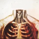
E. Body Cavities
Body Cavities: spaces within body that help protect, separate and support internal organs. Bones, muscles, ligaments and other structures also help.
cranial cavity: formed by cranial bones. Contains brain.
vertebral (spinal) cavity: formed by vertebral column. contains spinal cord.
- 3 layers of protective tissue line cranial and vertebral cavities called meninges.
thoracic cavity: chest, formed by ribs, chest muscles, sternum, and thoracic part of the vertebral column.
- pericardial cavity: fluid-filled space surrounding the heart
- pleural cavities: two located inside thoracic c. Each contain lung and a small amount of fluid
- mediastinum: central part of thoracic cavity between lungs; from sternum to vertebral column and from neck to the diaphragm. Contains all organs but the lungs.
* diaphragm: dome shaped muscle that separates thoracic cavity from - abdominopelvic cavity.
abdominopelvic cavity: extends from diaphragm to groin and encircled by abdominal wall and bones and pelvic muscles. divided into two portions.
- abdominal cavity: superior part containing stomach, spleen, liver, gall - bladder and intestine.
- pelvic cavity: inferior part, contains urinary bladder, parts of large - intestine and internal organs of the reproductive system.
- viscera: organs located inside thoracic and abdominopelvic cavity.
2. Thoracic and Abdominal Cavity Membranes
Serous Membrane: thin, slippery double layered membrane covering the viscera within thoracic and abdominal cavities and lines the walls of thorax and abdomen.
- parietal layer: lines walls of cavities.
- visceral layer: covers and adheres to the viscera within the cavities.
- serous fluid: between layers to reduce friction allowing viscera to slide - - around.
- pleura: serous membrane of pleural cavities
* visceral: clings to the surface of the lungs.
* parietal: anterior part. lines chest wall covering superior surface of - the diaphragm.
- pericardium: serous membrane of pericardial cavity.
* visceral: covers surface of the heart.
* parietal: lines chest wall. Between these layers is pericardial cavity.
- peritoneum: serous membrane of abdominal cavity.
* visceral: covers abdominal viscera.
* parietal: lines abdominal wall; covers inferior diaphragm. Between - parietal cavity.
* most abdominal organs located in peritoneal cavity.
- retroperitoneal: located between parietal peritoneum posterior - abdominal wall; ie. pancreas, kidneys, duodenum of small intestine.
- Other Cavities
* oral (tongue and teeth)
* nasal (nose)
* orbital (eyeballs)
* middle ear (small bones)
* synovial (freely movable joints with synovial fluid)
F. Abdominopelvic Regions and Quadrants
Abdominopelvic Regions: two horizontal and teo vvertical lines; tic-tac-toe pattern
subcostal line: top horizontal line; inferior to rib cage, across the inferior stomach
transtubercular line: bottom horizontal; inferior to tops of hip bones
left/right midclavicular lines: vertical lines. thorugh mid-points of clavicles, medical to nipples.
nine regions: [of the tic tac toe] right-hypochondriac, epigastric, left hypochondriac, right lumbar, umbilical, left lumbar, right invuinal (iliac) hypogastric (pubic), left inguinal (iliac)
quadrants: vertical and horizontal line passed through the umilicus (belly button)
- right upper, right lower, left upper, left lower
2. Uses
Regions used for anatomical studies
quadrants used for describing site of abdominopelvic pain, tumor abnormality
G. Medical Imaging
allow visualization of structures inside our body and helps for diagnosis of anatomical and physiological disorders.
Radiography
X rays pass through body producing image of interior structures.
Mammogram, bone desmiometry, angiography (blood vessels), urinary system (intravenous), gastrointestinal tract (barium contrast)
necessary to use contrast medium to make hollow/fluid filled structures visible
2. Magnetic Resonance Imaging (MRI)
exposure to high energy magnetic field causing protons in fluids to arrange themselves in the field
fine details for soft tissue, not for bone
detects tumors, artery-clogging fatty plaques, reveal brain abnormalities, etc.
Relatively safe, can't be used on patients with metal in their bodies.
3. Computed Tomography (CT)
X-ray traces an arc at multiple angles around a section
soft tissues and organs with much more detail than conventional radiographs.
Differing tissue densities show up as various shades of gray.
Screening for lung cancers, coronary artery disease and kidney cancers
4. Ultrasound Scanning
High-frequency sound waves produced by a handheld device reflect off body tissues and are detected by the same instrument. Image is called a sonogram.
Safe, noninvasive, painless and uses no dyes. Used to visualize the fetus during pregnancy.
Doppler Ultrasound: observe the size, location and actions of organs and blood flow through blood vessels.
5. Positron Emission Tomography (PET)
Substance emitting positrons (positively charged particles) injected into the body where it's absorbed by tissues.
Collision of positrons with electrons produces gamma rays. Computer receiving signals constructs a PET image.
Darker colors signal minimal activity, lighter colors are greater activity
Used to study the physiology of body structures such as metabolism in the heart or brain.
6. Radionuclide Scanning:
Radionuclide (radioactive substance) is introduced intravenously into the body and carried by blood to desired tissue
Gamma rays emitted by the radionuclide detected by the computer where an image is constructed.
Areas of intense color take up a lot of the raidonuclide and represent high activity
Single Photo Emission Computerized Tomography Scanning (SPECT): specialized type scanning useful for studying the brain, heart, lungs and liver.
Used to study the activity of a tissue or organ
7. Endoscopy
visual examination of the inside of body organs or cavities using a lighted instrument with lenses called an endoscope.
Colonscopy, laparoscopy, arthroscopy. Colonoscopy is used to examine the interior of the colon (part of the large intestine) .
Laparascopy: used to examine the organs within the abdominopelvic cavity.
Arthroscopy: used to examine the interior of a joint, usually the knee.
0 notes