
Last Seen Blogs

oblivionboy
jasper jo-lynn

incorrect-cbse
Fuck Ramlal and his wife

cinemadepoesies
cinéma poétique

crip-crap
im fucking tired

greenvflooringon-blog
Untitled
Text
MRI measurements of brainstem structures in patients with vascular parkinsonism, progressive supranuclear palsy, and Parkinson’s disease
Theoretical
Attractive reverberation (MR) estimations of brainstem structures have been accounted for to be helpful in separating patients with dynamic supranuclear paralysis (PSP) from those with Parkinson's sickness (PD). The point of this examination was to decide if quantitative estimations of brainstem structures on MR pictures can help separate vascular parkinsonism (VaP) from degenerative parkinsonism (PD and PSP). Regions of the midbrain and pons, and widths of the unrivaled cerebellar peduncle (SCP) and center cerebellar peduncle (MCP) were measured in 62 patients with PD, 25 patients with PSP (11 likely and 14 conceivable), and 24 patients with VaP on T1-weighted MR pictures. The midbrain-to-pons zone proportion (M/P proportion), MCP-to-SCP width proportion (MCP/SCP proportion), and MR parkinsonism file (MRPI; P/M × MCP/SCP) were computed. The midbrain territory and M/P proportion of patients with VaP (104 and 0.22 mm2, separately) were littler than those in patients with PD (121 and 0.24 mm2, individually) and bigger than those in patients with PSP (90 and 0.19 mm2, separately). The MRPI was fundamentally bigger in patients with PSP (13.6) in correlation with those with PD (10.1) and VaP (10.7). In any case, the MRPI of patients with VaP was not essentially not the same as patients with PD. Our investigation demonstrated that MRPI was valuable in separating PSP from VaP or PD. Consequently, MR imaging estimations of brainstem structures may assist separate patients with VaP from those with PD and PSP.
0 notes
Text
SUN/SPARC Boards for MRI and X-Ray systems
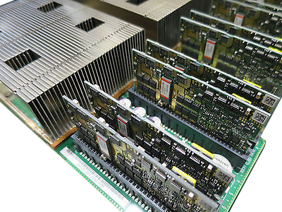
Prophet SUN/SPARC sheets for MRI and X-Ray workstations.
The Oracle SUN/SPARC sheets are a crucial piece of a therapeutic imaging framework because of the significance of the correct operation inside workstations and servers with the MRI and X-beam frameworks. medical imaging parts It is imperative that for the preparing of pictures and touchy patient information, these are dependably in appropriate operation. We as of now have an extensive variety of motherboards for substitution and get together of its workstations for gear of the Hitachi and Siemens brands.
A portion of the models we generally oversee are: SUN/SPARC Station 4 Motherboard, SUN/SPARC Station 5 Motherboard, SUN/SPARC Station 10, SUN/SPARC Station 10 Motherboard,SUN/SPARC Station 20 Motherboard, SUN/SPARC Server 1000 Motherboard, SUN/SPARC Server 1000E Motherboard, SUN/SPARC Server 1000 Motherboard among others.
Motherboard SUN/SPARC for Siemens and Hitachi Workstations.
SUN/SPARC sheets prepared to transport worldwideSUN/SPARC Motherboards for Siemens and Hitachi x-beam and MRI workstations prepared to send.
Culminate working condition
Universal transportation
Steady stock
Focused cost around the world
0 notes
Text
Ultrasound medical imaging parts
What is General Ultrasound Imaging?
Ultrasound is protected and easy, and produces photos of within the body utilizing sound waves. Ultrasound imaging, likewise called ultrasound examining or sonography, includes the utilization of a little transducer (test) and ultrasound gel set straightforwardly on the skin. High-recurrence sound waves are transmitted from the test through the gel into the body. The transducer gathers the sounds that skip back and a PC at that point utilizes those sound waves to make a picture. Ultrasound examinations don't utilize ionizing radiation (as utilized as a part of x-beams), in this way there is no radiation presentation to the patient. Since ultrasound pictures are caught continuously, they can demonstrate the structure and development of the body's inner organs, and in addition blood coursing through veins.
Ultrasound imaging is a noninvasive restorative test that enables doctors to analyze and treat therapeutic conditions.
Traditional ultrasound shows the pictures in thin, level areas of the body. Progressions in ultrasound innovation incorporate three-dimensional (3-D) ultrasound that configurations the sound wave information into 3-D pictures.
A Doppler ultrasound study might be a piece of a ultrasound examination.
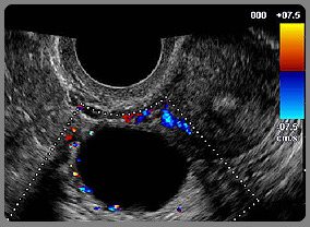
Doppler ultrasound, additionally called shading Doppler ultrasonography, is an exceptional ultrasound strategy that enables the doctor to see and assess blood course through corridors and veins in the midriff, arms, legs, neck as well as cerebrum (in newborn children and youngsters) or inside different body organs, for example, the liver or kidneys.
There are three sorts of Doppler ultrasound:
Shading Doppler utilizes a PC to change over Doppler estimations into a variety of hues to demonstrate the speed and heading of blood move through a vein.
Power Doppler is a more up to date method that is more touchy than shading Doppler and fit for giving more noteworthy detail of blood stream, medical imaging parts particularly when blood stream is pretty much nothing or insignificant. Power Doppler, nonetheless, does not enable the radiologist to decide the bearing of blood stream, which might be critical in a few circumstances.
Ghastly Doppler shows blood stream estimations graphically, as far as the separation voyaged per unit of time, as opposed to as a shading picture. It can likewise change over blood stream data into an unmistakable sound that can be heard with each pulse.
best of page
What are some normal employments of the strategy?
Ultrasound examinations can analyze an assortment of conditions and to survey organ harm following ailment.
Ultrasound is utilized to enable doctors to assess side effects, for example,
torment
swelling
contamination
Ultrasound is a helpful method for looking at a significant number of the body's inside organs, including however not restricted to the:
heart and veins, including the stomach aorta and its major branches
liver
gallbladder
spleen
pancreas
kidneys
bladder
uterus, ovaries, and unborn youngster (baby) in pregnant patients
eyes
thyroid and parathyroid organs
scrotum (balls)
cerebrum in newborn children
hips in newborn children
spine in newborn children
Ultrasound is likewise used to:
control systems, for example, needle biopsies, in which needles are utilized to test cells from a strange territory for research center testing.
picture the bosoms and guide biopsy of bosom growth (see the Ultrasound-Guided Breast Biopsy page.
analyze an assortment of heart conditions, including valve issues and congestive heart disappointment, and to evaluate harm after a heart assault. Ultrasound of the heart is generally called an "echocardiogram" or "reverberate" for short.
Doppler ultrasound pictures can help the doctor to see and assess:
blockages to blood stream, (for example, clumps)
narrowing of vessels
tumors and intrinsic vascular abnormalities
diminished or missing blood stream to different organs
more prominent than typical blood stream to various zones, which is once in a while found in contaminations
With information about the speed and volume of blood stream picked up from a Doppler ultrasound picture, the doctor can regularly decide if a patient is a decent possibility for a method like angioplasty.
best of page
By what means would it be advisable for me to get ready?
You should wear agreeable, baggy attire for your ultrasound exam. You may need to expel all dress and gems in the region to be inspected.
You might be made a request to wear an outfit amid the method.
Arrangement for the strategy will rely upon the kind of examination you will have. For a few outputs your specialist may teach you not to eat or drink for upwards of 12 hours before your arrangement. For others you might be made a request to drink up to six glasses of water two hours before your exam and abstain from urinating with the goal that your bladder is full when the output starts.
best of page
What does the hardware resemble?
Ultrasound scanners comprise of a support containing a PC and hardware, a video show screen and a transducer that is utilized to do the examining. The transducer is a little hand-held gadget that takes after a receiver, joined to the scanner by a rope. A few exams may utilize diverse transducers (with various capacities) amid a solitary exam. The transducer conveys high-recurrence sound waves (that the human ear can't hear) into the body and after that tunes in for the returning echoes from the tissues in the body. The standards are like sonar utilized by water crafts and submarines.
The ultrasound picture is instantly obvious on a video show screen that resembles a PC or TV screen. The picture is made in light of the plentifulness (uproar), recurrence (pitch) and time it takes for the ultrasound flag to come back from the zone inside the patient that is being inspected to the transducer (the gadget put on the patient's skin to send and get the returning sound waves), and in addition the kind of body structure and creation of body tissue through which the sound ventures. A little measure of gel is put on the skin to enable the sound waves to fly out from the transducer to the inspected territory inside the body and afterward back once more. Ultrasound is a fantastic methodology for a few territories of the body while different zones, particularly air-filled lungs, are ineffectively suited for ultrasound.
best of page
How does the strategy function?
Ultrasound imaging depends on similar standards associated with the sonar utilized by bats, ships and anglers. At the point when a sound wave strikes a question, it bobs back, or echoes. By measuring these reverberate waves, it is conceivable to decide how far away the protest is and additionally the question's size, shape and consistency (regardless of whether the protest is strong or loaded with liquid).
In medication, ultrasound is utilized to recognize changes in appearance, size or shape of organs, tissues, and vessels or to identify anomalous masses, for example, tumors.
In a ultrasound examination, a transducer both sends the sound waves into the body and gets the resounding waves. At the point when the transducer is squeezed against the skin, it coordinates little beats of indiscernible, high-recurrence sound waves into the body. As the sound waves bob off inner organs, liquids and tissues, the delicate collector in the transducer records small changes in the sound's pitch and heading. These mark waves are in a split second measured and showed by a PC, which thus makes a continuous picture on the screen. At least one casings of the moving pictures are ordinarily caught as still pictures. Short video circles of the pictures may likewise be spared.
Doppler ultrasound, a unique utilization of ultrasound, measures the bearing and speed of platelets as they travel through vessels. The development of platelets causes an adjustment in pitch of the reflected sound waves (called the Doppler impact). A PC gathers and procedures the sounds and makes charts or shading pictures that speak to the stream of blood through the veins.
best of page
How is the technique performed?
For most ultrasound exams, you will be situated lying face-up on an examination table that can be tilted or moved. Patients might be swung to either side to enhance the nature of the pictures.
After you are situated on the examination table, the radiologist (a doctor particularly prepared to direct and decipher radiology examinations) or sonographer will apply a warm water-based gel to the range of the body being contemplated. The gel will enable the transducer to reach the body and take out air pockets between the transducer and the skin that can obstruct the sound waves from going into your body. The transducer is put on the body and moved forward and backward finished the territory of enthusiasm until the point when the coveted pictures are caught.
There is normally no inconvenience from weight as the transducer is squeezed against the range being analyzed. In any case, if examining is performed over a zone of delicacy, you may feel weight or minor agony from the transducer.
Doppler sonography is performed utilizing a similar transducer.
0 notes
Text
Toshiba Medical Parts

Medical Imaging Parts
Meditegic® supplies fresh out of the plastic new, utilized and repaired Toshiba therapeutic imaging parts guaranteeing quality, conveyance time and focused cost.
We know every client has exceptional prerequisites who need to fit and that is the reason we manufacture custom programming to go after Toshiba restorative imaging save parts far and wide. We can supply from our stock's distribution center, stockrooms from business accomplices or find and find suspended from more than 600 providers.
Our parts incorporate a few modalities in Medical Imaging parts and Bio-prescription/Telemetry is likewise secured.
Toshiba Healthcare parts
Spic and span save parts
Utilized and restored parts
Finish units and area administrations
Aggressive cost around the world
We approach parts suspended by the producer and from more seasoned models. In the event that you require a difficult to-get part, get in touch with us.
0 notes
Text
Dental Radiovisograph CR
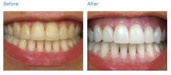
What Are Dental Savings Plans?
Everybody's dental wellbeing needs are exceptional. That is the reason DentalPlans.com offers you choices in addition to investment funds with more than 40 dental intends to look over. medical imaging parts Regardless of whether you need to keep your teeth solid or reestablish your delightful grin, we're here to offer assistance.
Conventional dental protection doesn't address everybody's issues. With a dental arrangement – the moderate contrasting option to dental protection - you pay one yearly expense and access fundamentally decreased rates offered by 100,000+ dental specialists across the country throughout the entire year. You'll get the quality care you merit, at a value you can manage.
Key Differences Between
Dental Savings Plans and Dental Insurance
Advantages Dental Savings Plans Dental Insurance
Speedy Plan Activation
No Health Restrictions
No Annual Limits
No Paperwork Hassles
No Waiting for Dental Care
Corrective Dentistry Savings*
0 notes
Text
How should I prepare for a PET and PET/CT scan?
Ladies ought to dependably educate their doctor or technologist if there is any plausibility that they are pregnant or in the event that they are breastfeeding. See the Safety page for more data about pregnancy and breastfeeding identified with atomic medication imaging.
You ought to advise your doctor and the technologist playing out your exam of any solutions you are taking, including vitamins and home grown supplements. You ought to likewise advise them on the off chance that you have any hypersensitivities and about late sicknesses or other therapeutic conditions.
You will get particular directions in light of the sort of PET output you are experiencing. Diabetic patients will get exceptional guidelines to plan for this exam.
In the event that you are breastfeeding at the season of the exam, you ought to ask your radiologist or the specialist requesting the exam how to continue. It might pump bosom drain early and keep it available for use after the PET radiopharmaceutical and CT differentiate material are no longer in your body.
Metal articles including adornments, eyeglasses, dentures and clasps may influence the CT pictures and ought to be left at home or expelled preceding your exam. You may likewise be made a request to evacuate amplifiers and removable dental work.
By and large, you will be approached not to eat anything for a few hours previously an entire body PET/CT filter since eating may change the appropriation of the PET tracer in your body and can prompt a problematic sweep. This could require the sweep to be rehashed on one more day, so following directions in regards to eating is imperative. medical imaging parts You ought not drink any fluids containing sugars or calories for a few hours previously the output. Rather, you are urged to drink water. On the off chance that you are diabetic, you might be given unique guidelines. You ought to educate your doctor of any pharmaceuticals you are taking and on the off chance that you have any sensitivities, particularly to differentiate materials, iodine, or fish.
You will be approached and checked for any conditions that you may have that may expand the danger of getting intravenous complexity material.
0 notes
Text
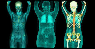
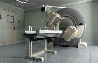
Positron Emission Tomography - Computed Tomography (PET/CT)
Positron emanation tomography (PET) utilizes little measures of radioactive materials called radiotracers, an exceptional camera and a PC to help assess your organ and tissue capacities. By recognizing body changes at the cell level, PET may distinguish the early beginning of malady before it is obvious on other imaging tests.
Tell your specialist if there's a plausibility you are pregnant or on the off chance that you are breastfeeding. Your specialist will teach you in light of the kind of exam to be performed. Examine any current ailments, restorative conditions, drugs you're taking and sensitivities – particularly to differentiate material. You will probably be advised not to eat anything and to drink just water a few hours previously your sweep. Leave adornments at home and wear free, open to apparel. medical imaging parts You might be made a request to wear an outfit.
What is Positron Emission Tomography – Computed Tomography (PET/CT) Scanning?
Positron outflow tomography, additionally called PET imaging or a PET sweep, is a sort of atomic pharmaceutical imaging.
Atomic solution is a branch of medicinal imaging that utilizations little measures of radioactive material to analyze and decide the seriousness of or treat an assortment of maladies, including many sorts of tumors, coronary illness, gastrointestinal, endocrine, neurological clutters and different anomalies inside the body. Since atomic prescription methodology can pinpoint sub-atomic action inside the body, they offer the possibility to recognize sickness in its most punctual stages and also a patient's quick reaction to restorative mediations.
Atomic medication imaging systems are noninvasive and, except for intravenous infusions, are typically easy therapeutic tests that assistance doctors analyze and assess medicinal conditions. These imaging examines utilize radioactive materials called radiopharmaceuticals or radiotracers.
Contingent upon the sort of atomic prescription exam, the radiotracer is either infused into the body, gulped or breathed in as a gas and in the end gathers in the organ or region of the body being inspected. Radioactive emanations from the radiotracer are identified by a unique camera or imaging gadget that produces pictures and gives sub-atomic data.
In many focuses, atomic drug pictures can be superimposed with figured tomography (CT) or attractive reverberation imaging (MRI) to create exceptional perspectives, a training known as picture combination or co-enlistment. These perspectives permit the data from two distinct exams to be associated and translated on one picture, prompting more exact data and precise analyses. Also, producers are presently making single photon outflow figured tomography/registered tomography (SPECT/CT) and positron discharge tomography/processed tomography (PET/CT) units that can perform both imaging exams in the meantime. A rising imaging innovation, yet not promptly accessible as of now is PET/MRI.
A PET output measures vital body capacities, for example, blood stream, oxygen utilize, and sugar (glucose) digestion, to enable specialists to assess how well organs and tissues are working.
CT imaging utilizes uncommon x-beam gear, and now and again a differentiation material, to create numerous pictures or photos of within the body. These pictures would then be able to be translated by a radiologist on a PC screen. CT imaging gives superb anatomic data.
Today, all PET sweeps are performed on instruments that are consolidated PET and CT scanners. The joined PET/CT examines give pictures that pinpoint the anatomic area of unusual metabolic movement inside the body. The joined outputs have been appeared to give more precise conclusions than the two sweeps performed independently.
0 notes
Text
Ultrasound parts for medical imaging
Portrayal
Ultrasound imaging (sonography) utilizes high-recurrence sound waves to see inside the body. Since ultrasound pictures are caught continuously, they can likewise indicate development of the body's inner organs and also blood coursing through the veins. Dissimilar to X-beam imaging, there is no ionizing radiation presentation related with ultrasound imaging.
In a ultrasound exam, a transducer (test) is set specifically on the skin or inside a body opening. A thin layer of gel is connected to the skin with the goal that the ultrasound waves are transmitted from the transducer through the gel into the body.
Photo of a transducer (test) utilized amid a ultrasound exam.
Photo of a transducer (test) utilized amid a ultrasound exam.
The ultrasound picture is created in light of the impression of the waves off of the body structures. medical imaging parts The quality (sufficiency) of the sound flag and the time it takes for the wave to go through the body give the data important to deliver a picture.
Back to Top Arrow
Employments
Ultrasound imaging is a therapeutic device that can enable a doctor to assess, analyze and treat restorative conditions. Regular ultrasound imaging systems include:
Stomach ultrasound (to imagine stomach tissues and organs)
Bone sonometry (to survey bone delicacy)
Bosom ultrasound (to picture bosom tissue)
Doppler fetal heart rate screens (to tune in to the fetal heart beat)
Doppler ultrasound (to picture blood move through a vein, organs, or different structures)
Echocardiogram (to see the heart)
Fetal ultrasound (to see the hatchling in pregnancy)
Ultrasound-guided biopsies (to gather a specimen of tissue)
Ophthalmic ultrasound (to picture visual structures
Ultrasound-guided needle situation (in veins or different tissues of intrigue)
Back to Top Arrow
Advantages/Risks
Ultrasound imaging has been utilized for more than 20 years and has an astounding security record. It depends on non-ionizing radiation, so it doesn't have an indistinguishable dangers from X-beams or different sorts of imaging frameworks that utilization ionizing radiation.
In spite of the fact that ultrasound imaging is for the most part thought to be protected when utilized wisely by properly prepared social insurance suppliers, ultrasound vitality can possibly deliver natural consequences for the body. Ultrasound waves can warm the tissues marginally. Sometimes, it can likewise deliver little pockets of gas in body liquids or tissues (cavitation). The long haul outcomes of these impacts are as yet obscure. As a result of the specific worry for impacts on the hatchling, associations, for example, the American Institute of Ultrasound in Medicine disclaimer iconhave upheld reasonable utilization of ultrasound imaging in pregnancy. Besides, the utilization of ultrasound exclusively for non-restorative purposes, for example, getting fetal 'token' recordings has been disheartened. Souvenir pictures or recordings are sensible on the off chance that they are delivered amid a restoratively demonstrated exam, and if no extra presentation is required.
Back to Top Arrow
Data for Patients including Expectant Mothers
For all medicinal imaging methodology, the FDA prescribes that patients converse with their human services supplier to comprehend the purpose behind the examination, the restorative data that will be acquired, the potential dangers, and how the outcomes will be utilized to deal with the therapeutic condition or pregnancy. Since ultrasound did not depend on ionizing radiation, it is especially helpful for ladies of tyke bearing age when CT or other imaging strategies would somehow or another outcome in introduction to radiation.
Eager Mothers
A pregnant lady getting a ultrasound from a doctor.Ultrasound is the most generally utilized therapeutic imaging technique for review the embryo amid pregnancy. Routine examinations are performed to survey and screen the wellbeing status of the baby and mother. Ultrasound examinations give guardians a significant chance to see and hear the pulse of the hatchling, bond with the unborn child, and catch pictures to impart to family and companions.
In fetal ultrasound, three-dimensional (3D) ultrasound permits the representation of some facial highlights and conceivably different parts, for example, fingers and toes of the baby. Four-dimensional (4D) ultrasound is 3D ultrasound in movement. While ultrasound is by and large thought to be protected with low dangers, the dangers may increment with pointless delayed presentation to ultrasound vitality, or when untrained clients work the gadget.
Eager moms ought to likewise know about acquiring over-the-counter fetal pulse observing frameworks (additionally called doptones). These gadgets should just be utilized via prepared social insurance suppliers when medicinally essential. Utilization of these gadgets by untrained people could open the embryo to delayed and perilous vitality levels, or could give data that is deciphered mistakenly by the client.
Extra assets on ultrasound imaging
Data for Facilities Conducting Ultrasound Imaging
Ultrasound practices ought to consider site and staff support in deliberate accreditation and affirmation programs that address both wellbeing and viability of the gadget following the standards of As Low As Reasonably Achievable (ALARA, for example, those offered by the American Institute of Ultrasound in Medicine disclaimer symbol and the American Registry of Diagnostic Medical Sonographers disclaimer symbol.
Any social insurance office utilizing ultrasound should direct customary quality control tests to guarantee that gear is working legitimately.
0 notes
Text
C-arm parts and refurbished intensifiers
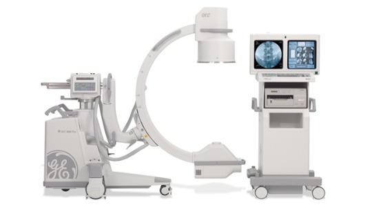
The OEC 9800 Super-C is a typical and prevalent choice on the revamped C-arm showcase, yet when first-time C-arm purchasers run over it in our stock, they frequently ask, "What precisely is so 'super' about this thing?"
All things considered, by and large, "super" means a bit of something all the more, something additional well beyond the normal; "Super-measuring" gets you more fries, the "Super Soaker" shoots a couple of feet more distant than your regular water gun.
Circular segment Depth
The essential contrast between a Super-C and standard units is the profundity of the circular segment of the C. Contrasted with a standard C-arm with a 9" picture intensifier (II), the Super-C has an additional 7". The Super C has 5" more in the bend contrasted with the 12" II setup. medical imaging parts This distinction in circular segment profundity offers Super-C clients 148 degrees of orbital turn rather than the 115 degrees managed standard C users.With the broadened revolution, Super-C clients can finish filters that beforehand would have required repositioning of the patient by essentially repositioning their c-arm. This offers more noteworthy adaptability in the OR and spares time.
L-Arm Design
Another distinction is the outline of the L-arm between the body of the unit and the circular segment of the C. The L-arm of the Super-C was abbreviated to suit the expanded measurements of the C. This change does not influence the side-to-side revolution of the Super-C.
General Size
In general size, the Super-C dwells between the somewhat littler standard 9" II design and the marginally bigger standard 12" setup. The distinction in either heading is 2" or less in both tallness and length. The screen trucks of every one of the three styles share indistinguishable measurements.
Cost
The last (and regularly asked after) distinction is in estimating. The purchaser hoping to buy a Super-C can hope to pay a normal of $3,000-$4,000 more than the cost of a standard C.
The Takeaway
Torment administration and heart centered doctors (and any other person utilizing the qblique see) will see a chose advantage from the additional turn of the Super-C. Different applications surely won't perceive any impediment from having a Super-C, yet may have the capacity to forego the additional cost.
Whatever your particular application, we're here to enable you to choose the best C-arm for your office. In case you're prepared to begin now, get in touch with us with the catch underneath. For those of you who'd jump at the chance to do somewhat more homework, there are joins for that as well.
Demand C-Arm Price
0 notes
Text
Telemetry parts and neonatal incubators (NICU MIC)
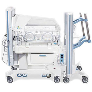
Suppliers utilize loads of gear in the NICU to enable take to care of your child. Each piece has an exceptional reason in your child's treatment. On the off chance that you have inquiries concerning any hardware used to treat your infant, ask his human services supplier or the attendants in the NICU.
Here's some gear suppliers frequently use in the NICU:
apnea screen — A machine that identifies when your child quits relaxing for a couple of moments. A caution heads out to give NICU a chance to staff know your child has quit relaxing.
blood vessel line — A thin tube that goes into your infant's corridor to check his circulatory strain and measure blood gasses. A corridor is a vein that conveys oxygen to all parts of your infant's body. Circulatory strain is the power of blood that pushes against the dividers of the veins. Blood gasses are corrosive, oxygen and carbon dioxide in your child's blood.
bililights — Bright lights over an infant's hatchery that treat jaundice. A hatchery is a reasonable plastic bed that keeps your child warm. Jaundice is the point at which an infant's eyes and skin look yellow. An infant has jaundice when his liver isn't completely created or isn't working. Treatment with bililights is likewise called phototherapy. Children can have this treatment for 3 to 7 days.
circulatory strain screen — A machine associated with a little pulse sleeve wrapped around your infant's arm or leg. The sleeve takes your child's circulatory strain at normal circumstances and showcases it on a screen. Circulatory strain is the power of blood that pushes against the dividers of the courses.
cardiopulmonary screen — A machine that tracks your child's heart and breathing rates. It's associated with your infant's chest with little sticky cushions called leads. Data from the screen shows on a screen and can be printed out. On the off chance that your child's heart or breathing rate turns out to be too quick or too moderate, an alert sounds.
focal line — A little plastic tube that goes into a huge vein. Your child gets pharmaceutical and liquids through the tube, and suppliers can coax blood out through the tube. One sort of focal line that is utilized a considerable measure is known as an incidentally embedded focal catheter (likewise called a PICC line).
constant positive aviation route weight (likewise called CPAP) — A machine that sends air and oxygen to your child's lungs through little tubes in his nose or windpipe (additionally called trachea).
cooling cover or top — A cover or top used to bring down your child's body temperature. They may help decrease or anticipate issues that can happen if your infant's mind doesn't get enough oxygen. The cover or top can cool your child's cerebrum and body to around 92 F (33.5 C). Your infant may get a cooling cover or top inside around 6 hours of birth and can utilize it for up to 3 days. From that point forward, your child is gradually warmed to a typical body temperature of 98.6 F (37 C) by expanding the temperature in the hatchery.
endotracheal tube — A little plastic tube that goes into an infant's nose or mouth and down to the windpipe (likewise called trachea) that sends air and oxygen to the lungs. The tube is appended to a machine called a mechanical ventilator to enable your infant to relax.
extracorporeal layer oxygenation (additionally called ECMO) — A machine that removes blood from your infant's body, places oxygen into the blood and after that sends the blood over into the body.
gastrostomy tube (additionally called g-tube or gastric encouraging tube) — A tube that goes into your infant's stomach for sustaining. Fluids experience the tube to nourish your infant. The tube is utilized for babies who can't take nourishment by mouth and need long haul help with encouraging.
high-recurrence ventilator — A machine that relaxes for your child at a quicker rate than different ventilators. Swaying and fly ventilators are cases of high-recurrence ventilators.
hatchery — A reasonable plastic bed that helps keep your child warm. You can touch your child through gaps (additionally called ports) in the sides of the hatchery. Sorts of hatcheries are Giraffe® and Isolette®.
intravenous line (likewise called IV) — A tube embedded with a needle into your child's vein. A vein is a vein that takes blood back to the heart. Your child can get liquids, prescription and blood through an IV.
mechanical ventilator — A machine that enables your infant to inhale or relaxes for him when he's not breathing without anyone else. It works by pushing warm air and oxygen into the lungs through a breathing tube called an endotracheal tube. The supplier sets the measure of oxygen, pneumatic force and number of breaths every moment for your child.
nasal cannula — Small plastic tubes that go into your child's nose. Air and oxygen experience the tubes into your child's lungs.
nasogastric tube (likewise called NG tube) — A sustaining tube that experiences your child's nose, down the throat and into the stomach. The throat is the tube in your child's body that conveys sustenance from the throat to the stomach. Your infant can get bosom drain, recipe and solution through the tube. At the point when your infant is bolstered bosom drain or recipe however a NG tube, it's called gavage encouraging.
orogastric tube (likewise called OG tube) — A nourishing tube that goes in your infant's mouth, down the throat and into the stomach. The throat is the tube in your child's body that conveys nourishment from the throat to the stomach. Your child can get bosom drain, recipe and solution through an OG tube. medical imaging parts At the point when a child is bolstered bosom drain or recipe however an OG tube, it's called gavage nourishing.
oxygen hood — A reasonable plastic box that fits over an infant's head and gives him oxygen. Suppliers utilize it with babies who can inhale without anyone else yet at the same time require some additional oxygen.
beat oximeter (additionally called a heartbeat bull) — A little gadget wrapped around your infant's foot or hand that measures the oxygen in her blood. It doesn't cause your child any agony. It enables suppliers to know whether your infant needs pretty much oxygen.
brilliant hotter — An open bed with overhead warming to help keep your infant warm. Suppliers may utilize a hotter rather than a hatchery if your infant should be taken care of a great deal. A hatchery is an unmistakable plastic bed that helps keep your infant warm.
tracheostomy tube — A bended plastic tube that goes in your child's windpipe (additionally called trachea) through a gap made in your infant's neck. The trachea is a piece of the aviation route framework that takes air to the lungs. Your child inhales through the tube rather than his nose and mouth. The tube doesn't go into your child's lungs.
umbilical catheter — A thin tube that goes into the supply routes in your child's umbilical line after the line is cut after birth. Suppliers can give liquids, blood, medication and supplements, similar to vitamins and minerals, to your child through the tube. They additionally utilize the tube to take blood from your infant. A little connection to the tube gives suppliers a chance to check your child's circulatory strain. Circulatory strain is the power of blood that pushes against the dividers of the courses.
urinary catheter — A thin tube that experiences the opening where pee goes out of your infant's body and into his bladder. Suppliers utilize it to gather pee for testing.
0 notes
Text
Telemetry parts and neonatal incubators (NICU MIC)
0 notes
Text
medical imaging parts
Atomic and SPECT parts available to be purchased around the world
Atomic parts new, utilized and repaired for tomography frameworks
In Meditegic we have new atomic parts for tomography frameworks in stock, utilized parts with the best focused costs and recreated parts with magnificent operation and usefulness. We have parts ceased by the maker and furthermore parts for more seasoned models. Our experience enables us to have associations with more than 500 providers and producers, we guarantee that each part for atomic tomography frameworks is precisely tried and completely certify.
The foundation we utilize enables us to act as a bi-national organization with an extent of worldwide supply, we offer any of the models reported on this site with a quick delivering administration to more than 28 nations, medical imaging parts this gives us the certainty to give quietness our customers telling them that they will be working without obstructions in a long haul.
We have a broad supply of atomic parts, particularly for frameworks, for example, GE, Philips, Siemens and Toshiba among others.
0 notes
Text
medical imaging parts
Restorative imaging and electromedicine parts

New parts for restorative frameworks
Quality in medical imaging parts
Parts for a wide assortment of frameworks
Framework
New, utilized and renovated parts
Partes para lightspeed 16 CT
At Meditegic we have in stock new parts for imaging gear, parts utilized at the best focused costs and completely practical revamped parts.
It is critical to consider visit upkeep and also a preventive administration to maintain a strategic distance from a leader repair.
Some support administrations require repair parts and when the disappointment is excessively basic, it is vital, making it impossible to do a sections substitution; we handle a high number of parts in stock with quick delivering to more than 28 nations.
We have a framework that enables us to fill in as a bi-national organization with supply of worldwide reach. Our experience enables us to have associations with more than 500 providers.
New and new parts for imaging frameworks:
We have new parts for therapeutic imaging gear, for example,
CT/PET Parts
X-ray Systems parts
Atomic/SPECT Parts
Parts for Ultrasound frameworks
Ultrasound tests and accesories
Parts for C-Arm Systems and Intensifiers
Telemetry/MIC parts and accesories
Biomedical Batteries
X-Ray Tubes
SUN/SPARC Boards
0 notes
Text
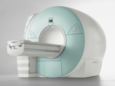
medical imaging parts
X-ray parts and arrangements around the world
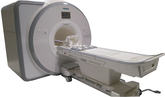
Attractive Resonance new parts, utilized and renovated
In Meditegic we offer new MRI parts, utilized at aggressive costs and completely utilitarian revamped parts. We approach suspended parts by the producer and furthermore for more seasoned frameworks. Our experience enables us to have associations with more than 500 providers and we affirm that each part for attractive reverberation frameworks is broadly and deliberately tried.
On account of our framework we can fill in as a bi-national organization with worldwide supply achieve, medical imaging parts we offer any model declared on this site with a quick conveyance administration to more than 28 nations.
We have a broad supply of attractive reverberation pieces (MRI parts) for frameworks, for example, GE, Hitachi, Philips, Siemens and Toshiba among others.
New, utilized and repaired MRI parts for the most asked for frameworks:
Siemens MRI Open/Closed
GE MRI Open/Closed
Philips MRI Open/Closed
Hitachi Closed MRI
Toshiba MRI Open/Closed
Bennet MRI Closed
X-ray Coils
X-ray Injectors
Furthest point MRI
Picker MRI Closed
Esaote MRI Closed
Fluid Helium (Dewar Helium)
0 notes
Text
Ct Scan
FULL TIME
If you’re looking for a long-term CT solution that truly fits your needs, no provider is more committed to your success than Shared Imaging®. For healthcare providers around the country, we provide full-time, permanent systems, including modular buildings, and in-house offerings, provided by the industry’s top manufacturers and configured for each client’s unique situation.
PART TIME
Shared Imaging can craft a part-time CT solution that will adapt to your evolving needs. Whether it is to assist you in reducing patient backlog, improving diagnostic capabilities or addressing lack of physical space for an in-house system, Shared Imaging part-time solutions will help you reach these goals.
INTERIM
Whether you need an interim rental for construction, reducing backlog or emergencies, Shared Imaging can provide equipment from all equipment manufacturers to address your specific needs. medical imaging parts Our flexible deal structures allow you to achieve the optimal level of service required at your facility.
0 notes
Link
We are a US bi-national company that supplies medical imaging parts to over 800 customers in more than 29 countries around the world. We have an extensive network of more than 500 suppliers and +500,000 part number.
With the support of some of the most prominent brands worldwide and years of experience and knowledge, you can always be sure that we will work hard to supply your medical parts needs including hard-to-get parts.
0 notes