
dovemed
Health Info For A Better Life
DoveMed is a trusted, physician-approved, and simple to understand, health information resource. http://www.dovemed.com
58 posts
Don't wanna be here? Send us removal request.
Last Seen Blogs

meemersthecat
Get Some

femboy-antalya
Trv.Antalya

iimaginekpop
♥Idols ♥


denizlitravestideniz
denizli travesti Deniz
Text
Classification of Venomous Snakes

Classification of venomous snakes and comprehensive information on snake bites, first aid, treatment, and prevention: (in alphabetical order)
Acanthophis antarcticus (Common Death Adder)
Agkistrodon bilineatus (Mexican Cantil)
Agkistrodon contortrix (Copperhead)
Agkistrodon laticinctus (Broad Banded Copperhead)
Agkistrodon piscivorus (Cottonmouth)
Agkistrodon taylori (Castellana)
Ahaetulla nasuta (Long Nosed Whip Snake)
Aipysurus apraefrontalis (Short-Nosed Sea Snake) - true sea snake; Timor Sea; inhabits shallow coral reef waters;
Aipysurus duboisii (Dubois' Sea Snake) - slightly aggressive; true sea snake; north of Australia and parts of southwestern Pacific Ocean; inhabits shallow reef waters and sandy bottoms;
Aipysurus eydouxii (Spine-Tailed Sea Snake) - not easily provoked; true sea snake; north of Australia and around tropical islands of Southeastern Asia; inhabits shallow bay waters and muddy estuarine bottoms;
Aipysurus foliosquama (Leaf-Scaled Sea Snake) - true sea snake; Timor Sea; inhabits shallow coral reef waters and seagrass bottoms;
Aipysurus fuscus (Dusky Sea Snake) - true sea snake; Timor Sea and Java Sea; inhabits shallow reef waters and sandy sea bottoms;
Aipysurus laevis (Olive Brown Sea Snake) - highly venomous and inquisitive; true sea snake; north of Australia and southwestern Pacific Ocean; inhabits coral reef waters;
Aipysurus pooleorum (Shark Bay Sea Snake) - highly venomous; true sea snake; Shark Bay, west of Australia; inhabits limestone reefs, seabed, and seagrass floor;
Amphiesma stolatum (Buff Striped Keelback)
Aspidelaps lubricus (Cape Coral Snake)
Aspidelaps scutatus (Shield Nose Snake)
Atheris squamigera (African Bush Viper)
Atractaspis bibronii (Bibron's Burrowing Asp)
Atractaspis dahomeyensis (Dahomey Burrowing Asp)
Atractaspis engaddensis (Palestinian Mole Viper)
Atractaspis microlepidota (Small Scaled Burrowing Asp)
Atropoides picadoi (Picado's Jumping Pitviper)
Austrelaps superbus (Lowland Copperhead)
Azemiops feae (Fea's Viper)
Bitis arietans (Puff Adder)
Bitis atropos (Cape Mountain Adder)
Bitis caudalis (Horned Puff Adder)
Bitis cornuta (Western Many Horned Adder)
Bitis gabonica (Central African Gaboon Viper)
Bitis nasicornis (Rhinoceros Viper)
Bitis parviocula (Ethiopian Mountain Adder)
Bitis rhinoceros (West African Gaboon Viper)
Boiga cyanea (Green Cat Snake)
Boiga dendrophila (Mangrove Snake)
Boiga irregularis (Brown Tree Snake)
Bothriechis lateralis (Side Striped Palm Viper)
Bothriechis nigroviridis (Black Speckled Palm Pitviper)
Bothriechis schlegelii (Eyelash Palm Pitviper)
Bothrops alternatus (Urutú)
Bothrops asper (Terciopelo)
Bothrops atrox (Common Lancehead)
Bothrops ayerbei (Ayerbe's Lancehead)
Bothrops caribbaeus (Saint Lucia Lancehead)
Bothrops cotiara (Cotiara)
Bothrops diporus (Chaco Lancehead)
Bothrops erythromelas (Caatinga Lancehead)
Bothrops fonsecai (Fonseca's Lancehead)
Bothrops insularis (Golden Lancehead Viper)
Bothrops itapetiningae (São Paulo Lancehead)
Bothrops jararaca (Jararaca)
Bothrops jararacussu (Jararacussu)
Bothrops lanceolatus (Martinique Lancehead)
Bothrops leucurus (Whitetail Lancehead)
Bothrops mattogrossensis (Mato Grosso Lanzenotter)
Bothrops moojeni (Brazilian Lancehead)
Bothrops neuwiedi (Neuwied's Lancehead)
Bothrops pauloensis (Black Faced Lancehead)
Bothrops taeniatus (Speckled Forest Pitviper)
Bungarus caeruleus (Indian Krait)
Bungarus candidus (Blue Krait)
Bungarus fasciatus (Banded Krait)
Bungarus flaviceps (Red Headed Krait)
Bungarus multicinctus (Many Banded Krait)
Calloselasma rhodostoma (Malayan Pitviper) - highly venomous and irritable; Southeastern Asia; terrestrial and usually nocturnal; often found near agricultural lands;
Causus rhombeatus (Common Night Adder)
Cerastes cerastes (Horned Viper)
Cerastes gasperettii (Arabian Horned Viper)
Cerastes vipera (Sahara Sand Viper)
Cerrophidion godmani (Godman's Montane Pitviper)
Cerrophidion sasai (Costa Rica Montane Pitviper)
Here is the full list of Venomous Snakes. https://www.dovemed.com/classification-disorders-and-tumors/classification-venomous-snakes/
#snake#snakes#lancehead#viper#venomoussnakes#classification#firstaid#health#who#worldhealthorganization#cobra#southamerica#asia#europe
10 notes
·
View notes
Text
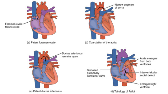
Congenital Heart Defects
What are the other Names for this Condition? (Also known as/Synonyms)
CHDs (Congenital Heart Defects)
Congenital Heart Anomalies
What are Congenital Heart Defects? (Definition/Background Information)
Congenital Heart Defects (CHDs) are relatively common birth defects that affect the function and structure of the heart. These include faulty heart valves, holes in the inside walls of the heart, and complex malformations. The exact cause of development of CHDs is not well-understood. These may develop from a combination of genetic and environmental factors. It is also believed that what the expectant mother consumes, such as food, drink, or even some medications, may be contributive.
Severe Congenital Heart Defects are easily diagnosed through the early manifestation of signs and symptoms. However, moderate to mild cases may be detected only much later (sometimes, even into adulthood). Since CHDs are congenital in nature, children are generally affected at birth.
The signs and symptoms noted, the complications that may develop, and the treatment (surgical) approach that is considered/planned, are all chiefly related to the specific type of heart defect noted in the affected individual. Managing a Congenital Heart Defect almost always involves surgically correcting the defect and providing long-term symptomatic treatment.
Presently, there are no available measures to prevent these defects. Detecting early signs and symptoms combined with a timely diagnosis, holds the key to managing the condition. A late recognition of Congenital Heart Defects and lack of appropriate treatment may lead to complications and adverse outcomes.
Congenital Heart Defects encompass a variety of specific conditions. Information is constantly being added through research, better documentation, and increased awareness. The following subtypes of CHDs are recognized:
Atrial Septal Defect (ASD)
Atrioventricular Septal Defect (AVSD)
Coarctation of the Aorta (CoA)
D-Transposition of the Great Arteries (d-TGA)
Hypoplastic Left Heart Syndrome (HLHS)
Pulmonary Valve Atresia
Tetralogy of Fallot (TOF)
Total Anomalous Pulmonary Venous Return (TAPVR)
Tricuspid Atresia
Truncus Arteriosus (TA)
Ventricular Septal Defect (VSD)
This article is a resource with links to other more specific conditions. Information on each Congenital Heart Defect may be viewed by clicking on the respective subtypes (above).
Information to join DoveMed’s patient forum called MyCIrcles to learn and manage the condition is also included. We are adding more information to this page periodically. Please bookmark this page for future reference and visit for updated content.
You can join Congenital Heart Defects MyCircles patient forum by visiting here: https://www.dovemed.com/mycircles/circles/all
#congenitalheartdefect#congenitaldisorders#chd#chds#congenital#heart#kids#defect#defects#children#signsandsymptoms
0 notes
Text
We are not White Mice, But...

To understand the scientific basis of what we ‘know’ about the medical world, we learn the database of centuries of observations, studies, successful and partially successful (as well as failed) treatments. This includes studies of laboratory animals, studies in Petri dishes and tissue cultures, and humans.
White-haired, pink-eyed mice are albinos, without melanin pigment. To test if ultraviolet (UV) light could cause squamous cell carcinomas of the skin, their delicate hair was shaved off, and a UV light source used to irradiate a patch of skin. Yes, this did cause local squamous cell carcinomas where the light shined.
To test if coal tar could cause squamous cell carcinoma of their skin, the hair was shaved off, and a liquid extract of crude coal tar was applied to the spot. Yes, the coal tar caused squamous cell carcinomas of the skin.
Coal is a soft, rock-like material derived from millennia-old plants subjected to heat and pressure underground. This causes the hydrocarbons formed by living plants: starch, lignin, sugars, etc. to form thousands of different, more complex chemicals. Coal burns readily and has been used as a home and industrial heat source for centuries. When coal burns, the more volatile, almost liquid chemicals within it are vaporized. As they cool, as when smoke carries them up a chimney, they form a tar-like material that condenses and coats the chimney wall. Coal tar is as flammable as coal, so a spark carried up on the draught can ignite a chimney fire, burning down a house or entire neighborhoods.
Enter the occupation of the chimney sweep. This was a very small person, usually a boy, a poor boy, who could shimmy up the chimney, using a scraper to remove the coal tar, and brushes to remove the dust. One of the major causes of death of chimney sweeps was scrotal carcinoma. The scrotum, like its analogous female tissue, the vulva, lacks a stratum corneum, the layer of skin that blocks the ready absorption of many chemicals. This provided an early observation that coal tar, and soot, contained cancer causing (carcinogenic) compounds.
Now let’s go back to the mouse study above. The mice that had their hair clipped off, coal tar extract applied, AND had ultraviolet light irradiation where the tar was, developed squamous cell carcinomas at the site much earlier than just UV light or coal tar, as well as having much more aggressive tumors.
Crude coal tar has been used extensively in dermatology for more than 100 years. Compounds with 2-4 % were often used for extensive psoriasis (PS) or eczema (atopic Dermatitis (AD). It often helped, and improved people’s overall health. A common psoriasis protocol, which I oversaw frequently in my training, consisted of hospitalization for 3 weeks, with daily applications of 4 % crude coal tar in petrolatum. The patient wore hospital scrubs, with a new set only given every week. That way the tar-soaked scrubs reapplied the tar with every movement. Ultraviolet B (UVB) light treatments were given Monday-Friday. Not everyone complained about the oily, tar-smelling course, because it cleared the PS reliably, often for up to a year. This was at a time that no other treatments were so successful, with a pretty good safety profile. Besides being available in petrolatum, coal tar was available in shampoo, bath oils, and soap. It still is available. If you buy it in California, each unit bears a sticker alerting the consumer that it contains known carcinogens.
Fifteen years ago, I started advising my patients not to use coal tar products. I had always wondered about why we continued to use coal tar. A patient came to see me with three separate whitish and pinkish patches about 5 mm each on his penis. They were all located on the areas that would have been covered by his foreskin, had he not been circumcised as a child. These tissues, like the scrotum, never gain the ability to exclude the absorption of chemicals. Each site on biopsy proved to be a squamous cell in situ of the penis. At this stage, the risk of spread outside the skin of the penis is close to zero. Yet if not definitively treated, or if recurrent, squamous cell of the penis can metastasize and be fatal or require amputation. I performed Mohs Micrographic Surgery on the lesions, with final clear margins, excellent healing, and no post-op disability. The day of surgery was not easy for him. He had no recurrence on 10-year follow-up.
Squamous Cell Carcinoma of the penis is sometimes associated with prior genital warts. He had no history of this. He had no history of ultraviolet light to the area through outdoor sun exposure or indoor tanning beds. He had no work exposure to potential carcinogens. Eventually he remembered that he had had extensive psoriasis as a teen and young adult. He had been clear of PS for a few decades. His treatment at the time was an hours-long soak in a bathtub with coal tar bath oil, 4-5 times weekly. We surmised that the coal tar exposure had led to his three squamous cell carcinomas.
Since then, I have advised all my patients using coal tar products to switch to pine tar, salicylic acid, or other active ingredients in their grooming.
“But I only use the shampoo” is a frequent response.
“Do you shampoo in the shower?”
“Yes, of course.”
“Where do the coal tar suds go before they go down the drain?”
“Oh. OK.”
I must point out that my personal opinion about the use of coal tar on the skin is just that, a personal opinion. Also, the coal tar, UV light treatment for psoriasis has NOT been found to increase the risk of skin cancer in patients so treated. My point is not to needlessly cause those patients to worry.
Science goes onward. Recently, the cell membrane receptor the coal tar activated to calm down PS and AD skin was identified. A non-coal-tar compound also activates the same receptor, giving us an opportunity to get the therapeutic effect with an even lower risk profile.
0 notes

Photo

Starbucks And A Car Door
I quit drinking coffee almost a year ago. Up to then I was a gold star premium member of the Starbucks club. Morning, noon and night was not unusual. I wouldn’t brew my own but I would pick up gas station coffee or Starbucks or Dunkin’ Donuts coffee at least once a day. Then one day I just decided the cost wasn’t worth the benefit. I lost interest in the taste. Then the smell. I use to like to music and atmosphere. Hundreds of blog notes were posted from Starbucks locations across the country.
Read More:
0 notes
Photo

Are You Claustrophobic Mr. Kaplan? A Fine Line Between Safety And Indolence
As part of my ongoing evaluation and treatment for heart failure that I have mentioned previously (see: http://tissuepathology.com/2017/11/05/the-clinic-door-chapter-2-the-emergency-room/#axzz52Q1JZUgB) I underwent a cardiac MRI recently.
Prior to scheduling the MRI, I was asked about pacemakers, defibrillators and artificial joints or shrapnel in my medical history. I was told to show up 30 minutes prior to the appointment and not to have any caffeine after midnight the night before the study. Standard questions and instructions having had other MRI for head and knee injuries playing hockey. They apparently have since added not having caffeine for cardiac specific studies.
Read More:
0 notes
Link
Any organ of the body can develop cancer. Benign cancers mostly remain confined to the primary site of origin, but malignant cancers can metastasize to other organs too. For example, lung cancer can metastasise to the bone, to form secondary cancer of bone. Such metastasis depends on many factors including the type of cancer, stage of cancer, organ involved, etc.
Bone is among the most common site for metastasis from advanced solid tumours. Mostly, the long bones are affected by such metastases. Vertebral column that protects the spinal cord is also a major site of metastasis. Up to 10% of all patients with advanced stage of cancer suffer from metastasis to the vertebras, leading to spinal cord compression. It is a difficult symptom to treat as important nerves are exposed to external pressure. Multi-dose radiation therapy to treat spinal cord compression symptom have been conventionally tried. Recently, researchers proposed that a single dose of radiation can be equally effective in treating the condition.
A recently conducted study involved 688 patients with advanced stages of metastatic cancer of the prostrate, lung, gastrointestinal tract, and breast. Researchers randomly divided them into two groups, one to receive single dose of 8 Gy external beam radiation therapy, and the other to receive 20 Gy external beam radiation therapy (split in five doses over five days). The patients were then evaluated for their ambulatory status. Ambulatory status was measured in a four-point scale; from 1 being good i.e., able to walk normally, to 4 being worse i.e., dependent on wheel chair.
The results of the study showed almost equal effect. After eight weeks of treatment, 69.5% of the patients receiving single dose radiotherapy had ambulatory status 1 or 2, compared to 73.3% in the five-dose group. There was no statistically significant difference between both the groups with respect to overall survival. The proportion of patients, who suffered from severe side effects, were almost similar (20.6% v/s 20.4%) in both the groups. Mild side effects were less frequent in the single dose group. Lesser side effects and lesser duration of treatment meant fewer hospital visits.
The lead author of the study Peter Hoskin, an oncologist at the Mount Vernon Cancer Centre in Middlesex, United Kingdom, said that “Our findings establish single-dose radiotherapy as the standard of care for metastatic spinal canal compression, at least for patients with a short life expectancy”. Regarding benefits of long-course radiotherapy he said that “Longer radiation may be more effective for preventing regrowth of metastases in the spine than single-dose radiation. Therefore, a longer course of radiation may still be better for patients with a longer life expectancy, but we need more research to confirm this”.
Reference:
1. A Single Radiation Treatment Sufficiently Relieves Spinal Cord Compression Symptoms [Internet]. ASCO. 2017 [cited 2017 Jul 11]. Available from: https://www.asco.org/about-asco/press-center/news-releases/single-radiation-treatment-sufficiently-relieves-spinal-cord
2. Metastatic Cancer [Internet]. National Cancer Institute. [cited 2017 Jul 11]. Available from: https://www.cancer.gov/types/metastatic-cancer
3. Understanding Advanced Cancer, Metastatic Cancer, and Bone Metastasis [Internet]. [cited 2017 Jul 11]. Available from: https://www.cancer.org/treatment/understanding-your-diagnosis/advanced-cancer/what-is.html
#cancer#radiationtherapy#radiation#therapy#symptoms#patients#radiotherapy#medicine#medicalschool#dovemed#protstatecancer#lungcancer
0 notes
Link
Breast cancer is the most common type of cancer diagnosed in women and is believed to be the second-most leading cause of death in women. It is a type of cancer in which certain cells in the breast become abnormal, grow uncontrollably, and form a malignant mass (tumor). Breast Cancer cells can also break away from the initial tumor, invade nearby tissues, and spread, or metastasize, to other regions of the body.
The presence of breast implants can cause significant difficulties in the diagnosis of breast cancer and its treatment, once a diagnosis is confirmed. But, it is important to note that current research shows that there is no correlation between increased incidences of breast cancer in women with breast implants. In other words, having breast implants does not increase one’s risk for getting breast cancer.
Mammograms are vital screening tools for the diagnosis of breast cancer. The interpretation of mammograms by radiologists can be challenging in the presence of breast implants. Also, with advances in technology, the sensitivity of detecting breast cancer in women with breast implants, compared to women without breast cancer is similar. In other words, one can diagnose suspicious area of breast cancer in mammograms of women with breast implants, compared to mammograms of women not having breast implants. Once a suspicious area is noted in a woman with a breast implant, a follow-up biopsy is essential. However, biopsies in women with breast implants can also be challenging. One of the complications of undertaking a biopsy in a woman with breast implants is rupture of the implant.
Once a diagnosis of breast cancer is confirmed, there may be treatment challenges associated with the presence of breast implant(s). Particular care is often needed while administering radiation therapy to women who have breast implants. Radiation therapy may induce radiation fibrosis around the implant causing significant cosmetic issues. The presence of breast implants can cause special challenges in the diagnosis and treatment of breast cancer. With advancing technologies and treatment modalities, it is getting easier to treat breast cancer in women with breast implants. Further research is being done to improve the experience of women with breast implants, during both the diagnosis and treatment of breast cancer.
References:
https://www.cancer.org/cancer/breast-cancer/screening-tests-and-early-detection/mammograms/mammograms-for-women-with-breast-implants.html (accessed on 9/25/2017)
https://www.mdanderson.org/publications/focused-on-health/december-2014/breast-implant-cancer.html (accessed on 9/25/2017)
https://www.fda.gov/MedicalDevices/ProductsandMedicalProcedures/ImplantsandProsthetics/BreastImplants/ucm259296.htm (accessed on 9/25/2017)
0 notes
Link
Breast cancer is the most common type of cancer diagnosed in women and is believed to be the second-most leading cause of death in women. It is a type of cancer in which certain cells in the breast become abnormal, grow uncontrollably, and form a malignant mass (tumor). According to research studies, the incidence of breast cancer in pregnant women is approximately 1.3 breast cancer cases per 10,000 live births. Detection of breast cancer during pregnancy can raise several diagnostic and treatment challenges. Studies have shown that breast cancers diagnosed during pregnancy, usually presents a larger tumor size and a higher incidence of metastasis to the lymph nodes. Most likely explanation of this phenomenon is that the breast cancer cells are responsive to the increased hormones during pregnancy. Studies show that the breast cancer diagnosed during a pregnancy are of higher grade, which means they are more poorly-differentiated. There also usually estrogen receptor negative, progesterone receptor negative and are occasionally, HER-2/neu positive tumors.
The presence of an enlarged breast during pregnancy can make the diagnosis of breast cancer difficult. Presence of a new breast mass in a pregnant woman is more likely to be associated with pregnancy changes. This often results in a delayed diagnosis. Diagnosing breast cancer during pregnancy causes significant challenges. Performing a mammography in a pregnant woman may be difficult because of potential radiation to the developing baby. Also, the mammograms are more difficult to interpret due to the increased breast density during pregnancy. However, ultrasonography of the breast and axillary lymph nodes may be very helpful in detecting the presence of suspicious areas. The presence of suspicious areas in the axillary lymph nodes may be used for performing a tissue biopsy. A core biopsy of the suspicious axillary or breast mass is often recommended.
Once a diagnosis of a breast cancer is confirmed on a tissue biopsy, the follow-up staging of breast cancer can also be challenging. Whole-body PET scan is often contraindicated in pregnant women with breast cancer. In such cases, undertaking an MRI scan of chest may be helpful. Treatment of breast cancer during pregnancy is challenging. It is generally recommended to avoid chemotherapy during the first trimester of pregnancy due to increased risk of fetal malformation. Chemotherapy during the second and third trimester may be given. The protocols are considered on a case-by-case basis.
Studies have shown that the incidence of fetal malformation in the second and third trimester is around 1.3% in women who received chemotherapy. This incidence is statistically not significant compared to the second and third-trimester pregnancy in women who have not received chemotherapy. The type of chemotherapy medication used is decided on a case-by-case basis. It is generally recommended that chemotherapy should not be given during week 35 of pregnancy, since this may lead to increased complications at the time of delivery. Further, radiation therapy and the use of endocrine therapy during pregnancy is contraindicated. Hence, a pregnant woman should not either receive endocrine therapy or radiation therapy. Such treatment measures should be withheld until the pregnancy is completed.
In summary, breast cancer during pregnancy can bring about diagnostic and treatment challenges. More and more studies are being reported regarding treatment of breast cancer in pregnancy. An experienced healthcare provider will be able to customize a treatment plan, based on the individual’s specific circumstances.
References:
https://www.cancer.org/cancer/breast-cancer/treatment/treating-breast-cancer-during-pregnancy.html (accessed on 9/25/2017)
http://ww5.komen.org/BreastCancer/TreatmentDuringPregnancy.html (accessed on 9/25/2017)
http://www.nationalbreastcancer.org/breast-cancer-and-pregnancy (accessed on 9/25/2017)
http://www.cancerresearchuk.org/about-cancer/breast-cancer/living-with/breast-cancer-during-pregnancy (9/25/2017)
0 notes
Link
Practicing brief sessions of Hatha yoga and mindfulness meditation can significantly improve brain function and energy levels, according to a new study from the University of Waterloo.
The study found that practicing just 25 minutes of Hatha yoga or mindfulness meditation per day can boost the brain's executive functions, cognitive abilities linked to goal-directed behavior and the ability to control knee-jerk emotional responses, habitual thinking patterns and actions. Read more at http://www.dovemed.com/current-medical-news/yoga-meditation-improve-brain-function-and-energy-levels-study-shows/
0 notes
Link
it is very important for individuals who have been diagnosed with melanoma to have regular follow-up visits. Any suspicious areas or new signs and symptoms should be brought to the attention of the healthcare provider.
0 notes
Link

September is Sports Eye Safety Month. Read an article on Eye Injuries in Sports.
0 notes
Link

This week is Malnutrition Awareness Week. Project Investigates Malnutrition In Children, Liver Impairments.
0 notes
Link

September is Thyroid Cancer Awareness Month. Read an article on Papillary Thyroid Cancer, which is the most common type of thyroid cancer.
0 notes
Photo

September is Fruits & Veggies—More Matters Month. Learn how certain fruits and vegetables can lower blood pressure.
0 notes
Photo

September is National ITP Awareness Month. Congenital Platelet Function Defects are a group of disorders characterized by various abnormalities in platelet function that is present at birth (these conditions are usually inherited). Read an article on these group of disorders.
0 notes
Link

#September is National Pediculosis Prevention Month-Head Lice Prevention Month. Read an article on #pediculosis, an infection caused by lice that live on the hairy parts of the body.
0 notes