Don't wanna be here? Send us removal request.
Statistics
We looked inside some of the posts by hengal and here's what we found interesting.
Average Info
Notes Per Post
0
Likes Per Post
0
Reblog Per Post
0
Reply Per Post
0
Time Between Posts
6 days
Number of Posts By Type
Text
17
Last Seen Tumblr Blogs


Fun Fact
70% of Tumblr users say the Dashboard is their favorite place to spend time online.
Text
The specific role of oral endoscopy in apical surgery
When the treatment effect of non-surgical treatment is not good, surgical treatment is required, that is, apical surgery. The use of magnifying devices such as magnifying glasses, microscopes, and
endoscopes
can improve the success rate of apical surgery.
During the operation, the continuous flushing and suction of the three-way handle of the endoscope can not only remove blood and debris, but also clean the lens in time, ensure the cleanness of the surgical field, clear the observation field, and improve the accuracy of the operation.
Compared with magnifying devices such as magnifying glasses and microscopes, the curved handle of the endoscope can ensure that the whole process of root apex preparation and filling can be monitored visually. The location and quantity of missing root canals, dentin microcracks, fractures, and aerosolized dentin.
Intraoral cameras have the following roles in apical surgery:
(1) Identify missing root canals and untreated isthmus of root canals
The isthmus of the root canal refers to the narrow and thin gap connecting two root canals. Studies have shown that the incidence of maxillary second molar isthmus is as high as 50%, so the detection and treatment of isthmus is crucial to improve the success rate of apical surgery.
(2) Auxiliary diagnosis of dentin microcracks.
The long-term impact of dentin microcracks on the clinical outcome of root canal therapy is unfavorable.
Some scholars suspect that dentin microcracks can cause micro-leakage and lead to the growth of bacteria. Other scholars have found that dentin microcracks have adverse effects on root filling materials during in vitro studies. Some studies have shown that teeth with dentin microcracks are chewing. The process of function is very likely to lead to the occurrence of the root column, which may be the reason for the failure of endodontic treatment.
The researchers compared the detection rate of microscopic (16x and 24x) and endoscopy (8x and 64x) dentin microcracks after excised tooth apicectomy and found that the 64x endoscope was more sensitive sex. However, because the distance between the lens and the root surface at 64x is too close, it is possible to mistake the scratches left in the undercut process as crack lines, resulting in a false positive rate. During clinical operation, it should be carefully identified to reduce the false positive rate and improve the accuracy of the diagnosis of dentin microcracks.
(3) To clarify the distribution position of the atomized dentin.
Another important structure that can be found on the surface of the root incision end is the frosted dentine, which is mainly characterized by the transformation of transparent pale yellow dentin into opaque white dentin. Its essence is unhardened dentin, and its strength is lower than that of hardened dentin. There is also speculation that atomized dentin is more likely to cause root microcracks in the process of root tip to preparation, but this has not been experimentally confirmed.
(4) The gap between the root filling material and the dentin of the root canal wall was found.
Since the instruments and root canal filling materials in the process of endodontic treatment are all circular, and the cross-section of the root canal is not a standard circle, there are often gaps that instruments and materials cannot enter during the preparation and filling of the root canal. This gap is the primary area for microleakage, bacterial retention and growth, and ultimately failure of endodontic treatment. Compared with other magnifying devices, the diameter of the endoscopic probe is small and flexible, which can easily find this structure, improve the visibility of the operation, and thus improve the quality of the operation.
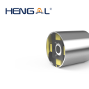
Shenzhen HenGal Innovation Technology Co.,Ltd. is a professional endoscope manufacturer with many years of production experience, advanced production technology and excellent product quality. 2.1mm OV6946M camera module with LED is one of our products,more details as follow:
Camera Model NO.
02A2101
Camera diameter
2.1 mm
Camera Module Dimension
2.1 mmx5.0 mm
Tube material
Stainless steel
Image device
1/18” High Color Sensor
Pixel Size
1.75µm x 1.75µm
Effective Picture Element
400(H)X400(V)
If you are interested in our products, please contact us as soon as possible.
Related news of endoscopes
Research progress and optimization direction of medical endoscope
Future prospect of medical endoscope device application
Application of oral endoscopy in the treatment of endodontics
0 notes
Text
Application of oral endoscopy in the treatment of endodontics

The most common diseases of endodontics are endodontic and periapical diseases. There are two types of modern endodontic treatment: non-surgical treatment and surgical treatment.
Non-surgical treatments mainly include root canal therapy and re-treatment. The complex structure of the root canal system, deep location, age-related changes, and chronic inflammatory stimulation will increase the difficulty of endodontic treatment and reduce the success rate. Therefore, the rational application of illumination and magnification equipment in endodontic treatment is very necessary.
The more mature magnification devices currently used in clinical practice include magnifying glass, microscope, and endoscope.
Some scholars have compared the clinical symptoms and imaging manifestations after endodontic treatment and found that the effective application of the three magnifying devices in the treatment process can significantly improve the success rate of treatment. Still, there is no significant statistical difference between the three. difference.
The disadvantage of the microscope in clinical application is that the operator's hands and instruments are often between the eyepiece and the surgical field, obscuring the operating field of view, resulting in inaccurate operations. Endoscopes can ultimately make up for this shortcoming. Its unique three-way handle and compact probe design determine that it has the following characteristics:
(1) Good vision and easy operation;
(2) Provide excellent visibility during surgery;
(3) Integrate magnification, irrigation/suction, and microsurgery equipment into one.
The development of oral endoscopy
In 1806, the first rigid medical endoscope, the light guide, appeared in Germany, and a new era of endoscope application in clinical medicine came. It was not until the end of the 20th century that the clinical endoscope was improved by adding a light source and magnifying equipment, and an endoscope specially used in the oral cavity, the oral mirror, appeared for the first time.
The oral mirror can effectively illuminate the inside of the root canal, observe the condition of the apical area, evaluate the filling effect of the root canal, and determine whether there is a root fracture. The development of microscopic instruments and equipment has continuously promoted the rapid development of microscopic endodontic treatment. The diameter of the anterior segment of the currently used fiberoptic endoscope is small, which improves the comfort of the patient in seeing a doctor. Endoscopy has played an increasingly important role in clinical work. One of the main directions of current research is to combine ultrasound technology, laser technology, confocal technology, etc. to form ultrasound endoscopy, laser endoscopy, and confocal endoscopy.
The use of endoscopes makes it possible to perform illumination, magnification, cleaning, and root canal shaping at the same time. At present, the most commonly used endoscope for endodontic treatment is a modular endoscope, which is a type of electronic endoscope. The modular endoscope consists of a mechanical system and an optical system.
The optical system includes eyepieces, endoscopes (objectives), tee-end handles that carry disposable instruments, and the like. The imaging principle is: the object is imaged on the photosensitive surface of the image sensor through the objective lens, the image sensor converts the optical signal into an electrical signal, and then transmits it to the image processing system through the optical fiber bundle, and obtains the final image after processing.
The most characteristic structure of the modular endoscope is its unique handle structure. Three access ports are installed at the end of the ergonomic handle, one for the ultra-fine fiber optic probe and two for the irrigation/suction system and other equipment. The effective connection of the irrigation/suction system is very important in the clinical application process. It can form an effective optical cavity in the root canal during the operation, which is more conducive to the observation of various structures during the operation.
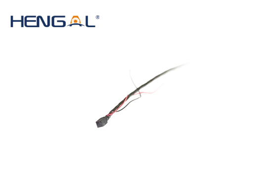
Shenzhen HenGal Innovation Technology Co., Ltd. is a professional endoscope manufacturer with many years of production experience, advanced production technology, and excellent product quality. 2.1mm OV6946M camera module with LED is one of our products, more details are as follow:
Camera Model NO.
02A2101
Tube material
Stainless steel
Image device
1/18” High Color Sensor
Pixel Size
1.75µm x 1.75µm
Effective Picture Element
400(H)X400(V)
If you are interested in our products, please contact us as soon as possible.
Related news of endoscopes
Research progress and optimization direction of medical endoscope
The operation process of using a bronchoscope during surgery
Future prospect of medical endoscope device application
0 notes
Text
Future prospect of medical endoscope device application

Medical endoscopes can improve diagnostic efficiency. The process of interventional diagnosis and treatment mainly depends on its design structure and imaging principle. The interventional process is prone to produce uncomfortable symptoms or affect the diagnosis and treatment effect. If the structure of traditional medical endoscopes can be improved and imaging technology can be improved, It will provide important help for medical endoscopic interventional diagnosis and treatment.
Analysis and research progress of key problems in medical endoscope devices
At present, medical endoscopes have been used in various departments of the hospital, from simple internal organ inspection to interventional treatment. Endoscopic minimally invasive technology has become an indispensable key technology for the diagnosis and treatment of systemic diseases such as digestion, breathing, urology, and ENT. However, there are still some problems in the use of medical endoscopes, such as small adjustment angle, fragile mirror body, poor imaging effect, complex structure, and defective illumination light sources, which restrict the further development of medical endoscopes.
With the combination of medical endoscopes with a series of high-tech technologies such as ultrasound technology and microscopic technology, more advanced medical endoscopes will be invented in the future. The future development of medical endoscopy is mainly divided into the following aspects:
1) Miniaturization
The size of the end of the medical endoscope conduit directly affects the degree of the trauma of the patient. By redesigning the medical endoscope operating part and other structures, the structure is compact, thereby reducing the contact caused by the small space of the patient's end cavity. Irritation and discomfort accelerated recovery.
2) Semi-automation of interventional diagnosis
The image processing methods supported by computer technology are integrated into medical endoscopes, and a computer-aided semi-automatic diagnosis system is developed to reduce the dependence on the doctor's experience and improve the accuracy of diagnosis and treatment.
3) High fidelity
Since the image quality of the endoscope is directly related to the process and trend of the entire operation, the improvement of the medical endoscope webcam and lens is used to solve the problem that the resolution affects the imaging of the affected area.
4) Improve imaging effect
In order to prevent the artifacts caused by the movement of the conduit during the diagnosis process, the image imaging rate of the endoscope is improved by redesigning the optical system of the medical endoscope.
5) Develop multimodal systems
Using a modular design scheme, various endoscopes can be used in combination with different converters to improve the accuracy of detection and tissue classification.
With the improvement of the mechanical structure of medical endoscopes, the development of imaging technology, and the continuous improvement of related technologies and theories, more mature medical endoscope devices are bound to appear in the future.
Shenzhen HenGal Innovation Technology Co., Ltd. is a professional endoscope manufacturer with many years of production experience, advanced production technology, and excellent product quality. 1.4mm OV6946M camera module without LED is one of our products, more details are as follow:
Camera Model NO.
02A1401
Camera Diameter
1.4mm
Camera Module Dimension
1.05mmx1.05mmx2.26mm
Image device
1/18” High Color Sensor
Pixel Size
1.75µm x 1.75µm
Effective Picture Element
400(H)X400(V)
If you are interested in our products, please contact us as soon as possible.
Related news of endoscopes
Research progress and optimization direction of medical endoscope
Application of endoscope in detecting leakage of landfill
Why are the prices of industrial endoscopes so different?
0 notes
Text
The operation process of using a bronchoscope during surgery

The medical endoscope mainly includes a power supply module, an acquisition module, a light source module, and a processing module.
A power supply module
The medical endoscope power supply module system mainly uses power management chips such as TPS70302PWP, which can eliminate the interference between different circuits and supply power to each module of the medical endoscope system;
An acquisition module
The medical endoscope acquisition module is mainly divided into two parts, namely the front-end adapter and the image sensor. The function of the front-end conversion head is to connect various soft and hard medical endoscopes with the host, and it contains a series of lens groups. , to ensure the integrity of the pictures collected by the camera at the front end of the catheter;
A light source module
The medical endoscope light source is mainly guided by the optical fiber inside the medical endoscope to provide illumination for the patient's diseased part. Due to the small size and low calorific value of LED lights, medical endoscopes currently use LEDs as the light source;
A processing module
The medical endoscope processing module is the most important module of the medical endoscope. The integration of the medical endoscope is improved by processing chips such as programmable logic gate arrays, thereby improving the imaging effect;
An image sensor
The image sensor mainly converts the collected optical signals into electrical signals that can be processed. Due to the high resolution and small size of the CCD sensor, the current medical endoscope mainly uses the CCD sensor as the image sensor.
In order to obtain the lesion information of the patient's internal organs and improve the treatment efficiency, doctors usually use medical history analysis, physical examination, and pathological examination to diagnose tissue lesions.
Among them, observing and taking out the diseased tissue from the patient's body for analysis is a commonly used diagnostic method in the process of medical endoscopy diagnosis and treatment. Taking a bronchoscope as an example, the patient being treated is in a state of sedation or anesthesia. The professional doctor holds the operation part of the medical endoscope with the left hand, and the catheter of the insertion part of the medical endoscope is placed into the lesion through the patient's orifice with the right hand. The light source and lens are used to capture the intratracheal image, transmit the image to the terminal computer, and display the patient's body image in real-time through the terminal computer display; the doctor changes the posture of the catheter in the patient's body by controlling the twisting, insertion, and removal of the catheter, etc. In this way, the contact and friction between the front end of the catheter, the tube wall, and the tracheal tissue are reduced, and the bending of the front end and the curved part of the catheter is controlled by the bending control of the operation part, so as to adapt to the curvature change of the tracheal path and the selection of the path at the bifurcation; the doctor can monitor the entire exploration process through the monitor. Observe the lesions of the tissue. When the front end of the medical endoscope lens reaches the lesion for corresponding treatment, the suction tube, puncture needle, biopsy forceps, etc. can be extended through the opening of the operating part along the catheter from the opening at the front end of the catheter. Depending on histopathology, doctors perform bronchial interventional diagnosis and treatment through biopsy forceps, basket dilation, and puncture needle puncture.
Shenzhen HenGal Innovation Technology Co., Ltd. is a professional endoscope manufacturer with many years of production experience, advanced production technology, and excellent product quality. 4.5mm medical endoscope camera module 4.5mm is one of our products, more details are as follow:
Camera Model NO
03D4506
Camera Module Dimension
4.5mmx18mm
Camera Direction
Front view ( 0°)
Tube material
Stainless steel
Effective Picture Element
640(H)X480(V)
If you are interested in our products, please contact us as soon as possible.
Related news of endoscopes
Research progress and optimization direction of medical endoscope
Application of endoscope in detecting leakage of landfill
Why are the prices of industrial endoscopes so different?
#medical endoscope processing module#medical endoscope power supply module#medical endoscope acquisition module
0 notes
Text
Research progress and optimization direction of medical endoscope
With the deepening of the world's aging trend and increasingly serious environmental problems, the demand for endoscopes continues to increase. Endoscopic technology has attracted widespread attention in the medical community due to its high accuracy of diagnosis and treatment, less trauma, less infection, quick recovery after surgery, and almost no scars, and has become one of the fastest growing products in the global medical device industry.
In recent years, although doctors can directly observe the morphology of human internal organs and lesions in the body through conventional medical endoscopes, the quality of surgery is still limited by the structure and imaging technology of medical endoscopes when doctors perform endoscopic diagnosis and treatment. , which may occur during the actual interventional operation. Medical endoscopes can effectively diagnose diseased organs in patients. In the process of medical endoscope interventional diagnosis and treatment, the diagnosis and treatment results mainly depend on its design structure and imaging principle. The interventional process is prone to produce uncomfortable symptoms or affect the diagnosis and treatment effect. The traditional medical endoscope structure and improved imaging technology will provide important help for medical endoscope interventional diagnosis and treatment.
Difficulties
1. Since the catheter of the medical endoscope needs to be inserted from the patient's orifice into the patient's body, there will inevitably be contact friction between the catheter and the patient's orifice tissue. If the resistance and torsional force on the catheter cannot be accurately fed back to the doctor, it will cause inconvenience to the patient. Necessary operational trauma.
2. When the endoscope is intervened, due to the narrow space of the patient's orifice, its movement in the patient's orifice is limited. When the doctor manipulates the endoscope, the movement of body joints such as the wrist and shoulder will cause the positional deviation and jitter of the endoscope catheter. , affecting the imaging effect and surgical accuracy.
3. At present, the operability of microscopic examination has high technical requirements for doctors. Therefore, clinicians generally need to be trained, and the learning cycle is long and difficult. As a result, there is an obvious supply and demand gap between the number of doctors operating microscopic examinations and the number of patients.
4. In the process of endoscopy, due to the patient's stress response, will cause damage to the internal fiber bundles of the medical endoscope, affecting the image quality and increasing the operation cost.
Prospects
Through the above analysis, there is still room for improvement in the design and use of medical endoscopes, and further integration and intelligence are needed. By improving the mechanical structure of medical endoscopes and optimizing imaging technology, the endoscope can be the purpose of moving into different cavities and obtaining high-definition images of lesions.
Shenzhen HenGal Innovation Technology Co., Ltd. is a professional endoscope manufacturer with many years of production experience, advanced production technology, and excellent product quality. 3.0mm side view endoscope camera diameter 3.0mm is one of our products, more details as follows:
Camera Model NO
02A3001
Camera Diameter
3.0 mm
Camera Module Dimension
3mmx11mm
Camera Direction
Side view ( 90°)
Tube material
Stainless steel
If you are interested in our products, please get in touch with us as soon as possible.
Related news of endoscopes
Application of endoscope in detecting leakage of landfill
Why are the prices of industrial endoscopes so different?
Application of industrial endoscope in the automotive industry
0 notes
Text
Application of endoscope in detecting leakage of landfill
HDPE film is often used to prevent leakage of domestic waste in landfills, and HDPE film may be damaged during production, transportation, and construction. In addition, during the operation of the landfill, the HDPE membrane will also be damaged due to various factors such as uneven subsidence of the foundation, plastic deformation, mechanical damage, etc. After the leakage of the anti-seepage system of the landfill occurs, the infiltrated leachate will pollute the soil and groundwater. Therefore, the accurate detection of the leakage point of the anti-seepage system has become an important prerequisite for the efficient repair of the anti-seepage membrane. The endoscopy method is applied to the landfill and combined with the previous investigation, the possible leakage range and the high probability leakage area can be inferred, which can effectively reduce the excavation scope and reduce the number of excavation works, which can be used for similar landfills. Leakage point exploration work provides reference.
Based on the experience of similar projects, the leakage of the landfill site with a greater impact mostly occurs at the corner of the anti-seepage system in the separation dam, pipe through the membrane, and near the dam. Based on the preliminary investigation results of this project, it is suggested to carry out local trial excavation in the 45-47 m area near the dam area.
Test indicators and analysis methods
The exploration objectives of each exploration pipe section are as follows:
(1) C1-C2 pipe section
Since the position of the pipe section is below the adjustment tank and is not connected to the bottom pipeline in the upstream landfill reservoir area, the actual exploration depth is about 48 m, which mainly reflects the horizontal anti-seepage situation at the bottom of the adjustment tank area.
(2) B0-B1 pipe section
The position of the pipe section is located below the adjustment tank and is connected to the upstream pipe section. The main reaction adjusts the horizontal anti-seepage and leakage situation at the bottom of the tank area.
(3) Blind ditch in A1-A2 segment
A1-A2 is a type A blind ditch, close to the second and third quadrants. From the design slope of the reservoir bottom, the groundwater in the blind ditch mainly comes from the second quadrant, and the groundwater in the third quadrant does not flow to A1-A2; therefore, the exploration results of this section mainly reflect the level of seepage prevention in the second quadrant of the first-phase landfill reservoir area. leakage.
(4) Blind ditch in the B1-O segment
B1-O belongs to the B-type blind ditch, close to the third and fourth quadrants. According to the design slope of the reservoir bottom, the groundwater of the blind ditch in the B1-O section comes from the third and fourth quadrants. Therefore, the exploration results of this section mainly reflect the leakage situation of the third and fourth quadrant horizontal anti-seepage.
(5) Blind ditch in the O-B2 segment
O-B2 belongs to the B-type blind ditch, close to the first and second quadrants. The slope line that parallels the blind ditch in the O-B2 segment is gentle (1.5%-2.0%). The slope line intersecting with it has a larger slope (4.4%~6.1%). Therefore, the exploration results of this section can reflect the leakage situation of the first and second quadrants.
Through the exploration and analysis of the leakage zone through the endoscope, the possible leakage range and the high probability leakage range are obtained through comprehensive analysis:
(1) Possible leakage range
The most prominent position of the leachate-contaminated groundwater pipe is located in the O-B2 section, and the overall pollution situation in the A1-A2 groundwater pipe section is good, indicating that the first quadrant and the second quadrant are close to the O-B2 area It is the possible leakage range; high-risk leakage area: in the 1st and 2nd quadrants, the area below the 48 m elevation of the anti-seepage layer is the possible leakage range.
(2) High probability leakage area
The area enclosed by the 46 m line, 48 m line, A-type blind ditch, and B-type blind ditch in the first quadrant is determined as the high probability leakage area. After considering the construction space, the floor area of the reservoir in this area is about 4,963 m2, and the excavated earthwork volume is estimated to be 216,600 m (calculated based on the average thickness of the three piles of 20 m).
This research is a practical study of leakage detection in sanitary landfills, and it has important reference significance for accurate detection of leakage points and effective repairs after leakage problems in sanitary landfills, and effective control of leachate pollution and diffusion.
Shenzhen HenGal Innovation Technology Co., Ltd. is a professional endoscope manufacturer with many years of production experience, advanced production technology, and excellent product quality. 2.1mm OV6946M camera module with LED is one of our products, more details are as follow:
Camera Model NO.
02A2101
Camera diameter
2.1 mm
Camera Module Dimension
2.1 mmx5.0 mm
Tube material
Stainless steel
Image device
1/18” High Color Sensor
If you are interested in our products, please contact us as soon as possible.
Related news of endoscopes
Why are the prices of industrial endoscopes so different?
Application of industrial endoscope in the automotive industry
Industrial endoscope waterproofing and sealing testings
0 notes
Text
Why are the prices of industrial endoscopes so different?
There are many types and functions of industrial endoscopes on the market, and the prices are also very different. Some are less than 100 yuan, and some are more than 10,000 yuan. Most of them are more than a few thousand yuan. The reason for the big price difference is that its detection is more refined. The image is clearer and the function is more complete. The industrial endoscope has a complex structure and consists of multiple parts such as a control power supply, host, control system, insertion tube, lens, and lighting source, among which the lens and host are very important. Due to the different materials and costs of some industrial endoscopes, the prices of different industrial endoscopes are also very different. So why are industrial endoscopes so expensive? We can learn from the following aspects.
1. Pipeline
The price of industrial endoscopes is mainly determined by the lens part, the pipeline part, the host part, etc. For different situations, customers will customize pipelines with different diameters, such as inspection pipelines. Generally, pipelines with small diameters are relatively thin. However, if you encounter objects with very small holes, you need to customize ultra-small diameter pipelines. The smaller the pipe diameter, the higher the process requirements and the more expensive the price.
2. Probe
The probe of the industrial video endoscope is the core component of the endoscope, which directly affects the speed of the detection effect and the efficiency of the calibration. The probe can be bent at a large angle. And the industrial endoscope with a guiding function can make detection easier. The inspector can adjust different degrees of tightness, and control the probe guide joint to bend in different directions through the change of the traction force, so as to realize the rotation of the probe in different directions and make the detection range more comprehensive. Therefore, the probe bending angle and guiding function greatly affect the price of industrial endoscopes.
3. Optional function
In addition to the basic configuration, many customers will also request to customize other functions, such as the c temperature warning function, infrared function, infrared thermal imaging function, VFT wireless function, recording function, and so on. Depending on the actual optional functions, the price of the tasks of the industrial endoscope will also increase. The price of industrial/endoscopes ranges from tens to hundreds or even tens of thousands. If it is only used to detect household pipes in daily life, then choosing ordinary industrial endoscopes is enough to meet the needs of users. If it is used to detect some high-tech and high-precision products, it is necessary to choose a clearer and smarter industrial endoscope.
Through analysis, you can see why industrial endoscopes are so expensive. You get what you pay for. Industrial endoscopes can detect the quality of precision instruments. Then industrial endoscopes must be more precise than precision instruments to ensure the detection effect. , Now the prices of industrial endoscopes on the market are different, and there are also relatively cost-effective industrial endoscopes. When choosing, try to choose the industrial endoscope products that suit you, not just the price.
Shenzhen HenGal Innovation Technology Co., Ltd. is a professional endoscope manufacturer with many years of production experience, advanced production technology, and excellent product quality. If you have any questions, please consult us, we will regularly share professional knowledge of industrial endoscopes to help you better understand industrial endoscopes.
Related news of industrial endoscopes
Solutions for blurred images of industrial endoscopes
Industrial endoscope waterproofing and sealing testings
Application of industrial endoscope in the automotive industry
0 notes
Text
What precautions should be taken before and after colonoscopy?
Colonoscopy, also known as an electronic colonoscopy, is a diagnostic method that doctors use to examine the interior of the large intestine and terminal ileum. A colonoscope is a slender, flexible instrument, about 1 cm in diameter, that is passed through the anus into the large intestine. The front end of the electronic colonoscope is equipped with a camera, and the image is transmitted to the display through the optical fiber, allowing the doctor to see the picture inside the intestine in real-time; it can also transmit the signal to the acquisition system to take a picture of the inside of the intestine. Usually, there is also a channel for placing operating instruments inside the colonoscopy, so that the tissue can be clamped under the colonoscope for pathological examination, and even operations such as resection of lesions can be performed.
In what situations do you need a colonoscopy?
1. Unexplained blood in the stool.
2. Changes in defecation habits: increased stool frequency or frequent bowel movements, but no stool discharge, or constipation.
3. Chronic diarrhea of unknown cause.
4. Mucus or pus and blood-like fluid in the stool.
5. Stool thinning. Flattened.
6. Abdominal mass, cannot rule out the large intestine and terminal ileum lesions.
7. Middle and lower abdominal pain of unknown cause.
8. Suspected benign or malignant colon tumor, which cannot be diagnosed by X-ray examination.
9. Suspected chronic intestinal inflammatory disease.
10. If an abnormality is found in barium enema or bowel system examination, it is necessary to further clarify the nature and scope of the lesions.
11. Determining the lesion range before the colon cancer surgery, re-examination, and curative effect follow-up after colon cancer polyps.
12. Abdominal distension. Abdominal pain and other unexplained low intestinal obstruction.
How to prepare for a colonoscopy?
The day before colonoscopy:
1. Only eat a slag-free semi-liquid diet, and do not eat vegetables and fruits containing fiber.
2. Take a laxative (such as 2 bisacodyl enteric-coated tablets) before going to bed at night.
Colonoscopy day:
1. Fasting before colonoscopy.
2.8:00 Start taking intestinal cleansers (such as polyethylene glycol electrolyte powder, see the video for how to take it). It is recommended not to go out at this time, so as not to find the toilet.
3. If necessary, take an antifoam (such as Simethicone Emulsion 30ml) one hour after taking the bowel cleanser. So, all the medicines have been taken.
4. Excretion will start about an hour after taking the medicine. When the excrement turns yellow or colorless water-like stool, the intestinal preparation is basically over.
What should I pay attention to after the inspection?
1. It is recommended that patients come to the hospital to be accompanied by their family members.
2. Stop taking iron grades (such as ferrous sulfate, polysaccharide iron, etc.) three days before the examination.
3. Long-term constipation patients should explain to the doctor in advance.
4. No food was eaten on the day of the colonoscopy, and hypoglycemic drugs before meals do not need to be taken.
5. Antihypertensive drugs should be taken one hour before bowel cleansing.
6. On the day of the colonoscopy, if there is dizziness and hypoglycemia without eating, you can take sugar water to replenish energy.
7. Be sure to inform your doctor about the long-term use of anticoagulants (such as aspirin, warfarin, clopidogrel, etc.).
8. If severe abdominal pain, abdominal distension, blood in the stool, etc. occurs 1-2 days after the examination, seek medical attention immediately.
9. Do not drive, ride, or work at heights within 8 hours after colonoscopy.
Shenzhen HenGal Innovation Technology Co., Ltd. is a professional endoscope manufacturer with many years of production experience, advanced production technology, and excellent product quality. If you are interested in our products, please contact us as soon as possible.
Related news of endoscopes
Factors Affecting the Price of Handheld Endoscopes
Uses of industrial video endoscopes
How to use the engine endoscope
0 notes
Text
Industrial endoscope waterproofing and sealing testings
Industrial endoscopescan be used for direct inspection and observation of harmful and narrow environments that cannot be seen by the naked eye, such as liquid environments, pipeline environments, etc.
The importance of waterproof test
Industrial endoscopes used to detect liquid environments, pipeline environments, etc. must have the functions of small size and strong protection ability. Waterproofing is one of the important functions. If the sealing measures are not in place, the lens and screen will enter the water, the function of the endoscope will be affected, and even short circuit, leakage, etc. will cause damage to the internal components. Therefore, in order to avoid unexpected situations, the endoscope must be tested to check its waterproof function after being built, and only in this way can the endoscope be used normally. So how is the waterproof industrial endoscopes testing done?
The specific steps of waterproof test
The sealing condition of the endoscope determines whether its waterproof function can be achieved. Therefore, the industrial endoscope sealing must be tested. Because the shape of the endoscope is irregular, it is necessary to make corresponding tooling fixtures according to the shape of the product. The endoscope is composed of multiple parts, the upper part is a rectangular lens, the lower part is a cylinder, and the end is also connected to the control and signal transmission harness. What is to be tested is the overall sealing except for the wiring harness, so the fixture is an integral sealing tool, and the wire grooves flow out of the fixture. Connect the jig to the sealing testing equipment, summarize and analyze the data of qualified products and unqualified products, and determine the effective parameters, such as detection time, leakage, etc., then the endoscope can be placed in the mold, the tooling fixture can be closed to complete the sealing, and finally Using the pressure drop method to obtain the waterproof test results of industrial endoscopes.
Single industrial endoscopes Suppliers - HENGAL
Shenzhen HENGAL Innovation Technology Co., Ltd. is a professional endoscope manufacturer with many years of production experience, advanced production technology and excellent product quality.Such as waterproof industrial endoscopes, PA Series Two-Way Articulation Industrial Endoscope is one of our products, more details as follow:
Insertion Probe Diameter
6.0mm
Insertion Probe material
Stainless steel braided wear-resistant tube
Camera Resolution
1280*720 Pixels
Filed of Viewing
110°
Light Source
LED
If you are interested in our products, please contact us as soon as possible.
Related news of industrial endoscopes camera
Solutions for blurred images of industrial endoscopes
Uses of industrial video endoscopes
Precautions for Measuring Defects in Industrial Endoscopes
0 notes
Text
Application of industrial endoscope in the automotive industry
This article introduces the specific application of industrial endoscopes camera in the automotive industry, which can help you better choose the right product
Definition of Industrial Endoscopy Camera
The industrial endoscope camera is a visual inspection tool that can perform non-destructive inspection. It can be used to view instruments with a narrow internal environment and expand the observation range. Because of its efficiency and convenience, it is now well known and widely used in more industries.
The specific application of industrial endoscope camera in the automotive industry
The importance of industrial endoscope cameras is rising with the trend towards non-destructive testing. In automobile manufacturing, industrial endoscope cameras are mainly used for assembly inspection of engines and transmissions. Engines and transmissions are the core components, and car manufacturers have strict requirements on their quality. Cylinder block, cylinder head, turbine, transmission valve plate and other components are the main detection objects to avoid pores, cracks and impurities. In auto repair, because of its simple and convenient operation, it has become an indispensable testing equipment in the auto repair industry. The internal parts of the car that are invisible to the naked eye can be directly displayed on the display screen, and the maintenance personnel can directly obtain the images of the engine, compressor, valve, gearbox and other parts, quickly find out the fault, and make the maintenance more efficient and convenient. The industrial endoscope camera has high resolution, clear images, and also has the functions of magnification and video recording, which can better analyze faults. This greatly improves work efficiency, and also avoids damage that occurs when components are loaded and unloaded during the inspection process.
Industrial endoscope Suppliers - HenGal
Shenzhen HenGal Innovation Technology Co., Ltd. is a professional endoscope manufacturer with many years of production experience, advanced production technology and excellent product quality.Such as camera module,3.0mm side view endoscope camera 3.0mm is one of our products,more details as follow:
Camera Diameter
3.0 mm
Pixel Size
1.75µm x 1.75µm
Effective Picture Element
400(H)X400(V)
Power input
DC 3.3V
Operating Temperature
-20°C to +70°C
If you are interested in our products, please contact us as soon as possible.
Related news of industrial endoscopes camera
How to use an industrial endoscope correctly
Solutions for blurred images of industrial endoscopes
Common auxiliary tools for industrial endoscopes
0 notes
Text
Inspection of mechanical equipment by industrial endoscope
If you want to use industrial endoscopes flexibly, you need to master enough skills to make the most of it. There are many reasons for wear and tear in various mechanical equipment wear fault detection, and the phenomena presented are also different. Let's take a look at how industrial endoscopes can detect wear and tear on a variety of mechanical equipment.
There are changes in the surface shape of mechanical products, and the degree of loss is low. Industrial endoscopes can be used to observe subtle changes on the surface of various mechanical equipment. If there are wrinkles and cracks, using the beam to illuminate the mechanical surface can find black shadows and lines, and the surface will be irregular. Because the reflection angle is different, the brightness presented by different illumination effects is also different. At the magnification of the surface, the change of the shape of the material can be clearly displayed, so the situation of cracks and wrinkles is better judged.
These are indistinguishable to the naked eye, but are revealed in the endoscope. The degree of wear of scratches is greater, and the shadow is deeper when inspected with an industrial endoscope.
Unevenness caused by external force can also be detected. Turn on the light source, there are depressions in bright places, and the depressed places will be farther from the light source. On the contrary, if there are shadows, it will be a convex place. This is because the convex place is very close to the light source, and there will be shadows. After turning on the beam, the boundaries of the uneven places will not be too obvious, but they can still be seen clearly.
The wear and tear of mechanical equipment that can be detected by industrial endoscopes is not only the change of shape and structure, but also the wear and tear after chemical reaction. , the surface is uneven, and many small shadows can be seen under the magnification of the endoscope, which is likely to be corroded.
In addition, with the help of industrial endoscopes, inspectors can easily find out whether there are omissions and redundant parts in the process of assembling parts of machinery and equipment. It is easy to move, and even large machinery can be inspected comprehensively.
Shenzhen HenGal Innovative Technology Co., Ltd. is a professional endoscope manufacturer. The company has been focusing on the design and application of endoscopic imaging systems since 2011. As a R&D-driven solutions company, we always adhere to the principle of "innovative and pragmatic" and put customers first. Contact us if you are interested in our products.
0 notes
Text
Industrial endoscope for non-destructive testing
Endoscope is a multi-disciplinary universal tool. Its function is to explore the depths of curved pipes, observe the parts that cannot be seen directly, observe the internal space structure and state in the sealed cavity, and realize long-distance observe and act. Endoscopic inspection is a method of using industrial endoscopes to inspect the interior of containers, pipelines, non-removable equipment, the inner surface of narrow gaps, the parts below the liquid level such as water and oil, and the areas that cannot be directly observed by human vision in special environments. Controlled visual inspection method.
Principles of nondestructive testing of industrial endoscopes
Industrial endoscopy is a branch of non-destructive testing, and it can also be said to be a specialized testing technology.
Due to its special size design, the industrial endoscope allows us to observe the internal surface structure or working state of the object easily and accurately without destroying the surface of the detected object.
Non-destructive testing requires the use of industrial endoscopes as testing tools, which are professionally designed and produced to meet complex industrial environments. Endoscopic inspection is a kind of inspection technology that has been widely used in recent years with the development of endoscope manufacturing technology. According to the characteristics of the manufacturing process, we generally divide industrial endoscopes into three types: optical rigid tube mirrors, fiber optic mirrors, and video mirrors.
Industrial endoscopes can be used for inspection and observation of high temperature, toxic, nuclear radiation and places that cannot be directly observed by the human eye. They are mainly used in automobiles, aircraft engines, pipelines, mechanical parts, etc. Non-destructive testing can be realized when the operation is stopped. On the other hand, the industrial endoscope can also be connected with a camera, a video camera or an electronic computer to form a photographing, imaging and image processing system, so as to monitor, record, store and map the field of view objects. Image analysis. Provides a good guarantee for diagnosis and treatment.
Development Trend of Industrial Endoscopy
Industrial endoscope technology has unique advantages, it can extend people's sight distance, and can change the direction of sight arbitrarily, and accurately observe the real condition of the inner surface of the object, which cannot be replaced by other detection instruments. For example, in aviation applications, the industrial sight glass can be extended into the interior of the aircraft engine to directly observe the actual internal conditions after operation or the internal surface conditions of equipment parts; effectively detect the surface conditions of relatively concealed or narrow parts, while There is no need to disassemble equipment or components for destructive inspection. The method is simple and reliable, very intuitive and effective. Whether it is in the aerospace field, the electronics industry and other manufacturing and maintenance industries, the use of endoscopes for NDT detection has achieved good results, solved many difficult problems, ensured the normal operation of the entire set of equipment, and avoided some accidents.
Shenzhen HenGal Innovative Technology Co., Ltd. is a professional endoscope manufacturer. We have advanced technology and excellent product quality. As a R&D-driven solutions company, we always adhere to the principle of "innovative and pragmatic" and put customers first. We look forward to working with you.
0 notes
Text
The difference between electronic endoscope and fiberscope
The market of fiber endoscopes used a long time ago is now basically replaced by electronic endoscopes. So what are the differences between electronic endoscopes and fiber endoscopes? In terms of use and technology , What are the differences between electronic endoscopes and fiber endoscopes? Although they are both endoscopes, they all play a great role in medical inspection and other fields, but with the development of science and technology, electronic endoscope technology has nearly Quite mature, combined with other precision instrument manufacturing, it is developing towards miniaturization and intelligence.
The essential difference between electronic endoscope and fiber endoscope:
The structure of the electronic endoscope is basically the same as that of the fiber endoscope. It can be simply understood that the CCD replaces the image guide beam, and many functions cannot be achieved by the fiber endoscope. The biggest difference between the electronic endoscope and the fiber endoscope is that the optical fiber image transmission beam is replaced by a CCD device called a miniature image sensor.
During the use of the endoscope, the bending part and the insertion tube of the lens body need to continuously perform repeated bending movements. Each component inside the endoscope is then subjected to various stresses such as bending, friction, and extrusion. Bundles are no exception.
In terms of the ability to resist external stress, the fatigue strength of cables is much greater than that of optical glass fibers. If other factors affecting the service life of endoscopes are not considered for the time being, the theoretical service life of electronic endoscopes is much longer than that of fiber endoscopes from the fact that the optical fiber is easily broken under the action of external force.
Another technical problem is the image clarity of the endoscope or the resolution of the endoscope. As far as the current mature technology is concerned, the resolution of the fiber endoscope is far less than that of the electronic endoscope. The resolution depends on the diameter of the optical fiber monofilament, and the space for improving the resolution by reducing the diameter of the optical fiber monofilament is quite small. The products of some manufacturers can be said to be close to the limit, which is only equivalent to the early low-end electronic endoscopes s level.
To sum up, the fiber endoscope has basically reached its technical limit. At present, it has been eliminated by the market without breaking the technical bottleneck, and the use of electronic endoscope has become an objective demand of the market.
Shenzhen HenGal Innovative Technology Co., Ltd. is a professional endoscope manufacturer. We have advanced technology and excellent product quality. As a R&D-driven solution company, we always adhere to the principle of "innovation and pragmatism", bringing customers Put it first. If you are interested in our products, please contact us as soon as possible, we will provide you with the best service.
0 notes
Text
Classification of Bronchoscopy
1. Rigid Bronchoscopy
structure
The modern rigid endoscope is a hollow stainless steel tube with a uniform diameter and a wall thickness of 2-3mm, which is similar to the rigid endoscope designed by Dr. Jackson 100 years ago. The diameter of the adult hard endoscope is 9-14mm, the length is about 40cm, and the distal end is beveled, so that it can pass through the glottis and airway stenosis, and it is also beneficial to remove the tumor on the airway wall. With a side hole, it is convenient to maintain ventilation of the contralateral airway when the scope enters the main bronchus of one side.
The operating end of the rigid mirror has multiple interfaces, including ventilator interface, light source interface, suction tube and laser fiber interface. The proximal end of the opening can be closed or open to facilitate passage of viewing eyepieces and other facilities.
2. Flexible bronchoscope:
Flexible bronchoscopy, also known as flexible bronchoscopy, is divided into fiberoptic bronchoscopy and electronic bronchoscopy.
1) Bronchofiberoscope (Bronchofibroscope)
Fiberoptic bronchoscopy consists of two parts, optical and non-optical. Its basic components are glass-fiber bundles. Each bundle of fibers has a diameter of 8-12 μm, and is covered with a second layer of glass, that is, the outer membrane. The light is constantly reflected back and forth by the beam wall, from the lens to the eyepiece in the operator's hand at a rate of 10,000 reflections per minute, and the outer membrane helps maintain image stability during the process. There is also an access channel in the cable that extends from the distal end to the handle. This channel can be used for suction, insufflation of oxygen, instillation of irrigation fluids or drugs. However, fiberoptic bronchoscopes with an outer diameter of <2 mm have no operating channel. Two wires from the handle lever run in the fiber optic cable and can control the movement of the tip in the sagittal plane. The metal "jacket" protects the entire length of the inserted fiber optic cable up to the level of the distal end, and the end of the fiber optic cable is hinged. Movement in the coronal plane is achieved by controlling the lever and rotating the entire fiberoptic bronchoscope from the handle to the distal end. The final component is the light source, 1 or 2 separate fiberglass bundles that transmit light from the shank to the distal end to illuminate the target. A "universal" fiber optic cable from the handle can be connected to the medical endoscope light source.
2) Electronic bronchoscopy
The electronic bronchoscope is connected to a miniature charge coupled device (CCD) interface at the eyepiece of the fiberoptic endoscope, which can convert the image into a digital signal, and then display it on the TV system, which can provide timely image capture, video recording, editing, etc. Function. This CCD can convert light energy into electrical energy, and then undergo video processing, that is, perform a series of processing on the image and store and regenerate the image in various ways, and finally display it on the TV screen, with clear images, vivid colors, It has the advantages of high resolution, magnification, photography, video recording, computer processing, data storage, easy operation, more safety and easy disinfection.
Shenzhen HenGal Innovative Technology Co., Ltd. is a professional endoscope manufacturer. We have advanced technology and excellent product quality. In the industrial field, we have developed a variety of handheld endoscope series, which are sold well in the market. It is a professional non-destructive testing tool in the fields of aerospace, automobile production and maintenance, power production and construction units, precision casting and machinery manufacturing, petroleum, chemical industry, pressure vessels, national defense and military industries. If you are interested in our products, please contact us.
0 notes
Text
Basic structure and working advantages of industrial endoscope
Recently, technology is marching toward breadth and depth, and industrial machinery is no exception. The mechanical structure is becoming more and more complex, and visual inspection, as a common form of non-destructive testing, is difficult to meet the inspection needs of fine machinery. The aid of scientific instruments is preferred. Industrial endoscope is a kind of non-destructive testing equipment that is widely used in industrial manufacturing and maintenance fields, breaking the limitation of the naked eye and can quickly detect mechanical details.
Industrial endoscope is a non-destructive testing instrument integrating electricity, light and machine. The structure of traditional industrial endoscope is composed of five parts: bending joint, snake tube, host, camera and display screen. The inspector holds the host, extends the snake tube into the area to be detected, and controls the bending joint to make the camera obtain the sound and image of the detected object.
Industrial endoscopes include three types, namely rigid endoscopes, fiber endoscope series and electronic video endoscopes.
Rigid endoscopes use relay lens optical technology to transmit images by providing light-transmitting illumination through optical fibers. When working, insert the effective stainless steel of the speculum into the relay lens system of the optical element, and the built-in optical fiber transmits the light to be photographed from the independent cold light source to the working position. This endoscope is mostly used in the inspection of the main body frame, turbine blades, and combustion chambers in the aerospace industry.
Fiberscope is to observe the image transmitted by the image transmission fiber through the eyepiece. The fiberscope is inserted into the front end, the bending part and the soft part, the eyepiece part and the operation part form the image transmission fiber bundle, and the steel wires for adjusting the front end swinging angle are all built-in. Observing the image can be obtained, tens of thousands of glass fiber monofilaments group an imaging bundle, each monofilament is an imaging element. Under the same diameter, the more monofilaments in an image beam, the clearer the transmitted image and the longer the product life. In the machinery manufacturing and maintenance industries, fiber endoscopes can be used to observe the cavity of large equipment or components.
Electronic video endoscopes are still mainly fiber endoscopes and rigid endoscopes due to the high price of this product and the small number of domestic use.
Due to the special structure, the advantages of industrial endoscopes. Without disassembling the equipment, it extends the visual distance of people to the inside of the object to be measured, and can change any direction of sight, breaking through the blind spot of visual observation, deepening the sense of spatial depth of the photographic picture, and accurately observing the wear and tear in the object. , damage, surface cracks, burrs, abnormal attachments, etc. Reduce the damage of internal parts, easy operation and high inspection efficiency.
Now industrial endoscopes have been widely used in printing technology, document management, computer image processing, image measurement and analysis, and completed the capture, storage, processing and analysis of endoscope images, and writing standardized reports.
Shenzhen HenGal Innovative Technology Co., Ltd. is a professional endoscope manufacturer. As a R&D-driven solution company, we always adhere to the principle of "innovative and pragmatic" and put customers first. If you are interested in our products, please contact us as soon as possible. We will provide you with the best service.
0 notes
Text
The development of gastroscope
The development of digestive endoscopy has gone through 4 periods:
rigid endoscope
In 1805, the German medical doctor Philipp Bozzini first proposed the idea of endoscopy.
In 1826, French urologist Pierre Segalas successfully developed cystoscope and esophagoscope.
In 1868, German doctor Adolph Kussmaul made the first esophagogastroscope.
semi-curved gastroscope
In 1932, the German technician Georg Wolf and the German doctor Rudolf Sc.hincller jointly developed it successfully. Most of the stomach can be observed. The patient can take the left side position, which makes the gastroscope reach a more practical stage. significance.
fiber endoscopy
In 1958, American gastroenterologist Basil Hirschowitz made the first fiber endoscope, and the endoscope entered the stage of fiber optics.
Electronic endoscopy
In 1983, the American WeIoh Allyn Company first developed the world's first electronic gastroscope, which was the third milestone in the history of endoscopy.
In 2000, Israel developed the first medical camera that continuously transmits images to the outside of the body. This type of endoscope is completely different from the above-mentioned types of endoscopes in terms of appearance and operation. It can be automatically recorded and excreted with gastrointestinal peristalsis without the need for a doctor to operate. It opens up a new avenue for endoscopy.
China Development
The development of endoscopy in China is relatively late. Since the 1950s, some large hospitals have carried out rigid endoscopy (or semi-curved endoscopy), but the number of endoscopy examinations per hospital rarely exceeds 50 times a year.
In the early 1970s, China began to introduce fiberoptic endoscopy, so that gastroscopy, colonoscopy, and endoscopic ret-rograde cholangio-pancreatography (ERCP) were gradually carried out.
Since the 1980s, it has developed rapidly. Electronic gastroscopy, ERCP examination, and endoscopic interventional therapy have basically been in line with international standards. By the 1990s, endoscopy has been popularized in primary hospitals across the country.
China began to develop fiberoptic endoscopes in 1966. In 1973, Shanghai Medical Optical Instrument Factory produced the first XW-I fiberoptic gastroscope, realizing the localization of fiberoptic endoscopes.
Shenzhen HenGal Innovative Technology Co., Ltd. is a professional endoscope manufacturer. The company has been focusing on the design and application of endoscopic imaging systems since 2011. We provide professional gastroscope solutions, if you are interested in our products, please contact us as soon as possible.
0 notes
Text
Precautions for gastroscopy
1. In order to prevent hepatitis infection, separate gastroscopy from patients with hepatitis and those without hepatitis, and perform liver function and hepatitis B surface antigen examination before gastroscopic examination.
2. In order to clearly see the mucous membrane of the digestive tract, the inspected part must be clean, that is, there is no food or blood clot remaining. Such as gastroscopy in the morning, after 8:00 pm the day before the examination, no food and beverages, no smoking. Eat low-residue, easily digestible food the night before. Even if the patient drinks a small amount of water, the color of the gastric mucosa can change. For example, in the natural lesions of significant atrophic gastritis, the gastric mucosa can turn red after drinking water, making the diagnosis wrong.
If gastroscopy is performed in the afternoon, the patient can drink some sugar water before 8:00 in the morning, but cannot eat other things, and not eat at noon. For patients with pyloric obstruction, gastric lavage must be performed the night before the examination to thoroughly wash out the gastric contents until the flushing fluid is clear. Before the gastric tube is withdrawn after gastric lavage, the patient is placed in a supine position with head down and feet up, so that the residual fluid in the stomach can be completely drained. Gastric lavage cannot be performed on the same day, as the color of the gastric mucosa can change after gastric lavage.
3. Local anesthesia is used for anesthesia, which is limited to the upper end of the throat and esophagus. Before taking any of the above medicines, tell your doctor about your history of allergies to medicines, that is, to what medicines you have been allergic to in the past.
4. The patient and the doctor should cooperate. Before the examination, the patient should go to urinate to empty the bladder. After entering the examination room, loosen the neckline and belt, remove the dentures and glasses, take the left lateral position, or change to other positions as needed.
5. After the examination, the patient sat up and spit out saliva. Although some air was injected during the examination, although it was sucked out when the mirror was withdrawn, some people still had a feeling of abdominal distension and a lot of belching. Because the anesthetic effect has not disappeared, it is easy for food to enter the trachea by eating too early, so 2 hours after the examination, try to eat liquid food after the effect of the pharyngeal anesthetic has disappeared.
Shenzhen HenGal Innovative Technology Co., Ltd. is a professional endoscope manufacturer. We have advanced technology and excellent product quality, and we are also a professional gastroscope solution supplier. If you are interested in our products, please contact us as soon as possible, we will provide you with the best service.
0 notes