
Don't wanna be here? Send us removal request.
Statistics
We looked inside some of the posts by pathologydiscussionforum and here's what we found interesting.
Average Info
Notes Per Post
2
Likes Per Post
2
Reblog Per Post
0
Reply Per Post
0
Time Between Posts
2 days
Number of Posts By Type
Text
17
Last Seen Tumblr Blogs


Fun Fact
Premium Tumblr themes are available from anywhere between $9 to $49.
Text

0 notes
Text
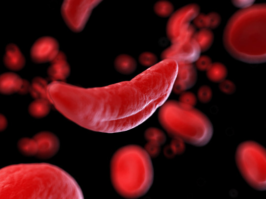
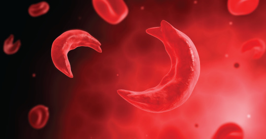
The sickle cell syndromes are caused by a mutation in the β-globin gene that changes the sixth amino acid from glutamic acid to valine.
HbS polymerizes reversibly when deoxygenated to form a gelatinous network of fibrous polymers that stiffen the RBC membrane, increase viscosity, and cause dehydration due to potassium leakage and calcium influx. These changes also produce the sickle shape. Sickled cells lose the pliability needed to traverse small capillaries. They possess altered “sticky” membranes that are abnormally adherent to the endothelium of small venules. These abnormalities provoke unpredictable episodes of microvascular vasoocclusion and premature RBC destruction (hemolytic anemia) in the liver and spleen. The rigid adherent cells also clog small capillaries and venules, causing tissue ischemia, acute pain, and gradual end- organ damage. This venoocclusive component usually dominates the clinical course. Prominent manifestations include episodes of ischemic pain (i.e., painful crises) and ischemic malfunction or frank infarction in the spleen, central nervous system, bones, joints, liver, kidneys etc
#hematology #hematologyoncology #hematologylab #hematologyforum #hematologygroup #hemato #hematologia #hematología #hematoloji #Pathology #pathologydiscussion #pathology #pathologynotes #pathology #patho #pathologist #pathologies #usmle #usmlestep1 #usmleprep #usmlestep2ck


1 note
·
View note
Text
Flea bitten appearance due to tiny petechial hemorrhage in malignant hypertension.
#path
#neet2021
#usmlestep
#renalpath
#malignanthypertension
Follow us for more updates!!!!
instagram
0 notes
Text
Causes of renal papillary necrosis: Diabetes (most common), obstructive uropathy, analgesic nephropathy and sickle cell disease.
#Pathology
#pathologydiscussion
#renalpath
#neetpgpreparation
#usmleprep

0 notes
Text
Commonest cause of pediatric glomerulonephritis is post strepotocccal glomerulonephritis. It is a type III hypersensitivity reaction.

0 notes
Text
RED CELL TRANSFUSION IN ANAEMIA:
Transfusion therapy is reserved for individuals who have symptoms of anemia, cardiovascular instability, and continued and excessive blood loss from whatever source and who require immediate inter-vention. The management of these patients is less related to the iron deficiency than it is to the consequences of the severe anemia. Not only do transfusions correct the anemia acutely, but the transfused red cells provide a source of iron for reutilization, assuming they are not lost through continued bleeding. Transfusion therapy will stabilize the patient while other options are reviewed.
#hematology
#pathology
#medicine
#anemia
#bloodtransfusion
0 notes
Text
The serum iron level represents the amount of circulating iron bound to transfer-rin. The TIBC is an indirect measure of the circulating transferrin. The normal range for the serum iron is 50–150 μg/dL; the normal range for TIBC is 300–360 μg/dL. Transferrin saturation, which is normally 25–50%, is obtained by the following formula: serum iron × 100 ÷ TIBC.
Iron-deficiency states are associated with saturation levels <20%. There is a diurnal variation in the serum iron. A transferrin saturation >50% indicates that a disproportionate amount of the iron bound to trans-ferrin is being delivered to nonerythroid tissues. If this persists for an extended time, tissue iron overload may occur.

0 notes
Text
• Curling ulcers refer to ulcers occurring in the proxi-mal duodenum in the context of severe burns or trauma.
• Cushing ulcers refer to gastric, duodenal, and esophageal ulcers arising in those with intracranial disease. They have an elevated risk of perforation
#pathology
#neetpg
0 notes
Text
Focal nodular hyperplasia:
Presents as a spontaneous mass llesion.Most frequently in young to middle-aged adults.
Female preponderance noted. Associated with long term use of anabolic hormones or ccontraceptives.
Typically, there is a central grey-white, depressed stellate scar from which fibrous septa radiate to the periphery. The central scar contains large vessels, usually arterial, that typically exhibit fibromuscular hyperplasia with eccentric or concentric narrowing of the lumen.
#Pathology #pathologydiscussionforum #neetpg2021 #medschoolstudent #liver

0 notes
Text
Causes of raised serum amylase other than pancreatitis:
-Upper gastrointestinal tract perforation
- Mesenteric infarction
- Torsion of an intra-abdominal viscus
- Retroperitoneal haematoma
- Ectopic pregnancy
- Macroamylasaemia
- Renal failure
- Salivary gland inflammation
#neetpg
#Pathology
#Pathologydiscussionforum
#usmle
0 notes
Text
Extranodal lymphomas can arise in virtually any tissue, they do so most commonly in the GI tract, particularly the stomach.
#NEETPG2021
#medschool
#pathology
0 notes
Text
Turcot syndrome:
Adenomatous colon polyposis + CNS tumors (medulloblastoma in 2/3rd and gliomas in 1/3rd patients).
0 notes
Text
Crohn's disease:
S–Skip lesions I–Ileum (MC affected site) S–Saccharomyces cerevisae antibody present T–Transmural involvement E–Extra fibrosis and fistula formation (as compared to ulcerative colitis) R–Radiological sign- String sign of Kantor, Rectum is usually spared.
1 note
·
View note
Text
Hallmark of Whipple’s disease had been presence of PAS positive macrophages containing the characteristic small bacilli. But, similar picture (PAS +ve macrophages with bacilli) can also be seen with M. avium complex (cause of diarrhea in AIDS). However, these organisms are acid fast whereas Tropheryma is not.
#Pathology #pathologydiscussionforum #medschool #neetpg2021 #histopathology #usmle
Follow us on Instagram, Facebook & Twitter
instagram
0 notes
Text
0 notes
Text
Kehr's sign:
Severe violent pain at the left shoulder due to irritation of the left dome of diaphragm in case of splenic rupture.
#kehrssign #Pathology #pathologynotes #medschool #medschoollife #medschoolstudent #pathologydiscussionforum
instagram
0 notes