#6. Thermal/Infrared Testing (IR)
Explore tagged Tumblr posts
Visit Tumblr Blog
Explore Tumblr blogs with no restrictions, modern design and the best experience.
Last Seen Tumblr Blogs


Fun Fact
Average visit duration of Tumblr.com is 10 mins and 25 secs.
Text
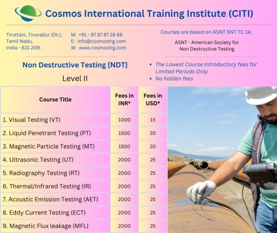
#𝗡𝗼𝗻 𝗗𝗲𝘀𝘁𝗿𝘂𝗰𝘁𝗶𝘃𝗲 𝗧𝗲𝘀𝘁𝗶𝗻𝗴 [𝗡𝗗𝗧] 𝐂𝐨𝐮𝐫𝐬𝐞𝐬#Level II#1. Visual Testing (VT)#2. Liquid Penetrant Testing (PT)#3. Magnetic Particle Testing (MT)#4. Ultrasonic Testing (UT)#5. Radiography Testing (RT)#6. Thermal/Infrared Testing (IR)#7. Acoustic Emission Testing (AET)#8. Eddy Current Testing (ECT)#9. Magnetic Flux leakage (MFL)#Contact Us:#Mob:#+919787872866#Email:#[email protected]#Web:#www.cosmostrg.com#WhatsApp Channel :#https://whatsapp.com/channel/0029VaHMYayGufJ45OH4Xo1n#WhatsApp Group :#https://chat.whatsapp.com/IunhDueyuLsEPc9SkB7dlK#NDT#ndtinspection#ndttraining#visualtesting#liquid#penetranttesting#Magnetic#magneticparticletesting
0 notes
Text
Comprehensive Energy Audit
Comprehensive Energy Audit Utilizing a comprehensive energy audit service is the most efficient way to find any issues affecting your home's energy efficiency and efficiency This audit is a thorough examination of your entire home, with a focus on any potential sources of energy loss A comprehensive energy audit examines factors such as insulation, HVAC systems, doors, windows, walls and roofing. This rating takes blower door and duct testing into account. If necessary, a thermal imaging camera is used to identify problem areas In a comprehensive report, our findings are given in detail along with recommendations for improvement
Examining the building's existing energy usage, identifying potential areas for energy savings, and presenting the findings are all goals of a building energy audit It is a vital tool for energy conservation, implementing energy efficiency measures, and lengthening the lifespan of electromechanical equipment in the construction industry This article underlines the relevance of a planned and well-structured energy audit methodology as the most important tool for the energy certificate of buildings as part of the execution
Both the energy demand of the building, which is primarily influenced by building use and the building envelope, and the energy consumption, which is primarily influenced by the machinery and type of energy used for heating, cooling, ventilating, lighting the building, and heating sanitary hot water, must be described in the data gathered during the energy audit from both observing/measuring the building, collecting data, and interviewing tenants
A well-structured survey should therefore contain at least the following data: 1 the position of the structure in relation to its surroundings and the determination of its size and volume; 2 Building materials for walls, roof and floor, as well as the type, size and number of windows and openings on each facade; 3 Occupancy and mode of operation; 4 Type of boiler for space heating and/or sanitary water heating and the fuel used and how it works; 5 type of air conditioning and how it works; 6 Are there energy savings in the building and 7 energy management systems;
Evaluation of complex energy audits The importance of a well-structured questionnaire for the energy performance of a building is discussed, as well as the context of one and the underlying data of the series. The need for continuous measurements is emphasized, including thermal and optical comfort assessment, IR thermography, power consumption measurements and gas emission measurements. To further emphasize the need for measurements, numerous practical examples are given, with emphasis on the thermographic method
In addition to the visual assessment of the building and its systems, the following measurements should be carried out during an energetic inventory:
1 With endoscopes, infrared thermography or heat flow and surface temperature sensors as well as building envelopes and distribution systems 2 can be identified, the latter giving a better idea of the thermal performance of the component; 3 Use of light meters, relative humidity sensors, air velocity sensors, air temperature sensors, and air and globe temperature sensors to ensure thermal and optical comfort; 4 Electricity consumption is measured with energy analyzers, and flue gas analyzers determine the efficiency of boiler systems
Need for Comprehensive Energy Audits The primary objective of the energy audit is to gather and measure data that will be useful for the building's energy evaluation, together with the electrical safety audit Its primary goals are to determine operation and maintenance procedures and to carry out a preliminary energy survey Its primary goals are to assess and analyze current energy demand and consumption, identify critical and essential changes to energy management, and, ideally, offer an energy management strategy
Performing a Comprehensive Energy Audit Service has the following advantages:
In order to provide consumers with access to real-time energy data, building systems' energy data must be gathered and evaluated as part of the ongoing monitoring of a facility's energy use or energy-consuming machinery With the aid of this information, facility managers may regulate resource usage and preserve optimum equipment performance
The following are the key advantages of energy monitoring: Unknown building systems and operational factors are measured and evaluated by Energy Analytics It has been shown that using interactive dashboards for facility employees or for public displays, like in-office monitors, can increase energy awareness among facility users and positively affect behavior, leading to decreased electricity costs .
System performance and energy consumption are continuously monitored by automated notifications, allowing for the early detection of problems or flaws Individualized alerts can be put up to reduce erratic or excessive intake.
With SAS Powertech's Comprehensive Energy Audit Service, your industry may become healthier and more energy-efficient Accurate energy audits are designed to look for elements affecting your industry's performance that you can't see or are unaware of We take a holistic approach to energy efficiency, so we can helpyou identify and fix problems with excessive summer heat in your office,excessive winter cold or drafts, or high humidity issues This can have a major impact on your energy costs.
Energy is one of the most important challenges of the future Businesses must strike a balance between operational efficiency and sustainable development With rising energy costs and depletion of natural resources, it makes financial sense to find ways to improve energy efficiency at all stages of the process, from planning to production to operations
One way to reduce energy costs is to improve energy efficiency by maximizing the use of existing resources and carefully planning investments in new technologies. This benefits the environment and reduces risk in addition to better customer service and cost control. Taking a formal stance in favor of sustainable development is another effective and efficient way to demonstrate social responsibility and adjust to shifting consumer needs
Benefits of using Comprehensive Energy Audit services:
1.For continuous monitoring of a facility's energy consumption or the energy consumption of energy-consuming equipment, energy data from building systems must be collected and analyzed This can provide clients with real-time energy information 2. Managers of facilities can use this information to make the best use of resources and guarantee optimal equipment performance 3. Energy Analytics measures and evaluates previously unrecognized data on building systems and operational aspects 4.It has been discovered that the use of interactive dashboards for facility staff and/or public displays, like in-office monitors, can increase energy awareness and positively influence people's behavior, resulting in reduced electricity costs 5. Automated Notifications continuously track energy usage and system activity, allowing for the early identification of problems or flaws
Best comprehensive energy audit service companies in India: Across a range of industries, SASOL has been providing Comprehensive Energy Audit services to its clients in South East Asia and India We are known for disclosing findings in an unbiased and open manner We provide the most affordable Comprehensive Energy Audit services and solutions, and we have helped clients achieve their goals
By implementing energy saving solutions, SAS Powertech is ready to help you minimize your energy costs Our energy services are aimed at all stakeholders, including building owners, manufacturers, energy suppliers, regulators and end users Explore the services below to learn more If you require a service that is not currently available, please contact our team
Conclusion: For more than a decade, SASOL has provided comprehensive energy audit options to clients in India and Southeast Asia. We are known for being open about our impact and providing balanced reporting. We have helped clients achieve their goals and offered the most cost-effective solutions for comprehensive energy audits.
0 notes
Text
Why is a Thermal Device So Expensive?
The revelation of 'infrared beams' dates to the start of the nineteenth century by Sir William Herschel, He closed through tests that there is an undetectable light that can warm articles. In 1963, after 163 years, the main warm camera was created. The warm imager doesn't depend on apparent light yet warm radiation from the item. It changes over it into Thermal Imaging Scope that is apparent to the unaided eye through a progression of sign handling.
It is at first restricted to military applications in light of its grouped use and excessive costs. Around 2000, following quite a while of advancement, warm imaging innovation has step by step developed and is broadly utilized for a considerable length of time. Notwithstanding, the cost of this innovation is still too far for most potential buyers today, and this article covers the reason why that is

While purchasing a warm gadget, we take a gander at the elements and the sticker price of the item and afterward contrast it and others on the lookout. Warm gadgets appear as cameras, riflescopes, optics, and so forth. Assuming you investigate the commercial center, you'll observe that the costs of these gadgets are somewhat high than standard day optics.
Presently, this could come as a shock from the get go, yet how about we investigate what makes these gadgets so expensive. Here are a few factors that conclude the sticker cost of various warm gear
Warm Imaging Detector
Germanium Lens
Field Programmable Gate Array (FPGA)
Warm Algorithm
Others (gathering, climate, tests, and so on.)
Cryogenic Cooler (for cooled warm imagers)
6 Reasons Why Thermal Devices are So Expensive
We should survey the elements referenced above in more detail.
1. Warm Detector
The indicator is essentially made out of a microbolometer and related circuits. It's answerable for getting warm radiation and changing over it into a computerized signal with radiation force data. The microbolometer is a matrix of hotness touchy materials on a comparing network of silicon. Infrared radiation from a particular scope of frequencies strikes heat-touchy materials, for example, vanadium oxide or shapeless silicon and changes its electrical obstruction. This opposition change is estimated and handled into temperatures which can be addressed graphically by related circuits. The microbolometer framework is normally planned and fabricated in three sizes, a 640×480 cluster, a 320×240 exhibit (384×288 nebulous silicon), or a more affordable 160×120 cluster.
The indicators cost a ton due to their weakness to different ecological circumstances, high power utilization, low large scale manufacturing strength, and reconciliation issues. Since the creation of identifiers requires high stage costs, the interest in innovation innovative work is additionally a sizeable consistent venture. The improvement of every boundary implies the redesign of the whole framework. There is still a lot of opportunity to get better in center markers, for example, NETD, pixel pitch, and amplification, which drives producers to proceed to research and move up to guarantee that they won't essentially fall behind their rivals.
2. Germanium Lens
A few top notch warm gadgets use germanium glass focal points. They have a high refractive record, permitting the IR scope of frequencies to go through and arrive at the sensor that recognizes an objective.
Due to the uncommonness of the germanium metal, it costs 1500 dollars for each kg; and that is only the expense in its crude structure. Whenever you add the cost of refining and handling germanium into an elite exhibition long-range warm reconnaissance focal point, the complete expense of the gear increments dramatically
Likewise, it relies upon the opening (the F-number of the gadget); the lower the F-number is, the more costly the focal point costs.
For more details, visit us :
Thermal Polaris 350RL
Thermal Imaging Sights
Thermal Scope for Sale Australia
Thermal Tracer 50 LRF
#Thermal Scope#Thermal Vision Australia#Thermal Scope For Sale#Thermal Monocular Australia#Thermal Tracer 50LRF
0 notes
Text
Application of Scanning Electron Microscopy and Fourier-Transform Infrared Spectroscopy to Study Surface Design and Chemical Group Components of Merino Wool, Paschmina and Angora Fibers - Juniper Publishers

Abstract
Textile fibrous materials are used for making fashion utility products in day to day use. These fibers find applications in polymer blends, composites, fiber reinforce plastics, bio-medical textiles and many such high end uses. However, fibers like merino wool, pashmina and angora fibers are some of the natural animal fibers which are obtained in nature and find wide applications if converted into usable fashion products. These natural fibers are characterized with specific microstructure which can be tapped to design and create innovative fashion products. The importance of understanding these surface and micro-structures helps in designing and engineering a particular fashion product in textile science and engineering. In this study we look into the finer detail application of SEM used to characterize the surface, inter surface and other dynamic properties of these fibers. Scanning electron microscope has the capability to image these fiber surfaces at different magnifications which thus facilitates the observations to be examined at varying conditions of usage. These SEM images also gives insight into the steps that can be used to manipulate the process and product development steps used for textile materials. On the other hand spectral analysis using Infra-red spectroscopy gives broad information on the qualitative and quantitative analysis of these fibers. FTIR helps in elucidation and identification of chemical groups and interfer structure property relationship of these natural fibers which are obtained from animal source.
Keywords: Scanning electron microscope; Textile fibers; Infra-red spectroscopy; Merinowool; Pashmina; Angora
Go to
Introduction
Wool is one of the protein fibers which find its application in many fashion products. Wool has excellent heat retention and moisture absorbance properties. Apart from this, other fibers like angora and pashmina do possess similar properties of wool fiber but outperform wool in terms of its surface properties. However, angora fibers are finer than wool and are used in making high quality yarns for hosiery and knitting garments. Pashmina is one of the well-known highest grade of animal fibers. Pashmina provides excellent warmth characteristics and is used to make clothing which is used in frigid conditions. Scanning electron microscopy is one of microscopy techniques used for investigation surface and morphological structure of fibers. Scanning electron micrographs [1], when coupled with digital imaging techniques will be able to give quantitative values of cuticle scale parameters like scale height, scale length, scale edge angle. Qualitative study and molecular structure analysis using Fourier transform infrared transmission spectra gives complete information on the presence of functional groups and its contribution to fiber properties. Quantitative study using Fourier transform infrared absorbance spectra data [2], throws light on the amount of light absorbed by the molecules present in the fiber. In this study field emission scanning electron microscopy is used for capturing the micrographs at different magnification for merino wool, angora and pashmina fibres. Perkin Elmer Fourier transform infra-red spectra are used for qualitative and quantitative analysis of these which include molecular structure and distribution of functional groups in protein fibres used in this study [3].
Go to
Experimental
Materials
In this study animal fibers like merino wool, angora and pashmina fibers were obtained from known sources. Images of samples of fibers used in this study are shown in Figure 1. Conditioning of the samples was carried out before subjecting them to testing on Scanning electron microscope and FTIR spectrometry to analyze their structural composition and characterization. Table 1 shows the list of fibers used in this study Merino wool (Figure 1) is a keratin based biopolymer which finds extensive application in fashion and apparel products. The molecular structure of wool fiber is composed of di-sulphide bonds which connects the peptide chains [4]. Merino wool is one of the protein fibers which possess excellent moisture and temperature regulation properties. The primary structure of wool is composed of 20 amino acids and secondary structure occurs when the sequence of amino acids are linked to hydrogen bonds. The regular configurations of protein macromolecules generally exist in three types like the alpha helix, secondly the beta pleated sheet and beta turn. The principle structural units in the native wool fiber are successive turns of the alpha helix. The intrinsic stability of the alpha helix, and thus the fiber results from intra molecular hydrogen bonds. The presence of disulphide bridges, H-bonds, electrovalent bonds, etc, in wool fibres form the hindrance to the movement of the molecular segments and thus to the secondary structure [5].
Angora fibers (Figure 2) are a keratin based protein material obtained from Angora goat which is usually long haired. Angora fibers are known for their lightness and outstanding thermal properties. Angora fibers have good potential for producing textiles with special properties. Angora fibers are extremely soft, antistatic, lustrous and durable, while giving high insulation and a warmer feeling to the garments due to its modulated hollow structure in the core. They have high heat retention and best moisture wicking properties of any natural fiber Pashmina wool (Figure 3) is one of the most valued animal fibers. The other name commonly used for pashmina is known as cashmere wool of highest grade category among protein fibers. Pashmina wool is known for its softest, most luxurious and used for making trendiest fashion fabrics. Pashmina wool is obtained from the underbelly of goat indigenous to the Himalayan region [6-8].
Field emission scanning electron microscope
Ultra 55 FE-SEM (Figure 4) is a general purpose ultra-high- resolution FE-SEM based on the unique GEMINI Technology was used in this study to capture images of fiber samples. Field emissions SEM are combined with analytical capabilities and excellent imaging properties thus making it suitable for a wide range of applications in materials science. Field emission scanning electron microscope work on the principle of electrons liberated from a field emission source and accelerated in a high electrical field gradient. The principle of working of a field emission electron microscope is shown in Figure 4. In this SEM experiment, samples were first prepared on a stub cleaned with acetone carefully to ensure better scanning. The stub was sputtered with gold coating approximately of 10nm for 100 seconds in a sputtering chamber [9]. After this process the samples were mounted on a SEM chamber. Images in this study were captured at a scale of 20|im (varying magnifications) and at high energy potential of 1000KV. The distance between the probe and the sample stub was adjusted at different levels keeping in view the image clarity and its sharpness. The samples were scanned under different magnification levels ranging from 100X to 3000X.
Working principle of FTIR spectroscopy
Infra-red (IR) spectra involve the study of interactions between matter and electromagnetic fields in the IR region. In this spectral region, the EM waves couple with the molecular vibrations. Molecules are excited to a higher vibrational state by absorbing IR radiation. The IR frequency when absorbed would actually interact with the molecule at a certain frequency. Hence, IR spectroscopy is a very powerful technique which provides fingerprint information on the chemical composition of the sample. Using IR Spectroscopy both qualitative as well as quantitative analysis of fiber samples can be carried out. FTIR spectrometry is found to be the most analytical type of techniques available in laboratories. FTIR works on the principle of Fourier transformation (Figure 5). Interferogram is determined experimentally in FTIR spectroscopy. The spectrum is plotted against the corresponding wave number against transmittance values [10-12]. This transformation is carried out automatically and the spectrum is displayed/infrared spectroscopy results in a positive identification (qualitative analysis) and the size of the peaks in the spectrum is a direct indication of the amount of material present. With the modern software, infrared is an excellent tool for quantitative analysis. Fourier Transform Infrared spectroscopy (FTIR) spectra were obtained by means of PerkinElmer Frontier spectrometer (Figure 6) working in the range 350 to 8300cm-1 with a resolution of 0.4cm-1 with a Ge- coated KBr optics and good signal to noise ratio [13].
The untreated samples (fibres) were directly mounted in the sample window and the data were collected with 100 scans per sample. The spectra were named as A, K, P and M for Angora, Kevlar, Pashmina and Merino respectively. Spectra were corrected to the base line blank and then plotted [14].
Go to
Results and Discussion
Figure 7 & 8 is the SEM micrographs taken on Zeiss field emission scanning electron microscopes at different magnifications of merino wool sample to characterize the surface properties of merino wool fibres. They were taken at 100X, 200X, 300X, 500X, 100KX and 300 KX magnifications. The surface structures of merino wool fibers are composed of scales are very prominently seen in all the images. However, the length of the scale in the fibers varied consistently along the fiber axis. At higher magnifications from 100KX and 500KX the scales were prominently visible. In these images the surface of merino wool fiber scale length is distributed randomly at times evenly spaced and some places merging with the previous scales. This is one of the important observations that image results reveal for merino wool fiber. The height of the scale is not well defined at some points since the scale is not exactly in the shape of a rectangle. The distribution of even scale height would influence the dynamic frictional properties of wool fibers. From the images, observation can be made on the scale frequency which is expressed as the number of cuticle scales in a 100micron field of view normally known as mean scale frequency. The scale frequency is found to be more uniform in case of image taken at 10|im and 300 magnifications. A lower mean scale frequency indicates a longer cuticle scale and a higher mean scale frequency indicates a series of shorter scales. The images of scale edges reveal that they form an obtuse angle and are arranged haphazardly which contributes to uneven locking of fibers at different spots on the fiber surface. A very precise way to measure the scale height, scale length and scale edge angle is to subject the images to digital imaging software [15].
Figure 9 & 10 are Imaging results of Scanning electron microscope on pashmina fiber. The images were taken at different magnification levels. However, the scales are clearly observed on the surface of the fiber only at 300KX magnification. In the images, the scale height is more as compared to merino wool fiber. The scales are longer, showing more curvature at the edges. The variation in scale height is more visible from the SEM images. The length of the scale is more as compared to merino wool fiber [16].
Figure 11 & 12 are scanning electron micrographs of angora fibers which are characterized with high luster. The images were captured at different magnifications. These SEM images of angora fibers reveal detail information on variation in scale height, scale length and scale edge angle when compared to merino wool and pashmina fibers. The images prove that fact that angora fibers have relatively large surface cuticle scales and low cuticle edge height relative to merino and pashmina fibers. The scales of angora fibers are more regular and prominently visible in the micrographs obtained through SEM. The scales are closely overlapped thus resulting in distribution of more uniform curvature on the fiber surface.
Effect of scales on felting property
The scaliness of the wool fibre is partly responsible for felting phenomena which is one of the dynamic properties of these fibres under discussion. The presence of scales exhibits a higher friction as compared to the smooth surface due to scales or any other crimps on the fiber surface. Merino wool does not contain exceptional property (lustre) that is found like in angora, hence it is found to be dull. The longer and fewer scales on the surface of these fibers result in having a smooth surface. However, the surface is rough if the scales are more and short, arranged very close to each other. The longer the distance between these scales result in more shiny and lustrous fiber.
FTIR results
Results of FTIR spectra for different fibers used in this study in the form of spectra are Figure 13-17. The Spectra in the range 600-4000cm-1 showed clearly dominant peaks around 1510 and 1600cm-1. The band observed at 1635cm-1 is assigned to the broad OH bending modes due to bound water. The band around 1590cm-1 is assigned to the hydrogen bonded carbonyl stretching and O-C-O stretching of carbonate ion [12].
The results of the observations of peaks in spectra are shown in Table 2-4 for merino wool, angora and pashmina fibers. The spectra of the fibers plotted give a qualitative analysis of the presence of functional groups which are identified with specific wavelengths [7]. However the quantitative values of spectra are obtained by plotting absorption values against wavelength for different fibers. The absorption spectra for merino wool, angora and pashmina fibers are shown in Figures 18-20.
The quantitative analysis of the molecular distribution in the fiber structure was explored using absorbance spectra for merino wool, angora and pashmina fibers. The amount of light absorbed indicates the concentration of molecules that absorb the light. The observations were found to be in conformance with Beer- lambert law. For all the fibers the absorbance indicated peaks in the range of 450-800cm-1 substantiating the presence and concentration of more carbon molecules. At this wave number the spectra shows a significant difference when compared at other points. The absorbance is found to be weak as the wave number increased for all the fibers [17].
Conclusion
The scanning electron microscopic study on merino wool, angora and pashminas fibers show significant difference in surface structural features. The surface structural features like scale length, frequency, edge height and edge angle are clearly captured using scanning electron microscope which has helped to discuss and understand the effect of scaly surface on dynamic properties like friction and felting properties supporting various theories proposed by eminent researchers. The preliminary characterization runs for merino, angora and pashmina fibers using FTIR spectroscopy throws light on many interesting aspects of molecular structure and composition of these fibers. The peaks corresponding to molecular vibrations have been assigned which is a qualitative output of FTIR technique. The Transmittance and absorbance spectra for these fibers have also been studied into order to make an attempt to quantify the distribution and concentration of functional groups in these fibers. In conclusion, the surface and molecular structure properties of these fibers largely contribute for dynamic properties and overall performance of these fibers when used to make fashion clothing items using these fibers.
Acknowledgment
The Authors wish to acknowledge Dr. Ramesh KP, Associate Professor, Department of Physics, Indian Institute of Science, Bangalore - 12 for providing testing facilities on FTIR study, Nagabhushan Patil, Research scholar, Material research centre, Indian Institute of Science, Bangalore for providing testing facilities of field emission spectrometry, Dinesh Bhatia, research scholar, Department of Textile Technology, National Institute of Technology, Jalandhar for providing fibrous materials for this study. The author also wishes to express thanks to one and all who are involved in this experimental work.
To know more about Journal of Fashion Technology-https://juniperpublishers.com/ctftte/index.php
To know more about open access journals Publishers click on Juniper Publishers
#Juniper Publishers#Juniper Publishers group#Fashion Technology#Textile Engineering#Juniper Open access Journals
0 notes
Text
Liquid Crystal Thermography Provides Precise Heat Maps
It is critical for the engineers to accurately determine temperature of a design, whether it is at the chip, component, board, or system level, to ensure that the design will function properly and maintain its optimal performance over its expected lifetime and meet its specified mean time between failures (MTBF). Thermal management of electronics is unarguably a critical component of the design phase.
To optimize thermal management, it is also critical to get an accurate picture of the heat distribution across a device or a board. One of the most precise methods for mapping temperature is liquid crystal thermography (LCT). LCT uses thermochromic liquid crystals (TLC) to give engineers a visual representation of the heat distribution based on the changing colors of the TLC when heated.

Liquid crystal thermography systems give a visual representation of the heat on a device, board, or system using thermochromic liquid crystals, high-resolution cameras, and an LED light source. (Advanced Thermal Solutions, Inc.)
LCT technology has been around since the 1950s and has been used in the electronics industry since the 1980s. In 1975, researchers published a review of LCT and it applications in the study of convective heat transfer that determined the method provided “both qualitative and quantitative heat transfer and fluid flow information to be obtained on heated objects placed in forced convection environments.” [1]
The authors added, “In addition to yielding precise quantitative heat transfer information, the liquid crystal thermographic technique afforded the opportunity to visually observe the effects of flow separation, the separation bubble region, the turbulent boundary layer, and the turbulent wake on the surface temperature of the heated cylinder.”
What are Thermochromic Liquid Crystals?
Thermochromic liquid crystals are the key to the LCT process. Rather than changing from a solid to a liquid when heated, TLC have an intermediate liquid-crystal phase and the temperature in which this phase-change takes place is precisely defined depending on the composition of the crystals. The phase-change causes the TLC, which starts as transparent, to reflect different wavelengths of light, represented visually as different colors.

TLC readings using thermVIEW Lite software from ATS. The TLC shows up as different colors depending on the heat applied, while the software is calibrated to give precise temperature readings. (Advanced Thermal Solutions, Inc.)
As explained by Dr. Bahman Tavassoli in an article for Laser Focus World, “Normally clear or slightly milky in appearance, liquid crystals change in appearance over a narrow range of temperature, called the color-play interval. This is the interval between the first (red) and last (blue) reflection. The displayed color is red at the low-temperature margin of the color-play interval and blue at the high end. Within the color-play interval, the colors change smoothly from red to blue as a function of rising temperature, with blue light corresponding to the clearing-point temperature.” [2]
To visualize the temperature response of the TLC, a bright and stable white light is required to remove the infrared and ultraviolet radiation from the output spectrum. Tavassoli explained, “Any IR energy present in the incident light spectrum will cause unwanted radiant heating of the test surface. Exposure to UV radiation can cause rapid deterioration of the TLC surface, which will result in unreliable color-temperature responses.”
Temperature ranges for the TLC material are established by the manufacturers. Narrow-band TLC have bandwidths below 1-2°C, while wide-band TLC range between 5-20°C. TLC are typically designated by a two-color/temperature system. For instance, R35C5W would indicate that the TLC would show up as red starting at 35°C and that the blue start temperature would be 5°C above the red, allowing engineers to see the estimated bandwith of the TLC.
TLC are inherently oily, and their thermal performance degrades from exposure to chemicals and UV radiation, so manufacturers have developed microencapsulation or polymer dispersion methods to make the materials easier to use in laboratory settings. Microencapsulation provides high resistance to contamination, but the polymer dispersion method provides a more brilliant color response. [3]
How do you use Liquid Crystal Thermography systems?
To obtain the most accurate results from LCT, a smooth and contamination-free surface is important. Test surfaces and calibration tools should be cleaned with alcohol, if possible, and dried before the process begins. A thin and uniform coating of black paint is applied to the test surface and dried using a hot air gun at low temperature. Once the surface is dry, the TLC materials can be applied to the test surface. [4]
A bright, stable, white light source is required to obtain an accurate reflected light intensity from the TLC-coated surface. As explained by Dr. Kaveh Azar in an introduction to LCT techniques, “Consistent light source settings and lighting-viewing arrangements between calibration and actual testing are essential to minimize color-temperature interpretation errors.”
Calibration is the key to using LCT. According to Dr. Tavossoli, the system for calibrating TLC is similar to calibrating the voltage-temperature response of a thermocouple. TLC is subjected to known temperature levels and the response is recorded with a color-sensitive camera. The response is recorded at different temperature levels on a test surface and the system develops calibration files that can later be used to interpret the response of TLC on the device being studied.
The following graph shows the relationship between temperature and color of a range of TLC:

While TLC response is visible to the naked eye, the LTC system uses a high-resolution, solid-state color camera and calibrated software to more precisely determine the temperature.
Advanced Thermal Solutions, Inc. (ATS) has recently added to its line of LCT systems to provide a cost-effective tool for temperature mapping studies. tvLYT™ is accurate, easy to assemble, and easy to use. It comes in a portable case containing the arm, high-resolution macroscopic optic camera, LED light source, black paint, and TLC material for the required temperature range. tvLYT™ can be quickly connected to a computer through a USB and uses thermVIEW™ Lite software (downloadable on the ATS website) to calibrate the readings and provide precise results.

The tvLYT™ liquid crystal thermography system from ATS provides highly accurate temperature mapping capability using TLC. (Advanced Thermal Solutions, Inc.)
For more information about tvLYT™, visit https://www.qats.com/Products/Instruments/Surface-Thermography/tvLYT.
What are the benefits of LCT?
There are several techniques for measuring temperature across a system, including the use of thermocouples and resistance thermometers. These are common techniques used in labs across the world, but there are challenges and limitations to that method.
As one report noted, “Traditional techniques employing sensors such as thermocouples and resistance thermometers can measure temperature at individual locations. Hence, a large number of sensors are required for complete mapping of the surface. Since physical sensors occupy space, the measurements are to be interpreted as spatial averages. This route may prove to be disadvantageous in regions of localized peaks and valleys of heat transfer. Liquid crystal thermography proves to be useful under these circumstances.” [5]
The tools for thermal imaging, which visualize IR emissions to show heat patterns, continue to improve, seemingly by the day, and several companies have released cameras that can be attached to a mobile phone to improve the mobility and accessibility of thermal imaging. [6] IR cameras can give a quick visualization of the heat emitted from a device and are excellent tools for finding hot spots at the system level, but they lack the precise temperature readings that LCT captures and accuracy counts in thermal management.

Comparison of LCT and IR systems. [4]
LCT gives engineers flexibility. It can be used in to measure temperature across micron-level electronic circuits or large-scale gas turbines. LCT is used in a wide range of applications from detecting lamination in composite polymer materials [7] to studying turbulent boundary layers in a water tunnel [8] to biomedical studies [9] such as testing for skin cancer, breast cancer, blood circulation, and more.
Because precision is the most important benefit to using LCT for temperature mapping studies, ATS has offered four free calibrations during the first year after purchasing tvLYT™ and also lifetime technical support.
For more information about ATS liquid crystal thermography systems, or thermochromic liquid crystal materials, visit https://www.qats.com/Products/Instruments/Surface-Thermography.
If you have questions about any ATS product or its consulting and design surfaces, contact ATS at [email protected].
References 1. http://heattransfer.asmedigitalcollection.asme.org/article.aspx?articleid=1436225 2. https://www.laserfocusworld.com/articles/print/volume-41/issue-12/features/thermal-imaging-liquid-crystal-thermography-characterizes-heat-issues.html 3. https://www.electronics-cooling.com/1995/10/making-surface-temperature-measurements-using-liquid-crystal-thermography/# 4. https://www.ewh.ieee.org/soc/cpmt/presentations/cpmt0201b.pdf 5. http://www.nptel.ac.in/courses/112104039/pdf_version/lecture35.pdf 6. https://www.thermal.com/compact-series.html 7. https://onlinelibrary.wiley.com/doi/pdf/10.1002/pc.20453 8. https://www.sciencedirect.com/science/article/pii/S0924424715300224 9. http://zm8pc.ippt.gov.pl/papers/JCPT_2014012610120554.pdf
0 notes
Text
Handheld IR Thermal Imaging Camera
Thermal imaging camera combines the functions of surface temperature and real-time thermal imaging.
Thermal imaging camera can turn thermal image into visional image, quickly find out the problem area.
The infrared imaging device is professional, precision and efficient.
Infrared thermal imager that can blend the visible and infrared images.
2.4 inches full angle high resolution color screen, easy to see the screen.
Resolution of infrared image/visible image is 32 x 32(1024 pixels)/0.3 mega pixels.
View options is 5 kinds of full infrared to the full visual, mixing visible and infrared.
Fits for car engine overhaul, transformer room line maintenance, air conditioner repair, etc.
Handheld thermal imaging camera is widely used in the electric power manufacturing industry, car industry, medical, transport, agriculture etc.
Color: Orange
Product size: 212 x 95 x 62mm
Display: 2.4 inches full angle high resolution color screen
Infrared image resolution: 32 x 32(1024 pixels)
Visible image resolution: 0.3 mega pixels
Field angle/minimum focal length: 33 x 33 / 0.5m
Thermal sensitivity: 0.15°C
Temperature test Range: -20°C ~ +300°C
Test measurement accuracy: +/-2% or +/-2°C(+/-2% or -4°F)
Emissivity: adjustable 0.1-1.0
Image capture frequency: 6Hz
Wavelength Range: 8-11.5um
Focal length: fixed focal length
Palette: iron red, rainbow, rainbow high contrast, gray(white glow), gray(black glow)
View options: 5 kinds of full infrared to the full visual, mixing visible and infrared
Memory card: small SD card
File format: bmp
Automatic shutdown time: 12 minutes
Operating temperature: -5°C to +40°C
Storage temperature: -20°C to +50°C
Relative temperature: 10%RH to 80%RH
Battery: 4 x AA battery(not included)
Battery Life: 6 Hours
Note:
Please take out the battery if the instrument is not used for a long time.
Do not use the instrument in explosive, flammable, damp or corrosive environment.
Package Included:
1 x HT-02D Handheld Thermal Imaging Camera
1 x Hanging Rope
1 x Packing Bag
1 x User Manual
0 notes
Text
How to Avoid Hearing, "You Have Breast Cancer…"
Breast cancer strikes fear in the hearts of all women. Most of us have known someone-a friend, a family member, a co-worker, a neighbor-who has been treated for breast cancer or worse, has died from the disease. Historically, breast cancer was diagnosed after a woman sought medical attention for soreness or a palpable mass. Annual mammograms became the “gold standard” for breast cancer screening; the goal has become to find tumors before they can be felt.
However, mammography is a late test. It does not prevent cancer; it detects cancer. Women place unreasonably high hopes on the ability of the technology to reduce their risk of cancer. A recent study, published in Annals of Internal Medicine, reported that women typically overestimate the risk reduction capability of a mammogram by more than 100-fold.(1)
Mammograms give a false sense of security about a clean bill of health. The sensitivity, or the number of women who have cancer and also have a positive mammogram, ranges from 54%-58% among women under age 40 to 81%-94% among women over 65. In women 40 to 65, the ability of a mammogram to identify a tumor depends on many variables: The size the lesion, hormone use, breast tissue density, the overall image quality and the interpretative skills of the radiologist.
Cancer is identified in only a small fraction of women (0.1%-0.5%, depending on age) who are screened. Studies have demonstrated that for each death prevented by screening, there are at least 200 false-positive results.(2) The number of follow-up procedures generated by cancer screening programs, such as repeat mammograms, ultrasounds and MRI, exponentially increases the cost of the disease. In the United States, biopsies that turn out to be benign cost the healthcare system more than $1 billion annually.(3)
Screening has another potentially harmful effect. Women are psychologically damaged by the stress of an abnormal mammogram that turns out to be a false alarm. A new survey developed by Brodersen and colleagues and released this month (7/07) measured six psychosocial dimensions: anxiety, behavioral impact, sense of dejection, impact on sleep, breast examination and sexuality. The survey showed that women who have an abnormal screening mammogram that is later confirmed to be a false-positive are negatively impacted in all six categories.
What else can be done?
Digital infrared imaging (digital IR), also called thermography, has been FDA-approved for breast health screenings since 1982. It is painless, non-invasive and uses no radiation. The scan detects and records thermal patterns on the skin not seen with any other tool.
Abnormal cells require large quantities of nutrients for rapid growth. Through a process called angiogenesis, new blood vessels are drawn in the direction of the atypical cell. Coupled with the release of cytokines and nitric oxide, additional blood flows into the area that increases heat in the tissues that can be detected by thermography.
Normal skin surface temperature is symmetrical and deviates less than 0.3C from side to side. New digital IR cameras can detect differences as small as 0.1C. Temperature variation of more than 1.0C suggests the presence of abnormal, possibly pre-cancerous, cells. These subtle changes can be detected seven to eight years before a mass becomes palpable or can be detected by mammography.
What is a thermogram procedure like?
After disrobing to the waist, approximately 15 minutes is needed to equilibrate to room temperature. An experienced technician performs three scans–front, left and right–with arms extended over the head. Both hands are then placed in cool water for exactly one minute. When the sympathetic nervous system is chilled, normal vessels constrict; abnormal vessels remain the same size or dilate. A second set of scans is taken for comparison. When the scans are completed, the exam is over.
Why isn’t everyone doing this test?
Why aren’t doctors routinely recommending this test? Why don’t they offer it in their office? An abnormal scan cannot be corrected with a drug. Conventional doctors are not trained to use vitamins, supplements and detoxification, essential tools for repairing damaged physiology. If a woman has a normal mammogram, the abnormal thermogram is dismissed by physicians as a false positive. This is a missed opportunity for true cancer prevention.
Despite the current status of breast thermography, it has been researched for over 30 years. More than 800 peer-reviewed studies on breast thermography exist in the index-medicus. The data base represents more than 250,000 women and the number of participants in many studies ranged from 37,000 to 118,000. Some of women who had a positive thermogram were followed for up to 12 years; 35 percent of those who had a positive thermogram went on to develop breast cancer.
What you can do
If an abnormality is identified on your thermogram, pro-active steps can be taken to heal the breast tissue and progress can be monitored non-invasively through serial scans. If you don’t have access to a physician who has digital IR equipment, you can take important steps toward breast health:
o Diet: Eat organically grown foods. Pesticides collect in fatty tissue, and the breast can become a reservoir for estrogenic chemicals. Eliminate all caffeine, chocolate, MSG, aspartame, and herbs that have estrogenic properties, especially soy. Studies found that soy supplements can increase breast cancer risk, especially in post-menopausal women.
o Exercise: Walking and arm swinging will get the lymphatics moving. Decongesting breast tissue will reduces the risk of cancer.
o Eliminate hormones: Discuss options with your physician to eliminate birth control pills and synthetic hormone replacement. The link between hormones and breast cancer has been confirmed.(5)
o Supplements: Many supplements have been shown to have a positive effect on breast tissue. Here are a few examples:
1. DIM (diindolylmethane) the phytochemical found in cruciferous vegetables, has anticancer activity and also works to repair damaged DNA.
2. Tumeric (curcumin) kills cancer cells and has strong properties against angiogenesis
3. Drinking green tea or taking green tea extract capsules has been associated with a reduced risk of breast cancer. All of these are available at your local health food store.
Women need to drive this industry and push conventional doctors to do their home work. The new cameras are digital and far superior than the cameras of 30 years ago. For women who are young, at high risk, have dense breasts or want to be more proactive about breast health, a breast digital scan is the way to go.
________________________________________
REFERENCES
(1) Elmore J., Choe J.Breast Cancer Screening for Women in Their 40s: Moving from Controversy about Data to Helping Individual Women. Ann Intern Med. Apr 3;146(7):529-31. 2007. PMID: 17404356
(2) Urbain JL.Breast cancer screening, diagnostic accuracy and health care policies. CMAJ. Jan 18;172(2):210-1. 2005. PMID: 15655243
(3) Ibid. Urbain JL
(4) “Survey Determines Impact Of False-positive Cancer Tests.” Science Daily. July 22, 2007.
(5) “Confirmed: A Link Between Breast Cancer and Hormone Therapy.” Scientific American.com July 26, 2007.
(6) Rahman KW. Gene expression profiling revealed surviving as a target of 3,3′-diindolylmethane-induced cell growth inhibition and apoptosis in breast cancer cells. Cancer Res. 2006 May 1;66(9):4952-60. PMID: 16651453
(7) Mosley CA Highly active anticancer curcumin analogues. Adv Exp Med Biol. 2007;595:77-103 PMID: 17569206 (8) Zhang M Green tea and the prevention of breast cancer: a case-control study in Southeast China. Carcinogenesis. 2007 May;28(5):1074-8. Epub 2006 Dec 20 PMID: 17183063
Source by Sherri Tenpenny, DO
from Home Solutions Forev https://homesolutionsforev.com/how-to-avoid-hearing-you-have-breast-cancer/ via Home Solutions on WordPress from Home Solutions FOREV https://homesolutionsforev.tumblr.com/post/186893491905 via Tim Clymer on Wordpress
0 notes
Text
How to Avoid Hearing, "You Have Breast Cancer…"
Breast cancer strikes fear in the hearts of all women. Most of us have known someone-a friend, a family member, a co-worker, a neighbor-who has been treated for breast cancer or worse, has died from the disease. Historically, breast cancer was diagnosed after a woman sought medical attention for soreness or a palpable mass. Annual mammograms became the “gold standard” for breast cancer screening; the goal has become to find tumors before they can be felt.
However, mammography is a late test. It does not prevent cancer; it detects cancer. Women place unreasonably high hopes on the ability of the technology to reduce their risk of cancer. A recent study, published in Annals of Internal Medicine, reported that women typically overestimate the risk reduction capability of a mammogram by more than 100-fold.(1)
Mammograms give a false sense of security about a clean bill of health. The sensitivity, or the number of women who have cancer and also have a positive mammogram, ranges from 54%-58% among women under age 40 to 81%-94% among women over 65. In women 40 to 65, the ability of a mammogram to identify a tumor depends on many variables: The size the lesion, hormone use, breast tissue density, the overall image quality and the interpretative skills of the radiologist.
Cancer is identified in only a small fraction of women (0.1%-0.5%, depending on age) who are screened. Studies have demonstrated that for each death prevented by screening, there are at least 200 false-positive results.(2) The number of follow-up procedures generated by cancer screening programs, such as repeat mammograms, ultrasounds and MRI, exponentially increases the cost of the disease. In the United States, biopsies that turn out to be benign cost the healthcare system more than $1 billion annually.(3)
Screening has another potentially harmful effect. Women are psychologically damaged by the stress of an abnormal mammogram that turns out to be a false alarm. A new survey developed by Brodersen and colleagues and released this month (7/07) measured six psychosocial dimensions: anxiety, behavioral impact, sense of dejection, impact on sleep, breast examination and sexuality. The survey showed that women who have an abnormal screening mammogram that is later confirmed to be a false-positive are negatively impacted in all six categories.
What else can be done?
Digital infrared imaging (digital IR), also called thermography, has been FDA-approved for breast health screenings since 1982. It is painless, non-invasive and uses no radiation. The scan detects and records thermal patterns on the skin not seen with any other tool.
Abnormal cells require large quantities of nutrients for rapid growth. Through a process called angiogenesis, new blood vessels are drawn in the direction of the atypical cell. Coupled with the release of cytokines and nitric oxide, additional blood flows into the area that increases heat in the tissues that can be detected by thermography.
Normal skin surface temperature is symmetrical and deviates less than 0.3C from side to side. New digital IR cameras can detect differences as small as 0.1C. Temperature variation of more than 1.0C suggests the presence of abnormal, possibly pre-cancerous, cells. These subtle changes can be detected seven to eight years before a mass becomes palpable or can be detected by mammography.
What is a thermogram procedure like?
After disrobing to the waist, approximately 15 minutes is needed to equilibrate to room temperature. An experienced technician performs three scans–front, left and right–with arms extended over the head. Both hands are then placed in cool water for exactly one minute. When the sympathetic nervous system is chilled, normal vessels constrict; abnormal vessels remain the same size or dilate. A second set of scans is taken for comparison. When the scans are completed, the exam is over.
Why isn’t everyone doing this test?
Why aren’t doctors routinely recommending this test? Why don’t they offer it in their office? An abnormal scan cannot be corrected with a drug. Conventional doctors are not trained to use vitamins, supplements and detoxification, essential tools for repairing damaged physiology. If a woman has a normal mammogram, the abnormal thermogram is dismissed by physicians as a false positive. This is a missed opportunity for true cancer prevention.
Despite the current status of breast thermography, it has been researched for over 30 years. More than 800 peer-reviewed studies on breast thermography exist in the index-medicus. The data base represents more than 250,000 women and the number of participants in many studies ranged from 37,000 to 118,000. Some of women who had a positive thermogram were followed for up to 12 years; 35 percent of those who had a positive thermogram went on to develop breast cancer.
What you can do
If an abnormality is identified on your thermogram, pro-active steps can be taken to heal the breast tissue and progress can be monitored non-invasively through serial scans. If you don’t have access to a physician who has digital IR equipment, you can take important steps toward breast health:
o Diet: Eat organically grown foods. Pesticides collect in fatty tissue, and the breast can become a reservoir for estrogenic chemicals. Eliminate all caffeine, chocolate, MSG, aspartame, and herbs that have estrogenic properties, especially soy. Studies found that soy supplements can increase breast cancer risk, especially in post-menopausal women.
o Exercise: Walking and arm swinging will get the lymphatics moving. Decongesting breast tissue will reduces the risk of cancer.
o Eliminate hormones: Discuss options with your physician to eliminate birth control pills and synthetic hormone replacement. The link between hormones and breast cancer has been confirmed.(5)
o Supplements: Many supplements have been shown to have a positive effect on breast tissue. Here are a few examples:
1. DIM (diindolylmethane) the phytochemical found in cruciferous vegetables, has anticancer activity and also works to repair damaged DNA.
2. Tumeric (curcumin) kills cancer cells and has strong properties against angiogenesis
3. Drinking green tea or taking green tea extract capsules has been associated with a reduced risk of breast cancer. All of these are available at your local health food store.
Women need to drive this industry and push conventional doctors to do their home work. The new cameras are digital and far superior than the cameras of 30 years ago. For women who are young, at high risk, have dense breasts or want to be more proactive about breast health, a breast digital scan is the way to go.
________________________________________
REFERENCES
(1) Elmore J., Choe J.Breast Cancer Screening for Women in Their 40s: Moving from Controversy about Data to Helping Individual Women. Ann Intern Med. Apr 3;146(7):529-31. 2007. PMID: 17404356
(2) Urbain JL.Breast cancer screening, diagnostic accuracy and health care policies. CMAJ. Jan 18;172(2):210-1. 2005. PMID: 15655243
(3) Ibid. Urbain JL
(4) “Survey Determines Impact Of False-positive Cancer Tests.” Science Daily. July 22, 2007.
(5) “Confirmed: A Link Between Breast Cancer and Hormone Therapy.” Scientific American.com July 26, 2007.
(6) Rahman KW. Gene expression profiling revealed surviving as a target of 3,3′-diindolylmethane-induced cell growth inhibition and apoptosis in breast cancer cells. Cancer Res. 2006 May 1;66(9):4952-60. PMID: 16651453
(7) Mosley CA Highly active anticancer curcumin analogues. Adv Exp Med Biol. 2007;595:77-103 PMID: 17569206 (8) Zhang M Green tea and the prevention of breast cancer: a case-control study in Southeast China. Carcinogenesis. 2007 May;28(5):1074-8. Epub 2006 Dec 20 PMID: 17183063
Source by Sherri Tenpenny, DO
from Home Solutions Forev https://homesolutionsforev.com/how-to-avoid-hearing-you-have-breast-cancer/ via Home Solutions on WordPress
0 notes
Text
Fluke – Thermal Imagers (Model: Ti 32)
Vui lòng liên hệ với chúng tôi– chúng tôi sẽ liên hệ lại với khách hàng bằng điện thoại hoặc email.
Lưu ý: Tham khảo ý kiến của nhân viên INO sẽ giúp bạn tiết kiệm được thời gian và chi phí khi cần mua sắm. Với sự tư vấn của chúng tôi, bạn sẽ không gặp khó khăn khi tìm hiểu về đặc tính của sản phẩm cần mua.
Superior image quality
Delivers the clear, crisp images needed to find problems fast with its 320×240 sensor.
Identify even the smallest temperature differences that could indicate problems with industry-leading thermal sensitivity (NETD).
Automatic alignment (parallax correction) of visual and infrared images with Fluke patented IR-Fusion®
Optional telephoto and wide angle lenses available for added versatility and special applications. (easily installable in the field)
Easy to use
Field replaceable batteries give you maximum flexibility no matter where your work takes you.
Intuitive, three-button menu is easy to use—simply navigate with the push of a thumb.
No need to carry pen and paper—record findings by speaking into the imager. Voice annotations can be recorded with every image you take. Voice comments are saved along with individual images for future reference.
One-handed focus capability, emissivity correction, reflected background temperature compensation, and transmission correction increase the accuracy of measurements in most situations.
Adjustable hand strap for left-or right-handed use.
Everything needed to get started is included.
Rugged
Optimized for field use in challenging work environments.
Engineered and tested to withstand a 2 m drop for the ultimate peace of mind – When was the last time you dropped a tool or piece of equipment?
Withstands dust and water—tested to an IP54 rating.
Specifications
Temperature Temperature measurement range (not calibrated below -10 °C) -20 °C to +600 °C (-4 °F to +1112 °F) Temperature measurement accuracy ± 2 °C or 2 % (at 25 °C nominal, whichever is greater) On-screen emissivity correction Yes On-screen reflected background temperature compensation Yes On-screen transmission correction Yes
Imaging performance Image capture frequency 9 Hz refresh rate or 60 Hz refresh rate depending upon model variation Detector type 320 X 240 Focal Plane Array, uncooled microbolometer Thermal sensitivity (NETD) ≤ 0.045 degrees C and 45 mK Total Pixels 76,800 Infrared spectral band 7.5 μm to 14 μm (long wave) Visual (visible light) camera Industrial performance 2.0 megapixel Minimum focus distance 46 cm (approx. 18 in) Standard infrared lens type
Field of view : 23 ° x 17 °
Spatial resolution (IFOV) : 1.25 mRad
Minimum focus distance : 15 cm (approx. 6 in)
Optional telephoto infrared lens type
Field of view : 11.5 ° x 8.7 °
Spatial resolution (IFOV) : 0.63 mRad
Minimum focus distance : 45 cm (approx. 18 in)
Optional wide-angle infrared lens type
Field of view : 46 ° x 34
Spatial resolution (IFOV) : 2.50 mRad
Minimum focus distance : 7.5 cm (approx. 3 in)
Focus mechanism Manual, one-handed Smart Focus capability
Image presentation Palettes
Standard: Ironbow, Blue-Red, High Contrast, Amber, Amber Inverted, Hot Metal, Grayscale, Grayscale Inverted
Ultra Contrast™: Ironbow Ultra, Blue-Red Ultra, High Contrast Ultra, Amber Ultra, Amber Inverted Ultra, Hot Metal Ultra, Grayscale Ultra, Grayscale Inverted Ultra
Level and span Smooth auto-scaling and manual scaling of level and span Fast auto toggle between manual and auto modes Yes Fast auto-rescale in manual mode Yes Minimum span (in manual mode) 2.5 °C (4.5 °F) Minimum span (in auto mode) 5 °C (9 °F)
IR-Fusion® information Automatically aligned (parallax corrected) visual and IR blending Yes Picture-In-Picture (PIP) Three levels of on-screen IR blending displayed in center of LCD Full screen infrared Three levels of on-screen IR blending displayed in center of LCD Color alarms (temperature alarms) High-temperature alarm (user-selectable)
Image capture and data storage Image capture, review, save mechanism The Ti32 allows user to adjust palette, blending, level, span, IR-Fusion® mode, emissivity, and reflected background temperature compensation, and transmission correction on a captured image before it is stored. Voice annotation 60 seconds maximum recording time per image; reviewable playback on imager Storage medium One-handed image capture, review, and save capability File formats SD Memory Card (2 GB memory card will store at least 1200 fully radiometric (.is2) IR and linked visual images each with 60 seconds voice annotations, or 3000 basic bitmap (.bmp) images, or 3000 jpeg (.jpeg) images; transferrable to PC via included multi-format USB card reader Export file formats w/SmartView® software
Non-radiometric (.bmp) or (.jpeg) or fully-radiometric (.is2)
No analysis software required for non-radiometric (.bmp and .jpeg) files
General specifications Operating temperature -10 °C to +50 °C (14 °F to 122 °F) Storage temperature -20 °C to +50 °C (-4 °F to 122 °F) without batteries Relative humidity 10 % to 95 % non-condensing Display 9.1 cm (3.7 in) diagonal landscape color VGA (640 x 480) LCD with backlight and clear protective cover Controls and adjustments
User selectable temperature scale (°C/°F)
Language selection
Time/Date set
Emissivity selection
Reflected background temperature compensation
Transmission correction
User selectable hot spot and cold spot, and center point on the image (other custom markers and shapes in SmartView® software)
High temperature alarm
User selectable backlight: “Full Bright or “Auto
Information display preference
Software SmartView® full analysis and reporting software included Batteries Two lithium ion rechargeable smart battery packs with five-segment LED display to show charge level Battery life Four+ hours continuous use per battery pack (assumes 50 % brightness of LCD) Battery charge time 2.5 hours to full charge AC battery charging Two-bay ac battery charger (110 V ac to 220 V ac, 50/60 Hz) (included), or in-imager charging. AC mains adapters included. Optional 12 V automotive charging adapter. AC operation AC operation with included power supply (110 V ac to 220 V ac, 50/60 Hz). AC mains adapters included.
Resource & Download
User Manual
Datasheet
Lưu ý: Nếu một thiết bị nào đó không được liệt kê ở đây, điều đó không có nghĩa rằng chúng tôi không hỗ trợ được bạn về thiết bị đó. Hãy liên hệ với chúng tôi để biết danh sách đầy đủ về thiết bị mà chúng tôi có thể hỗ trợ và cung cấp.
INO: Bán, Báo giá, tư vấn mua sắm và cung cấp, tư vấn sản phẩm thay thế; tương đương, hướng dẫn sử dụng, giá…VNĐ, …USD [email protected] | 02873000184 | Fluke – Thermal Imagers (Model: Ti 32).
Calibrator http://www.calibrator.vn/fluke/fluke-thermal-imagers-model-ti-32/
0 notes
Text
Formulation and Characterization of Rivastigmine Loaded Solid Lipid Nanoparticles
INTRODUCTION Alzheimer disease (AD) is most common prevalent neurodegenerative disorder. Today, it affects nearly 30 million people in the whole world. With each passing year about 4 million people in the world develop dementia. As the average population increases, the number of AD patients is expected to rise exponentially and about 110 million of patients are projected for 2050. There are some common features suggesting that in AD brain could be an acceleration of processes occurring in aged brain. Adult neurogenesis occurring in the dentate gyrus (DG), a process that decreases in aged mammals and that could be related with loss of memory, an important feature in AD. A loss in declarative memory has been found in patients with AD. In these patients, neurodegenerative at the hippocampal region takes place at the first steps of the disease. In normal ageing there is a mild cognitive impairment, but this impairment could be accelerated in AD.1 Alzheimer’s disease (AD) applied to a state of presenile dementia, extra-neuronal protein aggregations (plaques), and intraneuronal protein aggregations (tangles). Although it was recognized at the time that brains of persons with senile dementia could also manifest plaques and tangles, in the elderly this was not felt to represent an actual disease state.2 Alzheimer’s disease (AD) is a chronic and progressive neurodegenerative disorder that begins with cognitive and memory impairments, accompanied with behavioural disturbances such as aggression, depression, hallucination, delusion, anger and agitation and eventually progresses to dementia, physical impairment and death.3 Rivastigmine Tartrate Chemically Rivastigmine tartrate is N-Ethyl-N-methylcarbamic acid 3-phenyl ester (2R,3R)-2,3-dihydroxybutanedioate.4

Figure 1: Structure of Rivastigmine Tartrate Rivastigmine tartrate is a white to off-white powder.5 It is very soluble in water, soluble in ethanol and acetonitrile, slightly soluble in n-octanol and very slightly soluble in ethyl acetate. It has molecular formula C14H22N2O22.C4H6O6 having molecular weight 400.43 g/mol.6 Rivastigmine tartrate is a reversible (or pseudoirreversible because it separates too slowly from AChE) nonselective cholinesterase inhibitor which inhibits both AChE and BuChE in the central nervous system (CNS). It binds both esteratic and ionic sites of AChE just like a natural substrate, and it inhibits the metabolism of Ach. It is 4-6 times more effective on the G1 (monomeric) form of the enzyme, which is present at higher concentrations in the brain of AD patients. There is no affinity of rivastigmine tartrate for muscarinic, alpha- or beta-adrenergic, or dopamine receptors or opoid binding sites.7 Aim and objective of present research work Presently rivastigmine tartrate is available in the form of tablet, capsule containing 1.5mg, 3mg, 4.5mg, 6mg and the common side effects associated with oral administration (gastrointestinal) like vomiting, diarrhoea, increased acid secretion in stomach and reduced heart rates. Oral administration shows significant first-pass effect. Its half-life is about 1.5 hrs. Rivastigmine tartrate is also available in the form of transdermal patch containing 4.5mg, 9.5mg and the common side effects associated with transdermal route are allergic reactions such as hives, difficulty in breathing, swelling (face, lips, tongue or throat), pale skin, necessitating drug discontinuation. This inherent drawback of oral and transdermal rivastigmine tartrate administration warrants an alternative drug delivery system for rivastigmine tartrate. Hence in the present work an attempt is being made to provide an alternative colloidal drug delivery system for rivastigmine tartrate in the form of solid lipid nanoparticles which will have the following advantages Sites specificity and controlled drug release. Protection of drug against chemical degrada High drug pay loa Ease of manufacturing. In the present work an attempt has been made to develop SLN of rivastigmine tartrate by micro-emulsification method and evaluate it for the following; Preformulation studies on drug and polymer and to establish their compatibility in formulation using FT – IR. To prepare solid lipid nanoparticles of rivastigmine tartrate. Evaluation of the formulation for Physical characterization of the solid lipid nanoparticles which includes Particle size Analysis Determination of Particle shape and Surface morphology Percentage yield Drug entrapment efficiency In-vitro drug release study Release kinetics MATERIAL & METHODS Rivastigmine tartrate was purchased from Swapnroop Drugs & Pharmaceuticals, Aurangabad, Maharashtra, India. Stearic acid was purchased from Loba Chemicals, Mumbai. Poloxamer 188 (BASF, Germany) Supplied by RFCL limited, Mumbai. All other chemicals and solvents used were of analytical grade. Instrument Used UV-Visible double beam spectrophotometer Shimadzu UV1800 with 1cm matched quartz cells. Electronic Balance. IR Spectrophotometer, Magnetic Stirrer, High speed propeller, Particle size analyser, Scanning Electron Microscope, Differential Scanning Colorimetry, Zeta potential Preformulation Studies8 Preformulation testing is the first step in the rational development of dosage forms of the drug. The goals of preformulation studies are To establish its compatibility with different excipients. To establishment the necessary physicochemical characteristic of a new drug To determine its kinetic release rate profiles. Hence, preformulation studies carried out with pure sample of drug include physical tests (description, melting point & solubility) and compatibility studies (drug with excipients). Preparation of Calibration Curve 100 mg of rivastigmine tartrate was accurately weighed and dissolved in 100 ml water and methanol mixture (9:1) in volumetric flask, the resultant solution gives the concentration of 1mg/ml i.e.1000 µg/ml (stock solution-I). From this 10 ml solution was taken and then diluted up to 100 ml with the same solvent in a volumetric flask and then the concentration of this stock will be 100µg/ml (stock solution-II). From this stock solution-II10,20,30, 40, 50, 60, 70, 80, 90 and 100ml solutions were pippetted and volume was made to 100 ml using water to get concentrations of 10,20,30, 40, 50, 60, 70, 80, 90 and 100µg/ml respectively. The absorbance of these solutions was measured at 221 nm. Compatibility Studies A proper design and formulation of a dosage form requires considerations of the physical, chemical and biological characteristics of both drug and excipients used in fabrication of the product. Before producing the actual formulation, compatibility of rivastigmine tartrate with different polymers and other excipients were tested using the Infrared Spectroscopy (IR) technique and Differential Scanning Colorimetry (DSC). FTIR Spectroscopy9 IR spectra of rivastigmine tartrate alone and along with excipients, this final complex were determined by Fourier Transform Infrared spectrophotometer using KBr dispersion method. The base line correction was done using dried potassium bromide. Then obtained mixtures were taken in a diffuse reflectance sampler and spectra were recorded by scanning in the wavelength region of 500 to 5000 cm−1 in a FTIR Spectrophotometer. Differential Scanning Calorimetry (DSC) 10 DSC was performed in order to assess the thermo-tropic properties and thermal behaviour of the drug and the complex compacts prepared. A sample of 2-3mg was accurately weight was subjected to DSC run over the temperature range 40-350°C. Preparation of Solid Lipid Nanoparticles13 The SLNs were prepared by Microemulsion based method. A 32 full factorial design was utilized in the present study for the development SLNs. Rivastigmine loaded SLNs were prepared from a warm o/w microemulsion containing Stearic acid as internal phase, poloxamer188 as surfactant and sodium taurocholate as co-surfactant. Microemulsion prepared by melting lipid (stearic acid) at 50ºC with measured quantity of drug, followed by sonication. To this poloxamer 188 was added and stirred for 2 min. Aqueous phase containing co-surfactant (sodium taurocholate) heated at 50ºC and added to melted lipid phase with mechanical stirring for 10 to 15 min, results in o/w microemulsion. This microemulsion was then added carefully dropwise into ice cold water present in a beaker with continuous stirring. Factors such as rate of addition, distance of needle from the surface of the beaker, rate of stirring were standardized to reduce particle size. In order to obtain optimum microemulsion, the needle was placed 4cm from the surface of the water and mixture stirred at 3000 rpm. The SLN dispersion was further stirred for 3hr after the complete addition of micro-emulsion. After completion of stirring, the SLN dispersion was subjected to ultra-sonication for a period of 10 min. The nine batches (3x3) of SLN were prepared by varying the lipid concentration, surfactant concentration and co-surfactant concentration, using 32 factorial designs in three batches as shown in Table 1. Table 1: Formulation Table of Rivastigmine Tartrate Loaded Solid Lipid Nanoparticles Batch Formn Drug (mg) Stearic acid (mg) Poloxamer 188 (mg) Sodium taurocholate (mg) Batch 1 F1 50 250 150 30 F2 50 500 150 45 F3 50 750 150 60 Batch 2 F4 50 250 225 45 F5 50 500 225 68 F6 50 750 225 90 Batch 3 F7 50 250 300 60 F8 50 500 300 90 F9 50 750 300 120 Evaluation and Characterization of the Prepared Solid Lipid Nanoparticles12-17 Percentage Yield
The practical percentage yield was calculated from the weight of solid lipid nanoparticles recovered from each batch in relation to the sum of the initial weight of starting materials. The percentage yield was calculated using the following formula. Particle Size and Surface Morphology Analysis Particle size analysis was done by using particle size analyser. Surface morphology was done by using Scanning Electron Microscopy (SEM). Determination of Percentage Entrapment Efficiency
Entrapment efficiency of rivastigmine tartrate loaded solid lipid nanoparticles was estimated by centrifugation method. The prepared solid lipid nanoparticles were placed in centrifugation tube and centrifuged at 15000 rpm for 30 min. The supernatant (1ml) was withdrawn and diluted with water + methanol (9:1). The unentrapped rivastigmine tartrate was determined by UV spectrophotometer at 221 nm and calculated by following formula. In vitro Drug Release Studies Drug Release In vitro dissolution studies were carried out in 900 ml of phosphate buffer 7.4 as a medium using USP apparatus type II (basket type). The rotation speed was 50 rpm and a temperature of 37±0.5˚C was maintained. The samples were analyzed by UV double beam spectrophotometer at λ 221 nm. Cumulative percentages of drug dissolved from solid lipid nanoparticles were calculated and graphs were plotted. Release Kinetics The data of in-vitro study was fitted in to three different kinetic models namely zero order kinetic model, first order kinetic model, Higuchi’s classical kinetic model. The mechanism of drug release is defined statistically in terms of co-relation co-efficient the highest values of co-relation co-efficient signify the particular release mechanism. Zeta Potential Zeta potential is an important and useful tool to indicate particle surface charge. Zeta potential was carried for all formulations of Rivastigmine SLNs. RESULTS AND DISCUSSION Preformulation Studies The drug sample of rivastigmine tartrate was found to bewhite to off white powder having melting point 123 - 125°C and very soluble in water, soluble in ethanol and acetonitrile. Compatibility Study Physical Compatibility Study Table 2: Result of drug excipients physical compatibility study after 15 days at 37ºC±2°C / 75%RH± 5 % RH Sr. No. Drug + Excipients Initial Observation After 15days at 37ºC±2°C / 75%RH ±5 %RH 1 Drug: Rivastigmine tartrate White to off-white powder Compatible 2 Stearic acid A white to off white pellets Compatible 3 Poloxamer 188 White to off white powder or solid prill Compatible 4 Drug + Stearic acid A white powder Compatible 5 Drug +Stearic acid + poloxamer 188 A white to off white Creamy powder Compatible FTIR Compatibility Study
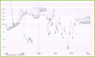
Figure 2: IR Spectra of Mixture of Rivastigmine Tartrate + Stearic Acid + Poloxamer 188 IR spectra of drug and polymer were obtained, which are depicted in Figure 2. All the characteristic peaks of rivastigmine tartrate were present in spectra at respective wavelengths (Table 3). Thus, indicating compatibility between drug and polymers. It shows that there was no significant change in the chemical integrity of the drug. Table 3: Peaks (Cm-1) And Functional Groups Present – Rivastigmine Tartrate + Steric Acid + Poloxamer 188 Sr. No. Peaks cm-1 Functional group 1 1597.06 C = C (Stre) 2 3172.90 C – H (Stre) 3 1849.73 C = O (Stre) 4 1294.24 C – O (Stre) 5 1544.98 Alkyl group DSC Compatibility Study
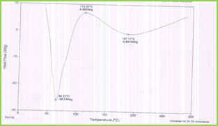
Figure 3: DSC Thermogram of Rivastigmine Tartrate + Stearic Acid + Poloxamer188
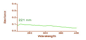
Figure 4: λmax of rivatigmine tartrate The results of DSC analysis showed that the melting temperature for rivastigmine tartrate was found to be 113.30°C. The details of thermograms are shown in Figure 3. There was no significant changes observed. Determination of λ max The λ max of rivastigmine tartrate was determined in water and methanol mixture (9:1) which was scanned between 200-400nm in the UV spectrometer. It was found to be 221nm. Standard Calibration Curve for Rivastigmine Tartrate Calibration curve for rivastigmine tartrate was constructed using water + methanol (9:1) as solvent at 221nm.The concentration selected was 10 – 90 µg/ml (Table 4, Figure 5). Table 4: Calibration data for rivastigmine tartrate Concentration (µg/ml) Absorbance (nm) 10 0.102 20 0.214 30 0.303 40 0.415 50 0.512 60 0.601 70 0.727 80 0.819 90 0.909
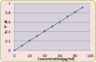
Figure 5: Standard Calibration Curve of Rivastigmine Tartrate A straight line was obtained at R2=0.999. Equation of straight line was found to be y= 0.010x Percentage Yield The percentage yields of all nine formulations were calculated and were affected by concentration of polymer and the ratio of the mixture of polymers. The increase in polymer concentration leads to increase in percentage yield. The percentage yields of all formulations are shown in Table 5. Table 5: Percentage Yield of Solid Lipid Nanoparticles of Rivastigmine Tartrate Formulation code Percentage yield (%) F1 55.66 F2 66.36 F3 72.8 F4 54 F5 64.72 F6 70.37 F7 50.33 F8 60.36 F9 67.37 Particle Size Analysis The mean particle size ranged from nm137 - 1300nm .The mean size was influenced by the concentration of lipid, surfactant and co-surfactant used in the formulations. This may be due to the less availability of amphiphiles during emulsion formation and may be partly due to more partitioning of surfactant into oil phase as the concentrations of aqueous phase was increased. The particle size of SLNs decreases with increase in the concentration of poloxamer188. An increase in the concentration of sodium taurocholate leads to decrease the particle size of SLNs. Sodium taurocholate has the ability to decrease the size of the particles. Mean particle size of all formulations are given in the Table 6 and its graphical representation were shown in Figure 6 to Figure 14. The average mean particle size of all formulations were shown in Figure 15. Table 6: Mean Particle Size and Polydispersity Index of Formulations Formulation Code Mean Particle Size (nm) PDI F1 1300 0.837 F2 194.7 0.980 F3 137.5 0.590 F4 531.0 0.938 F5 242.8 1.119 F6 212.2 1.279 F7 609.0 1.250 F8 191.0 0.890 F9 175.8 0.970

Figure 6
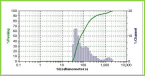
Figure 7

Figure 8
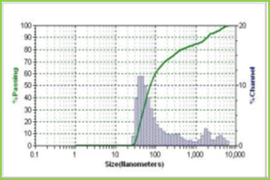
Figure 9

Figure 10

Figure 11

Figure 12
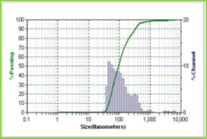
Figure 13
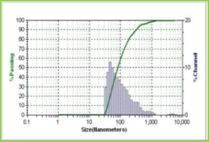
Figure 14
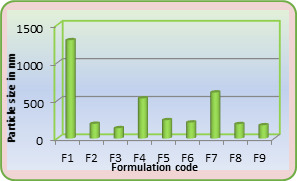
Figure 15: Average Particle Size
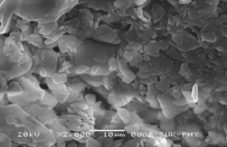
Figure 16: SEM Image of Rivastigmine Tartrate Loaded SLNs Shape and surface Morphology Solid lipid nanoparticles of rivastigmine tartrate were found to be spherical and irregular and their surface was smooth and devoid of cracks giving them good appearance. The SEM data obtained on the drug-loaded solid lipid nanoparticles of F9 shown in Figure 16. Drug Entrapment Efficiency The drug entrapment efficiency of a rivastigmine tartrate in sold lipid nanoparticles ranged from 93.26% to 99.80% (Table 7). It was observed that, when lipid concentration increased the entrapment efficiency was found to increase. Table 7: Drug Entrapment Efficiency of Different SLN Formulations Formulation code Entrapment efficiency (%) F1 93.26 F2 95.26 F3 97.82 F4 92.82 F5 96.04 F6 97.90 F7 93.35 F8 96.58 F9 99.80 Comparison of Formulations Table 8 and Figure 17 shows comparison of % yield, % entrapment efficiency and particle size. Table 8: Comparison of Percentage Yield, Drug Entrapment Efficiency, Particle Size of Solid Lipid Nanoparticles of Rivastigmine Tartrate Formulation code % Yield % Drug entrapment efficiency Particle size (nm) F1 55.66 93.26 1300 F2 66.36 95.62 194.7 F3 72.87 97.82 137.5 F4 54 92.98 531.0 F5 64.72 96.04 242.8 F6 70.37 97.90 212.2 F7 50.33 93.35 609.0 F8 60.36 96.58 191.0 F9 67.37 99.80 175.8
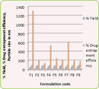
Figure 17: Comparison of % Yield, Particle Size and %Drug Entrapment Efficiency
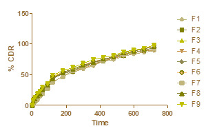
Figure 18: Zero order kinetic In-vitro Drug Release Release Kinetic Data for Solid Lipid Nanoparticle Formulations The data obtained from in vitro drug release studies were fitted to zero-order, first-order and Higuchi’s equations and is represented in Figure 18, 19 and 20 respectively. After performing statistical analysis for release study data the coefficient of correlation was found to favour Higuchi’s classical diffusion model.

Figure 19: First order kinetic
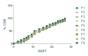
Figure 20: Higuchi’s diffusion model The values for regression coefficient shown in Table 9 for different kinetic models. From the results it is seen that the drug release mechanism from the formulation was found to follow Higuchi’s classical diffusion model. The rate of drug release is related to the rate of diffusion. The dissolution process is purely defined that the release rate is depends on the diffusion of drug from the lipid matrix, present in the developed formulation. Zeta Potential The zeta potential values obtained for the rivastigmine tartrate SLNs whichare given in Table 10 shows that the formulated rivastigmine tartrate SLNs are stable. F6 formulation was more stable than the other formulations. Table 10: Zeta Potential of Rivastigmine Loaded Solid Lipid Nanoparticles Formulation code Zeta Potential(mV) F1 -3.27 F2 -4.37 F3 -7.48 F4 -13.36 F5 -19.27 F6 -27.31 F7 -24.43 F8 -22.41 F9 -21.32 CONCLUSION In the present work, solid lipid nanoparticles of rivastigmine tartrate were formulated to deliver rivastigmine in a controlled manner. A satisfactory attempt was made to develop solid lipid nanoparticles of rivastigmine tartrate and evaluated for in vitro characterization studies. From the study following conclusions could be drawn. Rivastigmine loaded SLNs were prepared successfully, and the process parameters were optimized using 32 factorial design. The preformulation studies involving description, solubility, melting point of the drug were found to be comparable with the standard. Based on all the above preformulation studies, the drug rivastigmine tartrate was suitable for preparation of drug loaded solid lipid nanoparticles. Drug-polymer compatibility studies by FT-IR and DSC gave confirmation about their purity and showed no interaction between the drug and selected polymers. Practical and percentage yield increased as the concentration of lipid added increased. Particle size studies revealed that mean size of the prepared SLNs was in the size range of 137nm -1300nm and particles were spherical & irregular in shape. By varying the concentration of lipid, it was found that increase in lipid, surfactant (poloxamer188) and co-surfactant (sodium taurocholate) concentration in formulation leads to decrease in particle size, and increase in percentage entrapment efficiency and controlled release rate. By performing in vitro drug release study it was observed that the drug release from the formulations increases as the particle size of the formulation decreases. Rivastigmine tartrate release from all formulations followed Higuchi’s classical diffusion model kinetics. Zeta Potential shown that the F1 formulation was more stable than others. This outcome from release profiling strongly recommends that developed rivastigmine tartrate loaded solid lipid nanoparticles can be useful delivery carrier to deliver drug in controlled release manner. REFERENCES Jesus A, Ricardo I, and Joaquin R. Memory and neurogenesis in aging and Alzheimer’s disease. Aging and Disease. 2010; 1(1):30-36. Russell S, Khan M. A “mitochondrial cascade hypothesis” for sporadic Alzheimer’s disease. Medical Hypotheses. 2004; 63:8–20. https://doi.org/10.1016/j.mehy.2003.12.045, PMid:15193340 Brasnjevic I, M. Harry, Steinbusch, Christoph S, Pilar M. Delivery of peptide and protein drugs over the blood–brain barrier. Progress in Neurobiology. 2009; 87:212–51. https://doi.org/10.1016/j.pneurobio.2008.12.002, PMid:19395337 http://www.tocris.com/dispprod.php?ItemId=330410#.U-m2ZvQW1SQ http://www.mhra.gov.uk/home/groups/par/documents/websiteresources/con226931.pdf http://dailymed.nlm.nih.gov/dailymed/lookup.cfm?setid=0e20124c-725a-0145-996c-2ff908703162 N. Basaran, Zelihagul D. Bioavailability file: Rivastigmine tartrate. Journal of Pharmaceutical Science. 2005; 30: 150-57. Arthur I. Vogel. Elementary Practical Organic Chemistry. Part I: Small Scale Preparations. 2nd 76 Akhter S, Paul S, Ikramul H, Navid J, Syed S. and Reza S. Preparation, characterization and compatibility studies of naproxen loaded microspheres of cellulosic and polymethacrylic polymeric blend. Journal of Pharmaceutical Science. 2013; 12(1):11-21. https://doi.org/10.3329/dujps.v12i1.16295 Pintu K, Sahana B, and Soumen R. Enhancement of dissolution rate and stability study of ofloxacin solid dispersion. Der Pharmacia Sinica, 2011; 2(5):169-81. Vijaya, D. Kalyana. Formulation and Evaluation of Solid Lipid Nanoparticles of Prednisolone. International Journal of Pharmaceutical Research. 2012; 4(1):73-76. Gardouh A, Shadeed G, Ghonaim H. and Ghorab M. Preparation and characterization of glyceryl monostearate solidlipid nanoparticles by high shear homogenization. 1-26. Akifuddin, Z. Abbas, Marihal S, A. Ranadev, I. Santosh and Dr. Kulkarni R. Preparation, characterization and in-vitro evaluation of microcapsules for controlled release of diltiazem hydrochloride by ionotropic gelation technique. Journal of Applied Pharmaceutical Science. 2013; 3(04):35-42. Konwar R, Ahmad A. Nanoparticle: an overview of preparation, characterization and application. International Research Journal of Pharmacy. 2013; 4(4):47-57. https://doi.org/10.7897/2230-8407.04408 Silpa, R. Chakravarthi N, Yerram C, K. Hemant. Moxifloxacin loaded solid lipid nanoparticles (SLNs): Preparation and characterization. Asian Journal of Pharmacy and Research. 2012; 2(2) 105-12. Shinde S. and Hosmani A. Preparation and evaluation lipid nanoparticles of fenofibrate obtained by spray drying technique. Pharmacophore. 2014; 5(1):85-93. Khot V, Pillai M, Kininge P. Study of solid lipid nanoparticles as a carrier for bacoside. International Journal of Pharmacy and Biological Sciences.2013; 3(3): 414-26. Read the full article
0 notes
Text
Night time Imaginative and prescient Binoculars Wholesale
Whether you are looking at evening, deeply involved in the airsoft recreation, or simply want to see some critters in your yard, night vision goggles are an unbelievable piece of technology to have in your gear checklist.
Sadly, with the addition of MCP the worth of Gen 2 night imaginative and prescient scope has surged up. It's nonetheless extremely dark this early in the morning, and evening vision goggles show invaluable in this situation. It's my absolute go-to night imaginative and prescient machine among many a few of their recent reports.
The Armasight Ninox GEN 1+ Dual Tube Night Imaginative and prescient Goggles is the smartest determination that you'll ever make in buying a dual tube evening vision goggles. Night time imaginative and prescient devices of 2nd or third generation equipped with an IR illuminator ship a drastically improved commentary distance and picture decision.
Every of those can be trusted for producing top quality and affordable evening imaginative and prescient goggles. Should you determine to go forward with this explicit system…would you kindly tell us your opinion on how they evaluate with the Generation 1 Yukon you now have.
Affordability meets versatility in this particular product, as the Ghost Hunter incorporates a vary of options for evening vision use, as well as easy and comfortable arms-free operation. Fortuitously, my staff and I have scoured the evening imaginative and prescient world testing lots of of various models to bring you the top 6 evening vision units on the market in this price vary.
Do not expect Gen3 high quality kind Gen1 scope and you will be glad with this scope. The picture intensifier tubes permit for higher precision and unmatched efficiency. These Generations are evaluated on three frequent elements - the quantity of light amplification they provide, their system decision and the way sharp and clear the picture is.
The problem is although that not all evening vision gadgets are equal regardless of some folks pondering they're. For a multi-tasker, an evening imaginative and prescient monocular is ideal. Once you're on the field you don't want to get stuck in the tough mechanism and working programs that some evening vision scopes may need.
There are three,969 night imaginative and prescient goggles suppliers, mainly positioned in Asia. Once mild enter this tube, it is transformed into electrons because of the battery powered system. Otherwise, customers love the images and pictures these binoculars produce at an inexpensive price.
If you are unsure about what Evening Vision product to get, or certainly that of any daytime binocular or spotting scope and you cannot discover the information here on the BBR, please drop me a line here and I'll do my best to get again to you.
Though they can be utilized with out IR illumination, Gen 1 devices want some moonlight to function. Generally, the image intensifier amplifies mild is less than a thousand occasions with Gen 1 evening imaginative and prescient devices. Gen 1 equipment has lower light amplification (gain) and image quality (decision) when put next with Gen 2 and 3, especially on the perimeters of the display screen.
Like most Gen 3 units, the optics on this unit use Proshield coating. The Ghost Hunter 1×24 gives hunters, campers, tactical trainers and common out of doors fanatics with the proper solution for great nighttime imaginative and prescient and mobility. What does make a efficiency difference in Gen 1 merchandise is the quality of the lenses, the controls, and the gadgets' design.
The Evening Owl Professional Nexgen Night time Imaginative and prescient Binocular , combines 1st generation noctural viewing performance with a powerful 5x magnification stage for night time time outside viewing across lengthy distances. Now lights up your nights with the very useful and versatile Bushnell 260501 Equinox Z Digital Night Vision Binoculars.
And higher Digital Night time Vision units can provide you Gen 2 like image high quality at Gen 1 costs. I must pack fairly light and I am ready to spend 500 to one thousand dollars on nightvision. The Bresser Digital Evening Vision Scope comes with a sturdy bag, batteries and a comprehensive instruction handbook for those who don't know how one can use night imaginative and prescient goggles.
Because of the associated fee, most thermal items are monoculars somewhat than binoculars or goggles. Since producers of night imaginative and prescient goggles for teenagers use comparably low-cost optics, the prices of the goggles is affordable to virtually every American parent.
MCP boosts gentle gain of a picture tube by about a thousand times. All of the night imaginative and prescient scopes which can be sold available in the market run on batteries and don't have some other energy supply. Infrared illuminators throw out a beam of infra-purple lights that is close to invisible to the bare eye, however the NVD can see it. This allows you to use your binocular even in whole darkness.
In the event you disassemble the webcam and take away this filter, it's going to detect infrared gentle (at the price of decrease picture high quality). I am serious about a binocular set of night time vision goggles for underneath $1000. Right here as you possibly can see, the primary function is performed by the image intensifier tube which uses infrared illuminators.
Nonetheless the price of this high stage of Era 2 gets very close to some Gen 3 and with the for much longer life expectancy of Gen 3 picture tubes, the Era three is commonly the better selection. 12 In order for the webcam to work at night time, it can want an IR flashlight or different infrared light supply, however that is invisible to human vision.
Operating temperature is the temperature wherein you should use as well as store your night time imaginative and prescient scope. Additionally, you will have the ability to find a vary of night imaginative and prescient fashions which come with helpful accessories.

Whereas these are all era 1 evening vision units they are nonetheless extremely efficient and can serve usually most functions well. This web page involves plenty of Night time Vision Binoculars Wholesale, which are promoting at top quality and low cost value on our on-line store.
You will need to observe, that auto-gating might be positioned on previous version of night time vision and having this functionality does not essentially imply that the machine is classified as Gen three+. The monocular model of the Bestguarder Binoculars presents a much cheaper price with lots of the identical options.
0 notes
Text
Ranking the 10 Best Space Heaters
Space heaters are great for a little extra heat on your feet in a home office or to keep things from freezing over winter in your new RV. Some can even be used for camping or in a tent. They are a cheap, easy means of providing heat for smaller spaces without relying on excessive amounts of gas.
A space heater will be useful as long as you have a reliable electrical connection, but there are many to choose from. Our list of the Top Ten Space Heaters is designed to help you narrow down your options and our Buyer’s Guide will lay out some of the most important factors in selecting a new space heater.
1. Mr. Heater F232000 Buddy
Click here for the lowest price on Amazon
Made in the U.S., the F232000 Buddy from Mr.Heater is a 4,000 to 9,000-BTU radiant heater capable of providing heat for space up to 225 square feet. The Mr. Heater is ideal for camping and is safe for use in a tent. It’s approved for both indoor and outdoor spaces because it is clean burning and highly efficient. It may, however, shut off automatically if operated at altitudes over 7,000 feet above sea level.
This propane space heater boasts an auto-shut off if tipped over, if the pilot light goes out, or if it detects low oxygen levels. It also features a fold-down handle and swivel-out regulator that connects to a portable propane tank.
2. AmazonBasics Ceramic Space Heater
Click here for the lowest price on Amazon
The best selling space heater on Amazon is a ceramic heater with an adjustable thermostat that makes it ideal for home and office situations. It also offers a tip-over switch with auto shut-off and overheat protection to guarantee safety, but shouldn’t be used in bathrooms, laundry rooms, or other high-humidity areas.
This space heater features a 1500-watt high setting with three output options (low, high, and fan only). It also boasts a carrying handle for easy transport and a power indicator light that alerts you to when it’s plugged in and is receiving power.
3. Lasko 754200 Ceramic Heater
Click here for the lowest price on Amazon
This ceramic heater from Lasko is also great for homes and offices, but can also be used to provide heat for small RVs or garage apartments. It has an adjustable thermostat and doesn’t take up much floor space with dimensions of 7 x 6 x 9.5 inches.
The Lasko 754200 Ceramic Heater features high and low heat settings in addition to a fan only setting that’ll help to circulate air in a small space. It also boasts automatic overheat protection that provides peace of mind and convenient carry handles for easy portability.
4. Honeywell UberHeat
Click here for the lowest price on Amazon
The Honeywell UberHeat is designed to provide powerful, personal heating for small rooms and spaces. It offers a 3-year Limited Warranty (1) and can also be used on desks and tabletops in office settings.
This 1500-watt space heater features two heat settings with an adjustable thermostat to dial the temperature to your preference. It also boasts an auto-off tip-over switch, overheat protection and cool touch housing because, as Honeywell would say, “safety matters.”
5. Dr. Infrared Portable Space Heater
Click here for the lowest price on Amazon
The creators of the Dr. Infrared Portable Space Heater designed it to provide heat to large rooms without using an inefficient amount of energy. This 1500-watt heater is an auto energy saving model with high and low features, as well as tip-over and overheat protection.
This space heater also boasts dual heating systems with an infrared quartz tube and PTC (2). This system comes with a 12-hour automatic shut-off timer. It also features an IR remote control, high-pressure low nose blower, and an electronic thermostat that ranges from 50 to 86 degrees Fahrenheit.
6. Delonghi Full Room Radiant Heater
Click here for the lowest price on Amazon
This radiant heater from Delonghi is best used for medium to large rooms that require constant heat regulation throughout the colder months of the year. It offers 1500 watts of heating power, silent operation, and is constructed from high-quality, patented steel. It also contains a permanently sealed oil reservoir that never needs refilling.
The Full Room Radiant Heater features up to 40% more heat surface to heat rooms faster, a 24-hour on/off timer, and an adjustable thermostat with three heat settings. It also boasts patented thermal chimneys that are specifically engineered to maximize heat flow while still maintaining a low surface temperature.
7. Lasko Oscillating Space Heater (5409)
Click here for the lowest price on Amazon
Another great space heater from Lasko, the 5409 oscillating heater is made for tabletop or floor use, as it boasts easy-to-use, top-mounted controls, and an adjustable thermostat. This imported space heater offers combined plastic and metal construction.
This space heater will give you 1500 watts of comforting warmth with three quiet settings (high, low, and fan only). It comes fully assembled with an easy carry handle and won’t take up much floor or desk space with dimensions of 8.34” x 6.55” x 11.66”.
8. Vornado AVH10 Vortex Heater
Click here for the lowest price on Amazon
The Vornado AVH10 Vortex Heater is designed to provide gentle heat throughout an entire room to maintain an even, balanced room temperature. It offers two heat settings (1500/750-watt), as well as a fan-only option.
This space heater features a specially tuned airflow system (called ‘Vortex Action’) that effectively circulates heat through an entire room. It also boasts a cool-touch case, tip-over protection, and an automatic safety shut-off system. It is built to meet U.S. voltage requirements and is certified, safety-tested, and warrantied for use only in the U.S.
9. Lasko 751320 Ceramic Tower Heater
Click here for the lowest price on Amazon
This Ceramic Tower Heater from Lasko is designed to provide quiet, steady heat to reading nooks, home offices, and small rooms. It provides heat via an elongated ceramic element with penetrating air velocity that pushes warmth throughout the desired space.
This space heater comes with a multi-function remote control (batteries included) and has dimensions of 23.5 inches high, 7.4 inches wide, and 8.7 inches deep. It also features high (1500-watt) and low (900-watt) settings that allow you to dial heat for your comfort.
10. Optimus 2-Speed Fan Heater
Click here for the lowest price on Amazon
The Optimus H-1322 Fan Heater is also great for floor or desktop use in small rooms or offices. It is made in the USA or imported and comes with two heat settings, 750 and 1500 watts. It boasts an automatic thermostat control and four-position function switch (Off, Fan, Low, and High).
This space heater offers a thermal cut-off safety device and tip-over safety switch. It also features an internal, heat-limiting thermostat and a power indicator light that helps you be sure when it is powered on and receiving ample electricity.
—
Buyer’s Guide
Albert Marsh is widely recognized as “the father of the electrical heating industry” for his discovery of Chromel, which is an alloy made of one part Chromium and four parts Nickel (3). Before his discovery, low-cost cast iron radiators were the common method of heating homes with relative efficiency.
Since Marsh’s discovery at the beginning of the 20thcentury, we have continued to refine and improve home heating technology. Space heaters are just one example, but there are so many to choose from and it’s important to choose one that makes sense for the space you want to heat.
In an effort to aid in your decision, our Buyer’s Guide will focus on three main characteristics that help to distinguish heaters from one another. Those characteristics are size and coverage, fuel types, and important features.
Size and Coverage
The first and arguably most important consideration when selecting a new space heater is the size of the space you wish to heat. Large family rooms require more heating capacity than a small office or garage apartment. It’s also important to avoid buying a heater that’s overly large for a given space, as this will only lead to wasted energy and/or an unnecessarily high electric bill.
Generally, 1500-watt space heaters are great for heating rooms up to 175 square feet. On their low setting, when most of the heaters above put out 750-watts of heating capacity, they will heat 100 square foot space. The image below gives you a great idea of how much space you can heat with a given wattage or BTU rating.
Fuel Types
Space heaters can largely be separated into gas and electric options. Within the category of electric heaters, though, there are convection, infrared, panel, and radiation heaters. Convection heaters are generally regarded as the most energy efficient and commonly used means of heating an enclosed space.
Infrared heaters are great for providing direct heat to specific objects or people they are pointed at but don’t tend to disperse heat throughout a room as well. Panel heaters feature the best attributes of convection and infrared heaters while radiation heaters are best for leaving in a room for a long time because they utilize natural air circulation to disperse warmth.
Gas heaters are largely used with either liquid propane or natural gas. Liquid propane allows for the heater to remain portable and not connected to a gas line when not in use. Natural gas heaters typically require professional installation because they need to be connected to a dedicated gas line. The major benefit of gas space heaters is that they will continue to be useful during a power outage.
Important Features
The heaters detailed above offer a variety of features. Sorting through them to find the most important ones is an important part of the buying process. For starters, it’s hard to argue against buying a space heater that offers an auto shut-off feature, as safety should be the primary concern when buying any heating device. An auto shut-off will help to prevent overheating and fires caused by improper use.
Another important feature that many buyers don’t consider is the length of a heater’s power cord. Because it’s never safe to use a space heater with an extension cord, a heater with a longer cord will give you the freedom to place it exactly where you need it, especially in rooms that lack a good number of available power outlets.
The last feature we’ll mention today is a remote control. Although this isn’t necessarily an imperative feature, remote control operation will allow you to stay comfortably warm and snuggled in bed while adjusting or re-adjusting the settings of your space heater.
Frequently Asked Questions
Space heaters are extremely valuable for providing both concentrated heat on your cold toes in the winter or for dispersing heat throughout a room that otherwise wouldn’t have heat. They can, however, be dangerous if not used and cared for properly. This list of Frequently Asked Questions will help you gain more essential knowledge about space heaters.
What is ‘BTUs’?
BTUs are ‘British Thermal Units’. They measure how much heat a space-heating unit puts out. A heater’s BTU rating determines how much space it can heat safely and effectively.
Are space heaters dangerous?
Some statistics suggest that an average of 40% of home fires are caused by misuse of a space heater (4). The most common transgression is for space heaters to come in contact with flammable materials, so the simple answer is that, if used improperly, space heaters can absolutely be dangerous.
How do I ensure safety when using a space heater?
For starters, never operate your heater near flammable liquids (gasoline, paint, alcohol, etc.). Also be sure not to leave the heater unattended for long periods. Never place it near curtains or use an extension cord to connect a heater. Be sure to replace damaged parts on a heater rather than using it when it’s not “100 percent.”
Can you explain ‘convection heating’?
Convection heating is a common method used by space heaters. By circulating convection currents across a heating element (such as an electric coil, oil, or electric wire, these heaters create an overall warmth in your space.
Can I use a space heater as my main heat source?
In most homes, a single portable space heater won’t create enough warmth to heat the entire space. However, whether a space heater alone will be sufficient in your case will depend on environmental conditions, personal preferences, and housing construction.
Why does it smell like something is burning when I turn my heater on?
Many newer models will give off a slight burning odor when they are first used. This should go away after a while. Radiant heaters can sometimes burn up dust particles in the air, which will give off a noticeable scent. Additionally, some heaters can produce a burning odor as dust builds up on the heating element. These odors are generally not offensive, hardly noticeable, and typically temporary.
Summary
A quality space heater means the difference between uncomfortable winter nights and bearable conditions. In some situations, they can also spell the difference between frozen RV pipes and the ability to use all of your essential systems. We hope this article has helped you find a space heater that’s right for you and we wish you Happy Heater Shopping!
Via https://bestsurvival.org/best-space-heater/
source http://bestsurvivalus.weebly.com/blog/ranking-the-10-best-space-heaters
0 notes
Text
Airglow
Airglow
Airglow over the VLT platform. Airglow (also called nightglow) is a faint emission of light by a planetary atmosphere. In the case of Earth's atmosphere, this optical phenomenon causes the night sky never to be completely dark, even after the effects of starlight and diffused sunlight from the far side are removed.
Development
Airglow in Allier (FRANCE) during the night of 13 August 2015 The airglow phenomenon was first identified in 1868 by Swedish physicist Anders Ångström. Since then, it has been studied in the laboratory, and various chemical reactions have been observed to emit electromagnetic energy as part of the process. Scientists have identified some of those processes that would be present in Earth's atmosphere, and astronomers have verified that such emissions are present.
Description
Comet Lovejoy passing behind Earth's airglow on December 22, 2011. Airglow is caused by various processes in the upper atmosphere, such as the recombination of atoms which were photoionized by the sun during the day, luminescence caused by cosmic rays striking the upper atmosphere, and chemiluminescence caused mainly by oxygen and nitrogen reacting with hydroxyl ions at heights of a few hundred kilometres. It is not noticeable during the daytime because of the scattered light from the sun. Even at the best ground-based observatories, airglow limits the sensitivity of telescopes at visible wavelengths. Partly for this reason, space-based telescopes such as the Hubble Space Telescope can observe much fainter objects than current ground-based telescopes at visible wavelengths. The airglow at night may be bright enough to be noticed by an observer and is generally bluish in colour. Although airglow emission is fairly uniform across the atmosphere, to an observer on the ground it appears brightest at about 10 degrees above the horizon, because the lower one looks, the greater the depth of atmosphere one is looking through. Very low down, however, atmospheric extinction reduces the apparent brightness of the airglow. One airglow mechanism is when an atom of nitrogen combines with an atom of oxygen to form a molecule of nitric oxide (NO). In the process, a photon is emitted. This photon may have any of several different wavelengths characteristic of nitric oxide molecules. The free atoms are available for this process, because molecules of nitrogen (N2) and oxygen (O2) are dissociated by solar energy in the upper reaches of the atmosphere and may encounter each other to form NO. Other species that can create air glow in the atmosphere are hydroxyl (OH), atomic oxygen (O), sodium (Na) and lithium (Li). See Sodium layer. The sky brightness is typically quoted in units of astronomical magnitudes per square arcsecond of sky.
Calculation of the effects of airglow
The airglow above the horizon, captured from the ISS. File:Airglow ESO.webmPlay media Airglow. Two images of the sky over the HAARP Gakona facility using the NRL-cooled CCD imager at 557.7 nm. The field of view is approximately 38°. The left-hand image shows the background star field with the HF transmitter off. The right-hand image was taken 63 seconds later with the HF transmitter on. Structure is evident in the emission region. See also: Apparent magnitude In order to calculate the relative intensity of airglow, we need to convert apparent magnitudes into fluxes of photons; this clearly depends on the spectrum of the source, but we will ignore that initially. At visible wavelengths, we need the parameter S0(V), the power per square centimetre of aperture and per micrometre of wavelength produced by a zeroth-magnitude star, to convert apparent magnitudes into fluxes — S0(V) = 4.0×10⁻¹² W cm−2 µm−1. If we take the example of a V=28 star observed through a normal V band filter (B = 0.2 µm bandpass, frequency ν ~ 6×10¹⁴ Hz), the number of photons we receive per square centimeter of telescope aperture per second from the source is Ns: N_=10^\times {\frac {S_0(V)\times B}{h\nu }} (where h is Planck's constant; hν is the energy of a single photon of frequency ν). At V band, the emission from airglow is V = 22 per square arc-second at a high-altitude observatory on a moonless night; in excellent seeing conditions, the image of a star will be about 0.7 arc-second across with an area of 0.4 square arc-second, and so the emission from airglow over the area of the image corresponds to about V = 23. This gives the number of photons from airglow, Na: N_=10^\times {\frac {S_0(V)\times B}{h\nu }} The signal-to-noise for an ideal ground-based observation with a telescope of area A (ignoring losses and detector noise), arising from Poisson statistics, is only: S/N={\sqrt {A}}\times {\frac {N_}+N_}}}} If we assume a 10 m diameter ideal ground-based telescope and an unresolved star: every second, over a patch the size of the seeing-enlarged image of the star, 35 photons arrive from the star and 3500 from air-glow. So, over an hour, roughly 1.3×10⁷ arrive from the air-glow, and approximately 1.3×10⁵ arrive from the source; so the S/N ratio is about : {\displaystyle {\frac {1.3\times 10^{5}}{\sqrt {1.3\times 10^{7}}}}} = 36 We can compare this with "real" answers from exposure time calculators. For an 8 m unit Very Large Telescope telescope, according to the FORS exposure time calculator you need 40 hours of observing time to reach V = 28, while the 2.4 m Hubble only takes 4 hours according to the ACS exposure time calculator. A hypothetical 8 m Hubble telescope would take about 30 minutes. It should be clear from this calculation that reducing the view field size can make fainter objects more detectable against the airglow; unfortunately, adaptive optics techniques that reduce the diameter of the view field of an Earth-based telescope by an order of magnitude only as yet work in the infrared, where the sky is much brighter. A space telescope isn't restricted by the view field, since they are not impacted by airglow.
Induced airglow
SwissCube-1's first airglow image of the Earth (shifted to green from near IR) captured on March 3, 2011. Scientific experiments have been conducted to induce airglow by directing high-power radio emissions at the Earth's ionosphere. These radiowaves interact with the ionosphere to induce faint but visible optical light at specific wavelengths under certain conditions.
Experimental observation
SwissCube-1 is a Swiss satellite operated by Ecole Polytechnique Fédérale de Lausanne. The spacecraft is a single unit CubeSat, which was designed to conduct research into airglow within the Earth's atmosphere and to develop technology for future spacecraft. Though SwissCube-1 is rather small (10 x 10 x 10 cm) and weighs less than 1 kg, it carries a small telescope for obtaining images of the airglow. The first SwissCube-1 image came down on February 18, 2011 and was quite black with some thermal noise on it. The first airglow image came down on March 3, 2011. This image has been converted to the human optical range (green) from its near-infrared measurement. This image provides a measurement of the intensity of the airglow phenomenon in the near-infrared. The range measured is from 500 to 61400 photons, with a resolution of 500 photons.
Observation of airglow on other Solar System planets
The Venus Express spacecraft contains an infrared sensor which has detected near-IR emissions from the upper atmosphere of Venus. The emissions come from nitric oxide (NO) and from molecular oxygen. Scientists had previously determined in laboratory testing that during NO production, ultraviolet emissions and near-IR emissions were produced. The UV radiation has been detected in the atmosphere, but until this mission, the atmosphere-produced near-IR emissions were only theoretical. source - Wikipedia Dear friends, if you liked our post, please do not forget to share and comment like this. If you want to share your information with us, please send us your post with your name and photo at [email protected]. We will publish your post with your name and photo. thanks for joining us www.rbbox.in
from Blogger https://ift.tt/2HsHQ3K
0 notes
Text
Aerial site inspections can pinpoint problem areas during solar O&M
When it comes to solar O&M, site inspection is a key component. Whether through ground- or roof-level inspections or aerial assistance, understanding how a system is performing ensures a successful lifespan. Although inspection services by drones are on the rise, manned aircrafts are also excellent at providing precise information on a solar system’s performance.
Solar Power World talked to Heliolytics, a Canadian aerial inspection team specifically catered to the solar industry, about its services and how precisely its aircraft-mounted sensors can pinpoint system problem areas. Besides boosting performance, consistent aerial site inspections can also help with insurance claims in cases of extreme weather damage (like hurricane, tornado and flood events we described here). Heliolytics CEO Rob Andrews answered our questions.
SPW: What services does Heliolytics offer?
The view from one of Heliolytics’ manned aircrafts.
Andrews: Heliolytics provides aerial inspections and system performance analytics for solar PV systems. We have developed an aircraft-mounted sensor package and integrated artificial intelligence (AI) analytics package for the inspection of PV projects of all sizes. We have inspected over 6 GW of projects globally.
We use a manned fixed-wing aircraft to collect infrared (IR) and visible imagery of a site, then apply our analysis package to locate and classify every string- and module-level defect on a site that shows a thermal signature, indicating a reduction in energy production. We provide a digital “as-built” map of the site with all the fault locations and classifications, so clients and their technicians can easily identify what the issues are on the site and where to find them when they go to remediate the site.
SPW: What type of solar customer most benefits from your services?
Andrews: We work globally with customers on their distributed portfolios with sites ranging in size from 10 kW to over 700 MW. Our typical customers include system owners, O&M providers, EPC contractors, insurance providers and independent engineers. Our tools are aircraft-mounted, so we are able to service large portfolios of distributed rooftops as well as some of the largest ground-mount sites in the world.
SPW: How does Heliolytics’ method of inspection differ from others?
An IR image from a rooftop installation showing module issues.
Andrews: Our manned aircraft-mounted camera system and integrated AI analytics package is specifically designed for the inspection of PV arrays. We have built, from the ground up, a system specifically tuned to the needs of the PV community.
This translates to differences in the capture, classification and delivery of the data we collect. Our aircraft-based platform allows us to provide higher resolution visible and infrared imagery at a faster integration time and higher thermal sensitivity than drone imaging equipment. This allows us to detect much subtler defects, including individual cells and junction box heating. Because of our high speed of acquisition, we can collect data quickly—up to 30 minutes per 50 MW—which allows us to capture a clear thermal snapshot of a site that would normally take days or weeks to inspect.
On the processing side, because we have consistency in our dataset, we have developed new AI-based software specifically designed for the analysis of PV systems, and this provides consistency in detection across large portfolios. We have also leveraged this to provide digital delivery options, which allow the data collected from aerial inspections to be integrated directly into existing customer workflows.
SPW: Why is aerial inspection important on solar arrays?
Andrews: Aerial inspections can uniquely provide 100% visibility into DC system capacity, allowing faster and consistent remediation of string and other DC capacity losses. In one test, we compared our aerial inspection to ground-based string testing and saw that ground testing found only 20% of what we found from the air. The detection of these faults, which would not be found by traditional O&M procedures, provide a payback on services through improved site energy generation in addition to reduced labor costs.
After the scan, these inspections allow for remediation optimization, where a client can quickly asses where remediation is most critical and optimize repair schedules. Technicians can spend their time on site fixing problems rather than searching for them, and they’ll know exactly what type of faults they are dealing with prior to stepping on site.
SPW: How often would you suggest getting aerial inspections?
A close-up view of a few problem panels on a ground-mount installation.
Andrews: We usually recommend annual site scans to our clients. Annual site scans provide a big-picture understanding of how sites are performing and/or degrading over time. They also help our clients identify performance issues in particular parts of their systems, such as a specific module type that is prone to certain faults on their site, or the occurrence of distributed string faults on the site.
The most effective use of aerial inspections is coupled with advanced data analysis, where aerial data can provide the annual periodic true-up to data analytics, and the optimized performance models can be used throughout the year to better identify DC health issues between scans. It is important to note, however, that due to uncertainties in data collection, data analytics alone cannot identify all the defects identified by aerial inspection, and both provide a good complement to each other.
SPW: What are the most common faults or failures you’ve found? Do you suggest remedies?
Andrews: Aerial inspection can identify any defect which is causing a significant difference in performance between a module and its neighbors. Examples include major anomalies like string, combiner and module outages to more subtle defects like individual hot-spots, PID, module mismatch and MPPT errors. In addition, our visible imagery is critical for the detection of module breakage, dirt and fouling of modules, racking shifts and other visible anomalies.
Based on our data collected from over 6 GW of site inspections, we find that a given site has an energy weighted average DC loss of approximately 1.2%, and median weighted average DC loss of approximately 0.4%. This represents the level of loss which is being missed by current O&M practices and provides an opportunity for site optimization and energy improvement.
By energy impact, the most common fault we find are distributed string outages, which generally have too small of an energy impact to be detected by data analytics alone. The most common failure mode we find are sub-module failures which represent one-third of a crystalline module being deactivated, generally due to soldering issues or junction box damage.
Remediation recommendations depend on the level of energy impact associated with a particular fault, along with their distribution in a site. It is important to flag situations where groupings of failures may represent a serial module defect in a system.
SPW: How can aerial inspections help in extreme weather events?
Andrews: Annual aerial IR scans provide regular site assessment and benchmarking for solar PV sites, which can expedite the process of insurance claims when sites are damaged due to severe storms and other weather events. If we have performed consistent, annual scans for a client, we are able to assist them if one of their sites is affected by a damaging weather event. We create a differential report for this site, where we identify all the pre-existing defects on a site from our most recent aerial inspection before the weather event and what new/different defects have appeared on the site after the weather event from a post-event aerial inspection. This provides a clear picture of the site damage caused by the specific event for the client, including what damage can be claimed through insurance and what issues needs to be remediated on site by the client.
Solar Power World
0 notes
Text
Fluke TiR105 Building Diagnostics Thermal Imager, with IR-Fusion Technology, 9 Hz
Fluke TiR105 Building Diagnostics Thermal Imager, with IR-Fusion Technology, 9 Hz
Do not miss this chance to get this product on most inexpensive worth.
Pros:
Moveable thermal imager with an uncooled microbolometer sensor and a 160 x 120 FPA to seize infrared photos of thermal patterns, generally utilized in constructing diagnostic functions, together with power audits, constructing upkeep, evaluation, and restore
Detects temperatures from -20 to +150 levels C (-Four to +302 levels F), with an accuracy of + or – 2 levels C or 2%, and a thermal sensitivity of lower than or equal to zero.08 levels C (80mK) at 30 levels C
Digicam merges visible and thermal photos to pinpoint downside areas and supply a recognizable picture for ease of identification, and may show a full thermal, a full visible, or a picture-in-picture picture for flexibility in viewing
three.5-inch colour LCD display screen gives clear photos for ease of viewing and evaluation, and consumer selectable backlighting ranges assist remove gentle reflections and enhance distinction in low-light situations
Licensed to IP54 degree of safety in opposition to mud and splashing liquid
Get this Fluke TiR105 Constructing Diagnostics Thermal Imager, with IR-Fusion Know-how, 9 Hz
The Fluke TiR105 transportable thermal imager with an uncooled microbolometer sensor and a 160 x 120 focal airplane array (FPA) captures infrared photos of thermal patterns, and is often utilized in constructing diagnostic functions, together with power audits, constructing upkeep, evaluation, and restore. This thermal imager detects temperatures from -20 to +150 levels C (-Four to +302 levels F), with an accuracy of + or – 2 levels C or 2%, and a thermal sensitivity of lower than or equal to zero.08 diploma C (80 milliKelvins) at 30 levels C. It merges visible and thermal photos to pinpoint downside areas and supply a recognizable picture for ease of identification, and may show a full thermal, a full visible, or a picture-in-picture picture for flexibility in viewing. The three.5-inch colour LCD display screen gives clear photos for ease of viewing and evaluation, and consumer selectable backlighting ranges assist remove gentle reflections and enhance distinction in low-light situations. This thermal imager can output photos and knowledge to a pc for evaluation, comparability, annotation, and reporting. It has a laser pointer to assist goal objects throughout imaging, and the built-in flash and torch gentle assist seize photos in darkish environments. The big depth of area focus-free system works at a minimal of 48 inches for clear photos. This thermal imager can seize measurements when linked to as much as 5 suitable wi-fi modules for distant monitoring (offered individually). The thermal imager resists influence from drops as much as 2 meters, and is Ingress Safety licensed IP54 for cover in opposition to mud and splashing liquid. The European Conformity (CE) marking signifies compliance with relevant European Union (EU) directives. It comes with an AC energy adapter, a lithium ion good battery, a USB cable, a SD reminiscence card, a tough carrying case, a tender transport bag, an adjustable hand strap, directions, and SmartView reporting and evaluation software program on CD. This thermal imager is appropriate to be used in business constructing diagnostic functions.
Specs
Temperature vary-20 to +150 levels C (-Four to +302 levels F)Accuracy+ or – 2 levels C or + or – 2%, whichever is biggerThermal sensitivity< or = zero.08 diploma C (80mK) at 30 levels CDetector sort160 x 120 FPA, uncooled microbolometer sensorPicture seize frequency9HzSeen digital camera2 megapixel industrial grade digital cameraEmissivity correctionSureColour palettesGrayscale, blue crimson, ironbow, amberEnergy ProvideRechargable Li-Ion batteryWeightzero.726kg/1.6lbDimensions28.Four x eight.6 x 13.5cm/11.2 x three.Four x 5.three inches (H x W x D)
H is peak, the vertical distance from the bottom to highest level; W is width, the horizontal distance from left to proper; D is depth, the horizontal distance from entrance to again.
Thermal imagers, also referred to as thermal cameras, infrared imagers, or infrared cameras, seize photos of warmth patterns for examination and evaluation. Objects emit infrared gentle, and this gentle radiates warmth in various temperatures. A sensor inside a thermal imager detects radiant warmth and transforms it into patterns on a thermal imaging system. Thermal imagers have LCD screens to show a picture of the item in a variety of colour that corresponds with variations in temperature. Technicians can switch photos and video to a pc for course of monitoring and evaluation. Some thermal imagers can present visible photos together with thermal photos to make an object extra identifiable. Thermal imagers are utilized in an array of industries, together with building and constructing upkeep, manufacturing, firefighting, medication, regulation enforcement, and navy amongst others. A variety of functions embody constructing diagnostics to establish hidden moisture or warmth leaks, industrial and business upkeep to stop or establish electrical or mechanical tools failure, thermal cameras that restore visibility in hearth management, medical and veterinary diagnostic imaging to establish well being situations, and surveillance cameras for regulation enforcement and facility safety.
Fluke Company manufactures electrical testing instruments and software program for industrial and business functions. The corporate, based in 1948, is headquartered in Everett, WA.
What’s within the Field?
Fluke TiR105 transportable thermal imager
Rechargeable Li-Ion battery pack
AC adapter
USB cable
SD reminiscence card
SmartView reporting and evaluation software program on CD
Adjustable hand strap
Gentle case
Laborious-sided carrying case
Directions
It’s at all times higher to purchase Fluke TiR105 Constructing Diagnostics Thermal Imager, with IR-Fusion Know-how, 9 Hz from the the chief on this eCommerce trade. They’ve proved their capability in delivering high quality service after shopping for implausible merchandise from them.
YouTube Video
The post Fluke TiR105 Building Diagnostics Thermal Imager, with IR-Fusion Technology, 9 Hz appeared first on IPCAMVOX.
from Fluke TiR105 Building Diagnostics Thermal Imager, with IR-Fusion Technology, 9 Hz
0 notes