#C3R/CXL surgery
Photo
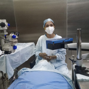
At Jehan Eye Clinic, Dr. Kareeshma Wadia, a leading cornea specialist in Mumbai, performs CXL / C3R surgery. When progression of Keratoconus is documented, either on Topography or increase in spectacle powers, this treatment called C3R Surgery or CXL needs to be done. She has successfully treated hundreds of patients Just a short term treatment for this may produce a significant improvement.
#cornea specialist#jehan eye clinic#dr.kareeshma wadia#cornea specialist in mumbai#C3R/CXL surgery#corneal cross linking
0 notes
Text
Keratoconus treatment in Pitampura
Keratoconus (corneal collagen crosslinking)
C3R is the term commonly used for the procedure of Corneal Cross Linking with Riboflavin ( hence the 3 Cs and R ). It is also sometimes known as CXL.
C3R is now the procedure of choice in preventing progression of keratoconus. Keratoconus is a condition in which there is gradual weakening and bulging of the cornea leading to progressive deterioration of vision. C3R is a non -invasive procedure that is a new ray of hope for keratoconus patients. Another indication of C3R is post LASIK ectasia.
It is a fairly simple procedure performed in the OT and does not require any admission. The cornea is treated with riboflavin ( vitamin B2 ) which is then activated by UV- A light. This leads to strengthening of the collagen fibres present in the cornea which arrests the further bulging of the cornea. In simple words it acts as a cementing force to the weak collagen fibres in patients of keratoconus. Various studies have documented that following C3R , the progression of keratoconus is reduced significantly. However, this procedure is just not a cure fir the disease and the patients still requires glasses or contact lenses to correct their vision. The procedure has practically no adverse effects and can be safely performed in all after consultation with your treating cornea specialist. It is a one time procedure and does not require frequent follow ups.

Dr. Rajiv Bajaj
MBBS, MS - Ophthalmology
Dr. Rajiv Bajaj is a renowned Ophthalmologist practising in Pitampura area of Delhi. He is the founder of Bajaj Eye Care Centre, a centre equipped with ultra modern facilities that provides solutions to a wide range of eye ailments. The centre known for its professional excellence is present on the panel of leading insurance companies, Govt. Organizations and majority of TPA's for Cashless Mediclaim Facilities.
Dr. Bajaj is an MBBS graduate from Maulana Azad Medical College, Delhi. He further pursued post graduation MS in Ophthalmology from Delhi University which involved the study of the anatomy, physiology and detailed study of the surgical problems of the eyes.
Prior to independent practice, Dr. Bajaj served at Safdarjang Hospital, Dr. Ram Manohar Lohia Hospital, Maharaja Agrasen Hospital and Sant Parmanand Hospital which have been his immense sources of experience in the ophthalmic field. He later established Bajaj Eye Care Centre which caters to patients from Delhi and NCR.
As a part of surgical education, Dr. Bajaj has demonstrated live surgery of MICS in national level conferences. He is among the first few doctors to incorporate advanced technology such as MICS, Phaco surgery and Lasik Surgery in his practice.
Call Now: 011-47024919, 011-27012054
Visit: www.bajajeyecarecentre.com
You can also search for:
Lasik treatment In Pitampura,
Lasik treatment Cost In Pitampura,
Lasik Eye Surgery In Pitampura,
Eye Care Hospital In Pitampura,
Eye Hospital In Pitampura,
Eye Care Centre In Pitampura,
Eye Specialist in Pitampura,
Eye Doctor in Pitampura,
Eye Clinic in Pitampura,
Best eye Doctor in Pitampura,
Cataract surgery in Pitampura,
Cataract surgery cost in Pitampura,
Cataract operation in Pitampura,
Keratoconus treatment in Pitampura,
Oculoplasty procedure in Pitampura,
Ophthalmology clinic in Pitampura,
#Lasik treatment In Pitampura#Lasik treatment Cost In Pitampura#Lasik Eye Surgery In Pitampura#Eye Care Hospital In Pitampura#Eye Hospital In Pitampura#Eye Care Centre In Pitampura#Eye Specialist in Pitampura#Eye Doctor in Pitampura#Eye Clinic in Pitampura#Best eye Doctor in Pitampura#Cataract surgery in Pitampura#Cataract surgery cost in Pitampura#Cataract operation in Pitampura#Keratoconus treatment in Pitampura#Oculoplasty procedure in Pitampura#Ophthalmology clinic in Pitampura
0 notes
Text
Keratoconus (corneal collagen crosslinking)
C3R is the term commonly used for the procedure of Corneal Cross Linking with Riboflavin ( hence the 3 Cs and R ). It is also sometimes known as CXL.
C3R is now the procedure of choice in preventing progression of keratoconus. Keratoconus is a condition in which there is gradual weakening and bulging of the cornea leading to progressive deterioration of vision. C3R is a non -invasive procedure that is a new ray of hope for keratoconus patients. Another indication of C3R is post LASIK ectasia.
It is a fairly simple procedure performed in the OT and does not require any admission. The cornea is treated with riboflavin ( vitamin B2 ) which is then activated by UV- A light. This leads to strengthening of the collagen fibres present in the cornea which arrests the further bulging of the cornea. In simple words it acts as a cementing force to the weak collagen fibres in patients of keratoconus. Various studies have documented that following C3R , the progression of keratoconus is reduced significantly. However, this procedure is just not a cure fir the disease and the patients still requires glasses or contact lenses to correct their vision. The procedure has practically no adverse effects and can be safely performed in all after consultation with your treating cornea specialist. It is a one time procedure and does not require frequent follow ups.
tags- Lasik treatment In Pitampura Lasik treatment Cost In Pitampura Lasik Eye Surgery In Pitampura
0 notes
Text
Treatment of Keratoconus by Collagen Cross-Linking with Riboflavin (C3R)
Dr. Neera Agrawal, Neera Eye Centre, Darya Ganj, New Delhi
Keratoconus is a bilateral non-inflammatory corneal ectasia (fig.1) with an incidence of approximately 1 per 2,000 in the general population. It normally presents in early teens or twenties and is known to be progressive. Despite intensive clinical and laboratory investigation, the aetiology of keratoconus remains unclear. Thinning of the corneal stroma, breaks in Bowman’s layer, and deposition of iron in the basal layers of the corneal epithelium comprise a triad of the classical histopathologic features found in keratoconus.

Fig. 1- Natural Progression of Keratoconus in a patient over 1.5 years
Till date, accepted methods of treatment have been contact lenses, intra-stromal corneal rings (INTACS), Photo-refractive keratectomy and cornea transplant. Cornea transplant is reserved for advanced cases in which vision can not be improved with contact lenses. Unfortunately none of these modalities prevent the progression of the disease. Now with the advent of Collagen Cross-Linking with Riboflavin and UVA, there is scope to arrest the progression of disease in these patients. Moreover, it is technically simple and less invasive than all other surgical therapies proposed for Keratoconus.
Mechanism of action
Similar to photo polymerization of polymers, Collagen Cross-Linking of Cornea using ultraviolet light and the Photo-sensitizer Riboflavin was developed to treat corneal thinning and ectasia by increasing the biomechanical strength of the tissue. This procedure is also addressed by various eponyms such as C3R, CXL and CCL.
First studies in Photobiology began in 1990’s, with attempts to identify biological glues that could be activated by heat or light to increase resistance of stromal collagen (2). It was discovered that the gluing effect was mediated by oxidative mechanism associated with hydroxyl radical release. A similar mechanism of natural hardening and thickening of collagen fibres has been demonstrated in corneal aging (3). Similarly, young diabetics never develop progressive keratoconus due to natural cross linking effect of glucose, which increases corneal resistance.

Fig. 2- Mechanism of action : C3R

Fig. 3- Mechanism of action : C3R
Collagen cross linking results in an increase in inter-fibrillar covalent bonds by photosensitized oxidation (fig.4), and causes bio-mechanical stabilization of cornea. A significant increase of 328.9% in the biomechanical rigidity of human corneas has been documented after collagen cross linking (5).

Fig.4- Effect of Collagen Cross Linking on Corneal Collagen

Fig. 5 – Effect of Cross-Linking on Cornea
Riboflavin plays a dual role in the procedure; it not only acts as a photo sensitizer for the photo-oxidative cross linking process but also has a barrier effect. It acts like a shield and prevents UV induced collateral damage to sensitive ocular structures like Corneal Endothelium, Lens and Retina.
Quality of Beam
All safety considerations regarding the Cross-Linking procedure assume an optically homogenous irradiation of the cornea. Optical in-homogeneities can lead to damage to corneal endothelium, which represents most endangered structure. Thus, having homogenizing optics and as well as pre-compensation for corneal curvature in the system is important (fig.6).

Fig. 6: Quality of beam for C3R
Indications
Most common indications are –
1. Mild to moderate Keratoconus – In a case of Keratoconus, the selection criteria for C3R are as follows -
· There should not be corneal scarring.
· There should be reasonably good acuity with glasses or contact lenses.
· Corneal thickness should be more than 450 microns (400 microns after epithelium removal) at thinnest point. This is based on the fact that UV light can penetrate cornea up to a thickness of 350 microns and will damage endothelium if enough corneal thickness is not there. That is why some people advocate doing specular microscopy before and after the procedure.
· Age should more than 16 yrs.
· Maximum K readings should be < 60Diopters.
· Keratoconus must be documented to be progressive. Progression of Keratoconus is ideally determined by Kaplan Meier or Fourier analysis which are based on Spherical equivalent power, Decentration component and Irregular astimatism component and calculation their yearly rate of progression. It is generally accepted that 1 dioptre increase in the power of cone per year is an indicator of progression of Keratoconus. It can also be seen as steepening of Keratometry readings (17)
2. Post Lasik Ectasia – Good results have been reported in treatment of post Lasik ectasia (13).
3. Progressive Hyperopia post RK – good results have been reported (12).
4. Pellucid Marginal Degeneration
5. Bullous Keratopathy – reduces Corneal oedema by increasing stromal compaction after Cross-linking. It may be combined with Intra-stromal administration of 0.1% Riboflavin with the help of Femtosecond laser (Can also be staged i.e. anterior and posterior separately)(16).
6. Infective Keratitis – to prevent Corneal Perforation by increasing resistance to the effect of collagen digesting enzymes.(16)
7. Scleral CXL for Glaucoma and Pathological Myopia (16)
Types of C3R
C3R surgery can be of several types depending on the technique used –
1. C3R Epi-off – this is the most commonly used technique where you remove the epithelium mechanically. Commonly follows Dresden Protocol which means that you instill Riboflavin for every 2 min for 30 min and cross link for 30 min at 3 MW energy. It needs min Corneal thickness of 400 microns after epithelium removal. It has been there for longest time and has proven its efficacy.
2. Trans-epithelial C3R or epi-on C3R – here we use special Riboflavin which can penetrate through epithelium and do not remove epithelium, rest of the protocol is same as above. However, I have noticed that despite leaving the epithelium in situ, healing is much slower after this procedure compared to epi-off C3R.
3. Rapid C3R – here Riboflavin can be of any of the above type but machine setting used are 9 MW for 10 min or 18 MW for 5 min. The efficacy and safety data on this type is still awaited.
4. Lasik Extra – In this treatment C3R is combined with Lasik surgery. After making the flap Riboflavin is directly put on the flap and cross linking is done with short duration of settings like 5 min or 2 min. It is said to be useful for suspected ectasia cases where you would like to do Lasik. However, there are no studies to prove its efficacy. In my opinion, it may help in preventing ectasia but will significantly alter the refractive result by causing flattening.
Types of Riboflavin in C3R-
1. Iso-tonic Riboflavin
2. Hypo-tonic Riboflavin
3. Trans-epithelial Riboflavin
4. Rapid Ribo-flavin
5. Riboflavin for Lasik Extra
Iso-tonic Riboflavin
It is of two types –
1. Riboflavin in Dextran
2. Riboflavin in HPMC
Iso-tonic Riboflavin with Dextran
It contains Riboflavin 0.1% in 20% Dxtran 500. It is time tested and gives predictable results. It causes better Corneal staining. Unfortunately not many companies produce this.
Iso-tonic Riboflavin with HPMC

Fig 7: Riboflavin with HPMC
Contains 0.1% Riboflavin with 1.1% HPMC.
Causes poor Corneal staining. I have noticed higher incidence of post operative Corneal oedema and Descemets folds after this.
Hypo-tonic Riboflavin
It is used for Corneas which are thin for Iso-osmolar Ribo-flavin. It is used after use of Iso-osmolar Riboflavin for 30 min. Therefore, you will need both types of Riboflavins for one case. It can swell Corneas upto 60 microns but the flip side is that effect lasts only for 10-30 minutes. You will keep repeating its use after repeating the per-op Pachymetry. That is why its use has gone down specially with advent of Trans-epithelial Riboflavin.
Trans-epithelial Riboflavin
Contains EDTA or BAK, which loosen the bonds in epithelium and allow Riboflavin to penetrate. Different companies have different composition.

Fig 8 :Trans-epithelial Riboflavin
Fig 9:Trans-epithelial Riboflavin
Trans-epithelium Riboflavin with Iontophoresis
Is a new modality. It reduces the diffusion time to 5 min.
Main advantage is that it is less toxic to the epithelium. It substantially reduces post operative pain and debility to the patient.
Rapid Riboflavin
Contains Riboflavin 0.1%, Saline and HPMC
Has a diffusion rate twice of normal Riboflain
Used only as epi-off .

Fig 10:Rapid Riboflavin
Riboflavin for Lasik Extra
Contains Riboflavin 0.22% in Saline and is applied to Stromal Bed.

Fig 11: Riboflavin for Lasik Extra
Technique ( Dresden Protocol)
The procedure is conducted in an operating room under sterile conditions. Topical anaesthesia is given by Proparacaine Hydrochloride (0.5%) eye drops & Xylocaine 4% drops. Central 7-8 mm of corneal epithelium is removed by mechanical debridement (gentle scraping with hockey stick knife) or Alcohol. My personal preference is mechanical debridement.
This is followed by instillation of 0.1% Riboflavin (3 mg Riboflavin - 5 Phosphate in 3 ml of Dextran-T-500 20% solution) eye drops at every 2 minute interval for 30 minutes (15 times).
To confirm adequate penetration of Riboflavin into the cornea, the patient may be examined on slit lamp. A greenish flare, similar to that seen with fluorescein dye instillation, may be seen in anterior chamber, signifying penetration through the thickness of cornea.

Fig.12 - C3R being done in a patient
There are studies available which say that epithelial debridement is not necessary. However, it’s mandatory to demonstrate the penetration of the riboflavin in the corneal stroma when performing the treatment without removing the epithelium.
Following this, the cornea is subjected to Ultraviolet A radiation (365nm) from a distance of 5 cm (Peschke Meditrade UVX system was used) for the next 30 minutes (fig.6). During this period, topical Riboflavin drops are again instilled at 5 minute intervals, to complete photo-sensitization and provide photo-protection by the ‘barrier’ effect. Thereby, a dose of 3 mW/ Sq cm (+ 0.3 Mw) (5.4 J/Sq cm) UV-A is delivered. The UV-A radiation lamp is checked for calibration with a UV- meter before and after the treatment. On completion of the treatment, eye is patched with antibiotic eye ointment or sterile bandage lens is applied.
Post-operative Care
Topical antibiotics along with lubricants and viscous tear substitute, mild steroids are prescribed 4-5 times a day for 3-4 weeks. Complete re-epithelization usually occurs in 3-4 days. Patients may experience mild pain and discomfort for first 2 days, for which oral analgesics are also required to keep them comfortable.
A mild epithelial haze with transient, mild stromal oedema may be seen in some cases after C3R, which usually disappears completely in a few weeks. There is no damage to the corneal endothelium.
Histopathological changes in cornea post C3R
C3R treatment leads to a dose-dependent keratocyte apoptosis (1) that can be expected in human corneas to a depth of 300 microns from the anterior surface utilising a surface UV-A dose of 5.4 J/ sq cm. In the first few weeks after the procedure, a ‘vertical transition line’ may be visible on the slit lamp, delineating the anterior cross-linked zone with the posterior unaffected stroma (fig.13).
Repopulation of corneal stroma may take up to 6 months. It is well documented that the corneal epithelium attains a regular morphology and density within 5 days after C3R. Initially, disappearance of the sub-epithelial stromal nerve fibres was observed in the treated area and initial re-innervation was seen one month after the procedure. Complete recolonization of the anterior sub-epithelial stroma by the keratocytes was observed in 6 months after C3R, with restoration of corneal sensitivity.

Figure 13- Effect of Cross-Linking on cornea
Histopathologically, it has been demonstrated that there is a significant increase in collagen fibre diameter after collagen cross linking. After Collagen Cross-Linking, the cornea shows an increase in thermo-mechanical stability as well as a markedly increased resistance to collagen digesting enzymes (5).
Results
· C3-R appears to stabilize corneas up to six months after treatment.
· In a 5 year study, it was noted that in all treated eyes, the progression of keratoconus was at least stopped. About 70% eyes show regression with a reduction of the maximal keratometry readings by 2 dioptres and of the refractive error by 1 D was found (14). Best-corrected visual acuity improved by 1.4 lines. I have personally experienced similar results in my 7 years experience with the procedure.
· The addition of C3-R to the INTACS procedure results in greater keratoconus improvements compared to INTACS alone (10).
There are reports available that surface ablation procedures could be used to correct refractive errors following C3R in Keratoconus patients but conclusive evidence is lacking.

Fig 14:About 5 D Corneal Flattening after C3R

Fig.15: About 4 D Corneal Flattening after C3R

Fig 16: Corneal Flattening after C3R in PMD
Safety
During corneal Cross-Linking, 95% of UVA energy is absorbed by only 350 microns of Riboflavin soaked Cornea (fig.17). As long as the treated cornea has a minimum thickness of 400 microns (after removal of epithelium), the corneal endothelium will not experience damage, nor will deeper structures such as lens and retina.

Fig. 17 - Absorption of UVA by Cornea
The light source should provide a homogenous irradiance, avoiding hot spots.
It has been seen that corneal and lens transparency, endothelial cell density, and intraocular pressure remained unchanged after the procedure (9).
Conclusion
C3R for ectatic Disorders of Cornea has revolutionised the treatment of Keratoconus and other ectatic disorders. Cornea Collagen Cross-linking with Riboflavin is a simple, safe and effective procedure in the management of early progressive ecstatic disorders of the cornea.
Different types of Riboflavin and machines have made this techniques useful in more number of patients.
Bibliography
1. Apoptosis After Corneal Collagen Cross-linking using Riboflavin/UVA treatment. Wollensak G. et al. Cornea 2004; 23: 43-49
2. Photodynamic biologic tissue glue. J. Khaderm, T. Truong, J.T. Ernest. Cornea 1994;13: 406-410
3. Collagen fibrils in the Human corneal stroma . structure and aging. A. Dover, K. Misof et al. Invest. Ophthalmol. Vis. Sc. 1998; 39: 644-648
4. Stress-strain measurements of human and porcine corneas after Riboflavin- ultraviolet-A induced cross-linking. Wollensak G.,Spoerl E., Seiler T. J Cataract Refract Surg. 2003;29:1780-1785
5. Increased resistance of cross-linked cornea against enzymatic digestion. Seiler T., Wollensak G., Spoerl E. Curr. Eye Res. 2004 July; 29(1): 35-40.
6. Thermo-mechanical behaviour of collagen cross linked porcine cornea. Spoerl E., Wollensak G., Dittert D.D., Seiler T., Ophthalmologica.2004, Mar-Apr; 218(2):136-140
7. C3-R Treatment Opens New Frontiers for Keratoconus and Corneal Ectasia. Roberto Pinelli, MD. Eyeworld 2007, 34-36
8. Safety of C3-R UVA at the Retinal Level and Compared to Outdoor UVA Exposure Leonard Yuen, MD and Brian S. Boxer Wachler, MD
9. Safety of UVA-Riboflavin Cross-Linking of the Cornea Eberhard Spoerl, PhD, Michael Mrochen, PhD, David Sliney PhD, Stephen Trokel, MD, Theo Seiler, MD, PhD, Cornea 2007; 26:385-389
10. The Effect of Inferior Segment Intacs with and without Corneal Collagen Crosslinking with Riboflavin (C3-R) on Keratoconus. Colin C.K. Chan, MD, FRANZCO, Munish Sharma, MD, Brian S. Boxer Wachler MD, J Cataract Refract Surg 2007;33:75-80
11. No Progression of Keratoconus 5 Years after C3-R. Wollensak G. Crosslinking treatment of progressive keratoconus: new hope. Curr Opin Ophthalmol. 2006 Aug; 17:356-60.
12. C3-R for Stabilization of Progressive Hyperopia (Farsightedness) after Radial Keratotomy and Laser Ablation. Presented at the American Academy of Ophthalmology Annual Meeting, 2006
13. Visual rehabilitation and outcomes of ectasia after corneal refractive surgery. Woodward MA, Randleman JB, Russel B, Lynn MJ, Ward MA, Stulting RD - J. cataract Ref. surg. 2008 mar; 34(3): 383-8
14. Riboflavin/UVA induced collagen cross-linking for treatment of Keratoconus. G. Wollensak, MD et al, Am. J. Ophthal. 2003 May; 135(2) : 620-627
15. Staged Intra-stromal Delivery of Riboflavin with UV-A Cross-linking in Advanced Bullous Keratopathy. Krueger RR, Ramos-Esteban JC, Kannelopoulos J – J. Refract. Surg. 2008; 730-736
16. Corneal Cross-linking for Different Corneal Diseases. Aylin Kilic Ertan – Cataract & Refractive Surgery Today Europe. April 2009; 25-28
17. Progression of Keratoconus Assessed by Fourier Analysis of Videokeratography Data. Tetsuro Oshika, Tatsuro Tanabe, Atsuo Tomidokoro, Shiro Amano – Ophthalmology 2002; 109:339-342
For Reprints request to -
Dr. Neera Agrawal
Neera Eye Centre
B-99, Bharat Ram Road
Opposite Commercial School
Darya Ganj
New Delhi- 110002
Phone: 23270775, 9810134653
0 notes
Text
Diagnosed with Keratoconus today. Been advised a C3R/CXL. Anxious and scared
I'm 21. And I'm in India. Have just completed undergrad studies and went for a regular eye check-up. The doctor got suspicious of the very irregular readings and suggested a scan. Cut to chase, she told me I have keratoconus in both eyes and should consider CXL asap. Right eye 428 micrometers and left eye 442. I'm scared of this process because, I don't know. The doctor advised surgery on the right eye asap and after the one month recovery consider the surgery on the left eye. I'm supposed to report for my first-job in the first week of July and I'm afraid the recovery period might be a problem for this. I can really use some advice in preparing for this surgery. Precautions, things I should know etc. Most importantly, I could use some reassurance because I'm scared of the negative possibility of the surgery. Thanks and apologies for repeating things if any. submitted by /u/rookie111 [link] [comments] https://www.reddit.com/r/Keratoconus/comments/8ljsur/diagnosed_with_keratoconus_today_been_advised_a/?utm_source=dlvr.it&utm_medium=tumblr
0 notes
Text
Info About Insurance Coverage for CXL / Refractive Surgery
Hello All, I want to make this post to potentially help anyone out who may be dealing with their insurance for treatment options for their KC. This shouldn't be considered medical advice or anything - just my experience. A while back, I had epi-on CXL (aka C3R) and Visian-ICL with Dr. Brian Boxer-Wachler. I know some people don't like his advertising strategies but after visiting a few other doctors, he was pretty knowledgable about KC and was very friendly. The other thing was, it was relatively easy to get an appointment ASAP to start treatment and his staff was very attentive (this is important and I will explain this later on). So anyways, as expected, the insurance co (Anthem) didn't cover anything. I'm a CA resident so we have the DMHC which regulates health insurance plans. The process is simple....first you have to request coverage for a procedure (in my case, C3R in both eyes and Visian ICL in the one eye). The insurance will likely deny it claiming "not medically necessary" or "investigational/experimental" - in my case, it was "investigational/experimental". Next step is to file a grievance / appeal by simply calling your insurance co and telling them you want to file a grievance against their decision. They'll probably also deny this citing the same reason - don't waste your time arguing - just move on and play their game. Now, once you get a letter stating your grievance was denied - simply go on your insurance regulator's website and file a complaint their and explain the situation. You will need your medical records (I got a PDF file of the topographies, visual-acuity measurement, the complaints about my vision, other eye scans etc). All you have to do is basically explain the problem and they'll have 3 independent ophthamologists review your claim and determine if your insurance co should pay for your surgery. Here's where your physician choice is important. Dr. Brian Boxer-Wachler's staff really helped me out big time by following-up with the DMHC about the procedure and also emailing me back my medical records promptly. They also provided all of the necessary documents and diagnosis paperwork (contact lens intolerance, keratoconus, etc). His treatment wasn't cheap by any means, however, after the insurance reimbursement, it was actually much cheaper than other doctors and I doubt another physician would have went through so much trouble. I was very fortunate that the DMHC found the Visian-ICL and C3R to be necessary. I didn't expect the Visian-ICL to be covered but it was a nice surprise. For anyone thinking about doing it, it really made a difference in my visual acuity (I no longer need contacts/glasses to function), however, the iridotomy that is required does cause some visual disturbances (called dysphotopsia) in some people. I had to have a corneal tattoo applied to minimize this and it's not as bad anymore. I may go for a second treatment to eliminate the dysphotopsia. If you can wait, I'd personally wait for the newer version of the Visian-ICL (toric) which corrects astigmatism and doesn't require the iridotomy. Here is a handy website for CA residents to see decisions made in the past: http://wpso.dmhc.ca.gov/imr/ Going forward, one eye is still uncorrected (not a candidate for ICL). I'm considering PRK/Intacs - one ophthalmologist said he doesn't do Intacs anymore because people don't notice much of a difference. Another told me that PRK is kind of risky. I'm still debating on what to do. in the meantime, I have started a process to see if I can get those approved. As always, YMMV depending on what state you live in and the severity of your KC. submitted by /u/Enferrari [link] [comments] https://www.reddit.com/r/Keratoconus/comments/7jqvxb/info_about_insurance_coverage_for_cxl_refractive/?utm_source=dlvr.it&utm_medium=tumblr
0 notes
Text
Update on Epi-on CXL with Dr. Boxer-Wachler
I just wanted to update this sub since I used some of the info from here to form a plan of action for myself. I went to Dr. Boxer Wachler in Beverly Hills for Epi-on CXL (aka Holcomb C3R). The procedure itself isn't really painful although I had ICL beforehand so I was on pain meds so that may have been a factor. Basically you have to stare into the UV lamp for 7.5 minutes, then theres a quick pause, then 7.5 minutes, quick pause, etc for a total of 30 minutes. The crosslinking wasn't painful or anything and the haziness subsided in about a day for me. I haven't noticed any changes in my vision yet but it's only been a week. As far as the ICL, I was a candidate in one eye so I had that done as well. The "1 day recovery" was an understatement since it's been about a week for me and still have some discomfort (but not much). The ICL definitely improved my vision. I was 20/30 the after surgery. I'm still hoping I can correct my right eye since it's a bit fuzzy. I was told intacs are not a guarantee so I passed for now (I heard some horror stories about glare etc). Soft contacts dont work unfortunately and I've never tried sclerals/RGPs. As far as pricing, it was expensive for sure but based on what people felt in this subreddit, they were happy with the results so I felt it was worth it. C3R was $3800/eye and ICL was $5600 eye. Only time will tell the rest. submitted by /u/Enferrari [link] [comments] https://www.reddit.com/r/Keratoconus/comments/5wuinp/update_on_epion_cxl_with_dr_boxerwachler/?utm_source=dlvr.it&utm_medium=tumblr
0 notes
Last Seen Blogs

talkzieyvette-blog
Talkzie-Learn Korean

minesenstiel
Minesenstiel

rfghytrd
lkjhedb

petitpapillonbebe
Petit Papillon Bebê & Criança

gag-art-blog
gagto57 art blog