#Focused Assessment for Sonography
Text

Find the best sonography centre near me thane
If you're searching for a sonography centre near me Thane, look no farther than Crystal Diagnostic. Sure for its phenomenal medical imaging affiliations, Crystal Diagnostic stands pulled out as the go-to objective for watchful and strong sonography. Utilizing arrangement setting advancement, the center gives epic standard pictures fundamental for wary end and treatment. Crystal Diagnostic qualities its high level workplaces and a party of fundamentally gifted specialists focused in on figuring out thought. The sonography affiliations offered harden a dumbfounding number of medical conditions, assisting with early straightforwardness and strong management. The immediate framework uses high-complement sound waves to convey point by point pictures of inside body structures, ensuring cautious and definite assessments. Truly coordinated in Thane, Crystal Diagnostic is really open, seeking after it an ideal choice for those searching for quality sonography benefits nearby. The center offers versatile appointment timetables to oblige the clamoring presences of its patients, ensuring unessential holding up times and brief help. Patients can expect a great and supporting experience, with the party of experienced radiologists and experts giving changed care and thought. Pick Crystal Diagnostic for your sonography needs in Thane, and experience the best demands for medical imaging and merciful thought. With a commitment to significance and patient fulfillment, Crystal Diagnostic is the reasonable sonography place near you in Thane.
0 notes
Text
Personalized Healthcare: Revolutionizing Patient Care with Tailored Solutions

In the realm of modern medicine, personalized healthcare has emerged as a transformative approach to patient care. This innovative model utilizes advanced technology and data analytics to tailor medical treatments and recommendations to individual patients. This article delves into how personalized healthcare is reshaping the industry and why it matters for you.
What is Personalized Healthcare?
Personalized healthcare, also known as precision medicine, focuses on customizing healthcare treatments and practices to individual characteristics. Unlike the traditional "one-size-fits-all" model, this approach considers genetic, environmental, and lifestyle factors to optimize patient care.
Key Components of Personalized Healthcare
Genetic Information: Using genetic testing to tailor treatments based on individual DNA profiles.
Lifestyle Data: Incorporating data on diet, exercise, and other lifestyle factors.
Advanced Diagnostics: Leveraging cutting-edge technologies such as 3D/4D ultrasound, MRI, and digital mammography to gain a comprehensive view of health.
Benefits of Personalized Healthcare
Enhanced Treatment Efficacy: By tailoring treatments to the individual's specific needs, personalized healthcare often results in better outcomes compared to standard treatments.
Reduced Adverse Effects: Personalized approaches minimize the risk of side effects by considering how individuals metabolize medications differently.
Improved Preventive Care: Identifying risk factors early through advanced diagnostics allows for proactive management and prevention of diseases.
Informed Decision-Making: Access to comprehensive data helps both patients and healthcare providers make well-informed decisions about treatments and lifestyle changes.
How Advanced Diagnostics Support Personalized Healthcare
Advanced diagnostic tools are crucial in the personalized healthcare model. They provide detailed insights into an individual's health, allowing for more accurate diagnoses and tailored treatment plans.
Key Diagnostic Tools in Personalized Healthcare
Digital Mammography: Offers high-resolution imaging for early breast cancer detection.
3D/4D Ultrasound: Provides detailed images for monitoring fetal development and diagnosing various conditions.
MRI: Produces detailed images of soft tissues to assist in diagnosing neurological and musculoskeletal conditions.
CT Scans: Utilized for detailed cross-sectional images of internal organs, aiding in precise diagnostics.
In Kota, several diagnostic centers offer these advanced tests, including digital mammography, MRI, and 3D/4D ultrasound. For example, the modern diagnostic center Kota provides comprehensive diagnostic services, including FNAC-Biopsy and Drainage Procedures and Barium studies.
Integrating Personalized Healthcare into Clinical Practice
Personalized healthcare requires the integration of various elements to be effective. Here’s how modern diagnostic centers in Kota support this approach:
Comprehensive Testing: Facilities like the 3D diagnostic center in Kota and Kota CT scan center provide a range of tests that contribute to a detailed health assessment.
Advanced Pathology: Labs such as the Advance Pathology Lab in Kota offer specialized tests including pathology, biochemistry, microbiology, and hormonal tests.
Holistic Monitoring: Centers like the Kota sonography center and MRI center in Kota ensure continuous monitoring and precise diagnostics.
The Role of Technology in Personalized Healthcare
Technological advancements have played a significant role in the evolution of personalized healthcare. Innovations such as digital X-ray, ECG tests, and ultrasound technologies contribute to a more nuanced understanding of patient health.
Innovations Driving Personalized Healthcare
Artificial Intelligence (AI): AI algorithms analyze vast amounts of data to predict health risks and suggest personalized treatments.
Wearable Devices: These devices track health metrics in real-time, providing valuable data for personalized care.
Commonly Asked Questions About Personalized Healthcare
1. How does personalized healthcare differ from traditional medicine?
Personalized healthcare tailors treatments based on individual genetic, environmental, and lifestyle factors, whereas traditional medicine often follows a standardized approach.
2. What are the benefits of personalized healthcare?
Personalized healthcare offers enhanced treatment efficacy, reduced adverse effects, improved preventive care, and better-informed decision-making.
3. How can advanced diagnostics improve personalized healthcare?
Advanced diagnostics provide detailed insights into an individual's health, enabling more accurate diagnoses and tailored treatment plans.
Conclusion: Embrace Personalized Healthcare with Dr. Vaaya’s Expertise
Personalized healthcare represents a significant advancement in medical science, offering tailored treatments and improved outcomes. With the support of modern diagnostic tools and expert guidance, you can benefit from this cutting-edge approach.
For top-tier personalized healthcare services in Kota, including FNAC-Biopsy and Drainage Procedures, digital mammography, and 3D/4D ultrasound, visit Dr. Vaaya's Diagnostic Center. Our state-of-the-art facilities and expert team ensure you receive the most accurate diagnostics and personalized care.
Contact Dr.Vaya’s Lab today at +91 8824 911 390 or visit Dr. Vaaya's for more information and schedule your appointment.
By integrating personalized healthcare practices with advanced diagnostic tools, we aim to enhance your overall health and well-being.
0 notes
Text
The Role of a Sonographer: A Vital Part of Modern Healthcare
A sonographer, also known as an ultrasound technician, plays a critical role in the medical field by using specialized equipment to create images of the body's internal structures. These professionals are essential in diagnosing and monitoring a wide range of medical conditions, from pregnancy to heart disease.
What Does a Sonographer Do?
Sonographers operate ultrasound machines that use high-frequency sound waves to capture images of organs, tissues, and blood flow inside the body. The images, called sonograms, provide valuable information that helps doctors diagnose and treat patients. Sonographers work closely with radiologists and other healthcare professionals, interpreting the images they capture and identifying any abnormalities.
Specializations Within Sonography
Sonography is a diverse field with several areas of specialization:
Obstetric and Gynecologic Sonography: Focuses on imaging the female reproductive system, including monitoring fetal development during pregnancy.
Abdominal Sonography: Involves imaging the abdominal organs, such as the liver, kidneys, and gallbladder.
Cardiac Sonography (Echocardiography): Specializes in imaging the heart and assessing its function and structure.
Vascular Sonography: Focuses on imaging blood vessels and evaluating blood flow and circulation.
Education and Training
To become a sonographer, one typically needs to complete an accredited educational program, which may result in an associate or bachelor’s degree in diagnostic medical sonography. These programs include both classroom instruction and hands-on clinical training. After completing their education, sonographers may also seek certification through organizations like the American Registry for Diagnostic Medical Sonography (ARDMS) to enhance their credentials and job prospects.
The Importance of Sonographers in Healthcare
Sonographers are crucial in the early detection and monitoring of diseases. Their ability to produce clear, accurate images allows doctors to make informed decisions about patient care. In addition, sonography is a non-invasive, painless procedure, making it a preferred diagnostic tool in many situations.
Conclusion
Sonographers are key players in the healthcare system, providing essential diagnostic information that aids in patient care. Their expertise in using ultrasound technology contributes to better health outcomes, making them an indispensable part of modern medicine. As technology advances, the role of sonographers will continue to evolve, further enhancing their impact on patient care and medical diagnostics.
0 notes
Text
Discover the Best Sonography Center in Vasai West: Your Health, Our Priority at Midas Care Clinic

Table of Contents
Introduction
What is Sonography?
Why Choose Midas Care Clinic?
Advanced Technology and Expertise
Services Offered
Patient Care and Comfort
How to Prepare for Your Sonography Appointment
Frequently Asked Questions (FAQ)
Conclusion
1. Introduction
At Midas Care Clinic, we prioritize your health and well-being. Our state-of-the-art sonography center in Vasai West is dedicated to providing accurate diagnostics and exceptional patient care. Discover why our clinic is the best choice for your sonography needs.
2. What is Sonography?
Sonography, also known as ultrasound imaging, uses high-frequency sound waves to create images of the inside of the body. It’s a safe, non-invasive, and widely used diagnostic tool that helps in assessing various medical conditions.
3. Why Choose Midas Care Clinic?
Choosing the right sonography center is crucial for accurate diagnosis and effective treatment. Midas Care Clinic stands out for several reasons:
Expert Radiologists: Our team comprises experienced radiologists who are experts in interpreting ultrasound images.
Cutting-Edge Technology: We use the latest ultrasound machines to ensure precise and detailed imaging.
Comprehensive Care: From initial consultation to follow-up, we provide holistic care tailored to each patient’s needs.
4. Advanced Technology and Expertise
Our clinic is equipped with advanced ultrasound machines that offer superior image quality and enhanced diagnostic capabilities. Our radiologists undergo continuous training to stay updated with the latest advancements in sonography.
5. Services Offered
At Midas Care Clinic, we offer a wide range of sonography services, including:
USG Neck/Thyroid: Our USG Neck/Thyroid service provides a detailed examination of the neck and thyroid gland. This ultrasound is valuable for identifying thyroid nodules, cysts, or other abnormalities.
USG Chest: Our USG Chest service provides detailed imaging of the chest area, aiding in the diagnosis of lung and heart conditions.
MSK Ultrasound: Our Musculoskeletal (MSK) Ultrasound service is designed to assess joints, muscles, and soft tissues, providing valuable information for the diagnosis and management of musculoskeletal conditions.
USG Upper Abdomen: Our USG Upper Abdomen service focuses on the upper part of the abdominal cavity, including the liver, gallbladder, and pancreas. It plays a crucial role in diagnosing liver diseases and assessing the overall health of these vital organs.
USG Whole Abdomen Pelvis: Unlock a comprehensive view of your abdominal and pelvic regions with our USG Whole Abdomen Pelvis service. This scan aids in detecting various conditions, from organ abnormalities to potential issues in the reproductive system.
USG KUB & Prostate: Our USG KUB & Prostate service combines kidney, ureter, and bladder imaging with a detailed examination of the prostate gland. This comprehensive approach aids in identifying conditions such as kidney stones and prostate abnormalities.
6. Patient Care and Comfort
We understand that medical appointments can be stressful. Our friendly and compassionate staff ensures a comfortable and reassuring experience for all our patients. We maintain a clean and welcoming environment, making your visit as pleasant as possible.
7. How to Prepare for Your Sonography Appointment
Proper preparation can help ensure accurate results. Here are some tips:
Follow Pre-Appointment Instructions: Depending on the type of ultrasound, you may need to fast or have a full bladder.
Wear Comfortable Clothing: Loose-fitting clothes make it easier to access the area being examined.
Bring Previous Medical Records: Any relevant medical history can assist in accurate diagnosis.
8. Frequently Asked Questions (FAQ)
Q1: Is sonography safe?
A: Yes, sonography is a safe, non-invasive procedure with no known risks.
Q2: How long does a sonography session take?
A: Typically, a sonography session lasts between 15 to 30 minutes, depending on the area being examined.
Q3: Do I need a referral for a sonography appointment?
A: While referrals are often recommended, you can also consult directly with our clinic to schedule an appointment.
Q4: Can I see the images during the ultrasound?
A: Yes, our radiologists often show and explain the images during the procedure.
Q5: When will I receive the results?
A: Results are usually available within a few hours to a day. Our radiologist will discuss the findings with you and your referring doctor.
9. Conclusion
Midas Care Clinic is your trusted partner for sonography in Vasai West. With advanced technology, expert radiologists, and a patient-centered approach, we ensure you receive the best care possible. Schedule your appointment today and take the first step towards better health.
For more information or to book an appointment, visit our website or contact us at [[email protected]].
#Dr Chandrakant#Midas Care Clinic#Best sonography in Vasai West#sonography in Vasai West#sonography Center in Vasai West
0 notes
Text
The Rise of Innovative Sonography Machines: Revolutionizing Medical Imaging

Sonography, the use of high-frequency sound waves to create real-time images of the body's internal structures, has become an indispensable tool in modern healthcare. As technology continues to advance, sonography machine providers are introducing innovative solutions that are transforming the field of medical imaging.
Advancements in Ultrasound Technology
One of the key drivers of this revolution is the development of more compact and portable sonography machines. Traditionally, these devices were bulky and stationary, limiting their accessibility and mobility. However, the emergence of handheld and wireless ultrasound systems has changed the game.
These new-generation sonography machines are equipped with features that enhance their versatility and ease of use. For instance, the ability to connect wirelessly to smartphones or tablets allows healthcare professionals to perform scans directly at the patient's bedside, streamlining the diagnostic process. Furthermore, the integration of advanced imaging algorithms and high-resolution transducers has resulted in improved image quality, enabling more accurate diagnoses.
Expanding Applications of Sonography
The versatility of modern sonography machines has led to their adoption in a wide range of medical specialties. Beyond the traditional applications in obstetrics and gynecology, these devices are now widely used in emergency medicine, critical care, and even point-of-care settings.
In emergency departments, for example, the portability and rapid deployment of handheld sonography machines have proven invaluable. Clinicians can quickly perform focused scans to assess for life-threatening conditions, such as internal bleeding or cardiac abnormalities, allowing for faster decision-making and improved patient outcomes.
Similarly, in the field of anaesthesiology, sonography has become an essential tool for guiding procedures like nerve blocks and vascular access. The real-time visualization provided by these machines helps anesthesiologists navigate complex anatomical structures, reducing the risk of complications and improving patient safety.
Enhancing Diagnostic Accuracy and Efficiency
One of the key advantages of modern sonography machines is their ability to enhance diagnostic accuracy and efficiency. By providing high-quality, real-time images, these devices enable healthcare professionals to make more informed clinical decisions, leading to better patient outcomes.
Furthermore, the integration of advanced features, such as Doppler imaging and elastography, allows for the assessment of blood flow and tissue stiffness, respectively. These capabilities expand the diagnostic capabilities of sonography, enabling the detection of a wider range of pathologies.
In addition to improved diagnostic accuracy, sonography machines are also contributing to increased efficiency in healthcare settings. The portability and ease of use of these devices allow for faster and more convenient examinations, reducing the time and resources required for traditional imaging modalities like CT or MRI scans.
The Future of Sonography: Artificial Intelligence and Telemedicine
As the field of sonography continues to evolve, the integration of emerging technologies, such as artificial intelligence (AI) and telemedicine, is poised to further transform the landscape.
AI-powered sonography machines can assist healthcare professionals by automating image analysis, detecting abnormalities, and providing decision support. This technology has the potential to enhance diagnostic accuracy, reduce variability, and improve workflow efficiency.
Moreover, the rise of telemedicine has opened up new possibilities for remote patient care. Sonography machines equipped with telehealth capabilities allow healthcare providers to perform examinations and consult with patients remotely, expanding access to specialized care, particularly in underserved or rural areas.
Conclusion
The advancements in sonography machine technology have revolutionized the field of medical imaging, offering healthcare professionals a powerful tool for diagnosis, treatment, and patient care. From the development of compact and portable devices to the integration of cutting-edge technologies, sonography machine providers are driving a transformation that is improving patient outcomes and enhancing the efficiency of healthcare delivery.
As the demand for innovative sonography solutions continues to grow, the future of this field looks bright, with the promise of even more advanced and versatile machines that will further redefine the boundaries of modern medicine.
0 notes
Text
Sonography Centre in Ahmedabad - Imaging World
Selecting the best Sonography Centre in Ahmedabad is a must if you want a reliable evaluation of your internal health. Imaging World is a premier diagnostic centre in Ahmedabad that is dedicated to providing excellent sonography services to its patients.
What is sonography?
Sonography, commonly referred to as imaging, is a medical imaging method that uses the power of sound to produce highly detailed images of tissues in the body, tissues, and blood flow. It is a painless, safe technique that is commonly utilized for:
Pregnancy Monitoring: Sonography is vital for pregnant patients because it helps doctors keep an eye on growth of the fetus, track the location of the developing baby, and spot any possible problems.
Internal Organ Examination: Sonography can be used to assess the liver, kidneys, ovaries, uterus, the liver, and other organs to look for defects or assess how healthy they are.
Evaluation of the Musculoskeletal Disorder System: Sonography Centre is useful in the diagnosis of soft tissue masses, joint injuries, and tissue tears.
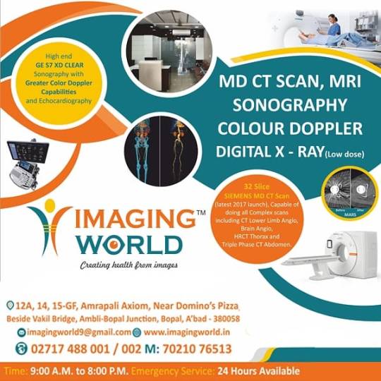
Imaging World is dedicated not only to excellence and patient care, but also to innovation and continued growth. Sonography in Bopal To keep up with the most current advances in medical imaging technology, we make regular investments in research and development. This allows us to provide our patients with access to innovative imaging methods and treatments that might not be available outside of in Ahmedabad.
Imaging World's Commitment to Your Well-being:-
At Imaging World, they understand that undergoing a medical procedure can be stressful. Their dedicated staff is committed to providing a comfortable and supportive environment throughout your visit. They will explain the procedure clearly, answer any questions you may have, and ensure you feel at ease during the examination.
The Concept of the the Mission to Your Health
Imaging World is aware of the stress related to undertaking treatments. During your stay, their committed team will work hard to provide a welcoming and helpful environment. They will make sure you are comfortable through the examination, address any concerns you have about it, and provide a comprehensive explanation of the process.
Finally, Imaging World is a shining example of excellence in Ahmedabad's diagnostic imaging business. With our advanced technology, qualified employees, focused on customer’s technique, and dedication to innovation, we are committed to providing excellent sonography services near me that raise the bar for healthcare and provide patients and healthcare providers alike more power. We at Imaging World are more than simply imaging specialists; we are customers on your path to improved overall well-being.
Visit our Sonography Centre in Ahmedabad for superior imaging services tailored to your needs For More Info: - https://www.imagingworld.in/sonography-ultrasound-in-ahmedabad.php
#Sonography Centre in Ahmedabad#sonography near me#Sonography Bopal#Sonography ahmedabad#Sonography in Bopal#Sonography centre in bopal
0 notes
Text
"Sonography: Understanding the Procedure and Pricing"

Introduction:
In the realm of diagnostic medicine, sonography stands out as a versatile and invaluable tool, offering a non-invasive glimpse into the intricate structures within our bodies. Whether it's monitoring fetal development during pregnancy or investigating potential health concerns in various organs, sonography has become an integral part of modern healthcare. This blog aims to delve into the world of sonography, exploring its procedures and shedding light on the crucial aspect of price considerations.
Understanding Sonography:
**Sonography, or ultrasound imaging, utilizes high-frequency sound waves to generate real-time images of the inside of the body.** The procedure involves a transducer, a small hand-held device, which emits sound waves and captures the echoes as they bounce back. These echoes are then translated into visual images, providing a detailed view of organs, tissues, and blood flow. One of the key advantages of sonography is its non-invasive nature, making it a safe and widely used diagnostic technique.
Common Types of Sonography Procedures:
1. Obstetric Sonography:
- This type of sonography is well-known for monitoring fetal development during pregnancy. It helps assess the health and growth of the fetus, detect any abnormalities, and determine the baby's gender.
2. Abdominal Sonography:
- Abdominal sonography is used to examine the organs in the abdominal cavity, such as the liver, gallbladder, kidneys, and pancreas. It aids in identifying abnormalities, including tumors or cysts.
3. Pelvic Sonography:
- Pelvic sonography is commonly employed to assess the health of the reproductive organs in both males and females. It can detect issues like ovarian cysts, fibroids, or prostate abnormalities.
4. Vascular Sonography:
- Focusing on blood vessels, vascular sonography helps in diagnosing conditions such as blood clots, aneurysms, or arterial blockages.
5. Cardiac Sonography:
- Also known as echocardiography, this type of sonography evaluates the structure and function of the heart. It aids in detecting heart conditions and assessing overall cardiac health.
**Factors Influencing Sonography Price:**
The cost of a sonography procedure can vary significantly, and understanding the factors influencing these prices is crucial for making informed decisions about healthcare expenditures.
1. Type of Sonography:
- Different types of sonography procedures come with varying levels of complexity. Obstetric sonography, for instance, may be more routine compared to a detailed vascular or cardiac sonogram, influencing the overall cost.
2. Geographical Location:
- The cost of healthcare services often varies based on the region. Urban areas and well-established medical facilities might have higher prices due to increased operational costs.
3. Facility Reputation:
- Renowned hospitals and diagnostic centers with a reputation for accuracy and reliability may charge higher prices. The assurance of quality service can be a significant factor for many individuals.
4. Technology and Equipment:
- Facilities equipped with state-of-the-art ultrasound machines and advanced technology may charge more for their services. However, the higher cost may be justified by the improved precision of the imaging.
5. Additional Services:
- Some sonography centers offer additional services, such as consultations with specialists, 3D imaging, or extended reports. These extras can contribute to an increase in overall test prices.
Navigating Sonography Prices:
1. Government Healthcare Facilities:
- Government hospitals and clinics often provide sonography services at subsidized rates or even for free. This makes them an accessible option for those on a tight budget.
2. Private Diagnostic Centers:
- Private diagnostic centers offer a range of sonography services, and their prices can vary. It's advisable to research multiple centers to find a balance between affordability and quality.
3. Hospital Sonography Departments:
- Many hospitals have dedicated sonography departments, providing integrated services with other medical specialties. While prices may be slightly higher, the convenience and comprehensive healthcare approach can be beneficial.
4. Specialized Sonography Clinics:
- Specialized clinics focusing solely on sonography may offer competitive prices, especially if they operate efficiently with lower overhead costs.
Srishti Hospital and Dr. Mamta Gupta for IVF Treatment
Srishti Hospital in Jaipur, Rajasthan is a leading fertility and IVF center under the direction of Gynecologist, Obstetrician and IVF Specialist Dr. Mamta Gupta. With over 21 years of experience helping couples conceive, Dr. Mamta Gupta offers comprehensive fertility treatments including IVF, IUI, ICSI, egg/embryo freezing, egg donation, and gestational surrogacy.
As both a gynecologist and infertility specialist, Dr. Gupta provides science-based care paired with emotional support throughout the challenges of infertility treatment. Srishti Hospital has state-of-the-art IVF labs and nursing staff to monitor progress every step of the way. From your first injection to your embryo transfer, you can trust Dr. Mamta Gupta and her team at Srishti Hospital for your best chance at successful conception through IVF.
0 notes
Text

Find the best sonography centre near me thane
If you're searching for a sonography centre near me Thane, look no farther than Crystal Diagnostic. Undeniable for its phenomenal medical imaging affiliations, Crystal Diagnostic stands pulled out as the go-to objective for cautious and strong sonography. Utilizing arrangement setting advancement, the center gives epic standard pictures fundamental for careful end and treatment. Crystal Diagnostic qualities its top tier workplaces and a party of fundamentally gifted specialists focused in on figuring out thought. The sonography affiliations offered harden an amazing number of medical conditions, assisting with early straightforwardness and strong management. The immediate framework uses high-accentuate sound waves to convey point by point pictures of inside body structures, ensuring mindful and precise assessments. Genuinely arranged in Thane, Crystal Diagnostic is truly open, seeking after it an ideal choice for those searching for quality sonography benefits nearby. The center offers flexible appointment timetables to oblige the clamoring presences of its patients, ensuring unessential holding up times and brief help. Patients can expect a splendid and supporting experience, with the party of experienced radiologists and experts giving changed care and thought. Pick Crystal Diagnostic for your sonography needs in Thane, and experience the best demands for medical imaging and merciful thought. With a commitment to significance and patient fulfillment, Crystal Diagnostic is the reasonable sonography place near you in Thane.
0 notes
Text
Exploring Cutting-Edge Ultrasound Services in Perth: Advancing Healthcare with Precision Imaging
In the realm of modern medicine, diagnostic imaging plays a crucial role in the detection, diagnosis, and treatment of various medical conditions. Among the most versatile and widely used imaging modalities is ultrasound, a non-invasive technique that utilizes sound waves to produce real-time images of internal structures and organs. In Perth, Western Australia, ultrasound services have become an essential component of healthcare, offering patients access to state-of-the-art technology and expertise for accurate diagnosis and personalized care. In this article, we delve into the world of ultrasound services perth, exploring their applications, benefits, and the advancements shaping the future of medical imaging in the region.
The Role of Ultrasound in Healthcare
Ultrasound imaging, also known as sonography, uses high-frequency sound waves to create detailed images of the body's internal structures, including organs, tissues, and blood vessels. Unlike other imaging modalities such as X-rays or CT scans, ultrasound does not involve radiation, making it safe for use in various patient populations, including pregnant women and children.
Ultrasound is commonly used for:
Diagnostic Imaging: Ultrasound imaging is used to visualize and assess various medical conditions affecting organs such as the heart, liver, kidneys, gallbladder, and reproductive organs.
Prenatal Care: Ultrasound is a routine part of prenatal care, allowing healthcare providers to monitor fetal development, assess fetal health, and detect any potential abnormalities or complications during pregnancy.
Guidance for Procedures: Ultrasound is used to guide minimally invasive procedures such as biopsies, needle aspirations, and injections, ensuring accuracy and precision during the procedure.
The Advantages of Ultrasound Services in Perth
Accessibility: Perth is home to a wide range of medical facilities and clinics offering mandurah ultrasound services, ensuring accessibility and convenience for patients across the region.
Advanced Technology: Many ultrasound facilities in Perth are equipped with state-of-the-art equipment and technology, including high-resolution imaging capabilities and advanced Doppler techniques for assessing blood flow.
Expertise and Experience: Ultrasound technicians and radiologists in Perth are highly trained and experienced professionals, ensuring accurate interpretation of imaging studies and personalized care for each patient.
Comprehensive Services: ultrasound Perth cover a wide range of medical specialties and applications, including obstetrics, gynecology, cardiology, abdominal imaging, musculoskeletal imaging, and more.
The Future of Ultrasound Imaging
Advancements in ultrasound technology continue to drive innovation and expand the capabilities of this versatile imaging modality. In Perth, ongoing research and development efforts are focused on:
Enhanced Image Quality: Improving image resolution and clarity to enhance diagnostic accuracy and visualization of anatomical structures.
Functional Imaging: Developing new techniques for functional and molecular imaging, allowing for the assessment of tissue perfusion, metabolism, and cellular activity.
Point-of-Care Ultrasound: Integrating ultrasound into various clinical settings, including emergency departments, intensive care units, and primary care clinics, for rapid diagnosis and bedside monitoring.
Conclusion
Ultrasound services play a vital role in healthcare delivery in Perth, providing patients with access to accurate diagnosis, personalized treatment, and compassionate care. With state-of-the-art technology, experienced professionals, and a commitment to excellence, ultrasound facilities in Perth are at the forefront of medical imaging, driving innovation and improving patient outcomes. As advancements in ultrasound baldivis technology continue to evolve, the future holds promising possibilities for further enhancing diagnostic capabilities, expanding clinical applications, and improving the delivery of healthcare services in the region.
Source Url:- https://sites.google.com/view/ohheybabycom111/home
0 notes
Text
Sonography: Understanding the Procedure and Pricing
Sonography: Understanding the Procedure and Pricing

Introduction:
In the realm of diagnostic medicine, sonography stands out as a versatile and invaluable tool, offering a non-invasive glimpse into the intricate structures within our bodies. Whether it's monitoring fetal development during pregnancy or investigating potential health concerns in various organs, sonography has become an integral part of modern healthcare. This blog aims to delve into the world of sonography, exploring its procedures and shedding light on the crucial aspect of price considerations.
Understanding Sonography:
**Sonography, or ultrasound imaging, utilizes high-frequency sound waves to generate real-time images of the inside of the body.** The procedure involves a transducer, a small hand-held device, which emits sound waves and captures the echoes as they bounce back. These echoes are then translated into visual images, providing a detailed view of organs, tissues, and blood flow. One of the key advantages of sonography is its non-invasive nature, making it a safe and widely used diagnostic technique.
Common Types of Sonography Procedures:
1. Obstetric Sonography:
- This type of sonography is well-known for monitoring fetal development during pregnancy. It helps assess the health and growth of the fetus, detect any abnormalities, and determine the baby's gender.
2. Abdominal Sonography:
- Abdominal sonography is used to examine the organs in the abdominal cavity, such as the liver, gallbladder, kidneys, and pancreas. It aids in identifying abnormalities, including tumors or cysts.
3. Pelvic Sonography:
- Pelvic sonography is commonly employed to assess the health of the reproductive organs in both males and females. It can detect issues like ovarian cysts, fibroids, or prostate abnormalities.
4. Vascular Sonography:
- Focusing on blood vessels, vascular sonography helps in diagnosing conditions such as blood clots, aneurysms, or arterial blockages.
5. Cardiac Sonography:
- Also known as echocardiography, this type of sonography evaluates the structure and function of the heart. It aids in detecting heart conditions and assessing overall cardiac health.
**Factors Influencing Sonography Price:**
The cost of a sonography procedure can vary significantly, and understanding the factors influencing these prices is crucial for making informed decisions about healthcare expenditures.
1. Type of Sonography:
- Different types of sonography procedures come with varying levels of complexity. Obstetric sonography, for instance, may be more routine compared to a detailed vascular or cardiac sonogram, influencing the overall cost.
2. Geographical Location:
- The cost of healthcare services often varies based on the region. Urban areas and well-established medical facilities might have higher prices due to increased operational costs.
3. Facility Reputation:
- Renowned hospitals and diagnostic centers with a reputation for accuracy and reliability may charge higher prices. The assurance of quality service can be a significant factor for many individuals.
4. Technology and Equipment:
- Facilities equipped with state-of-the-art ultrasound machines and advanced technology may charge more for their services. However, the higher cost may be justified by the improved precision of the imaging.
5. Additional Services:
- Some sonography centers offer additional services, such as consultations with specialists, 3D imaging, or extended reports. These extras can contribute to an increase in overall test prices.
Navigating Sonography Prices:
1. Government Healthcare Facilities:
- Government hospitals and clinics often provide sonography services at subsidized rates or even for free. This makes them an accessible option for those on a tight budget.
2. Private Diagnostic Centers:
- Private diagnostic centers offer a range of sonography services, and their prices can vary. It's advisable to research multiple centers to find a balance between affordability and quality.
3. Hospital Sonography Departments:
- Many hospitals have dedicated sonography departments, providing integrated services with other medical specialties. While prices may be slightly higher, the convenience and comprehensive healthcare approach can be beneficial.
4. Specialized Sonography Clinics:
- Specialized clinics focusing solely on sonography may offer competitive prices, especially if they operate efficiently with lower overhead costs.
Srishti Hospital and Dr. Mamta Gupta for IVF Treatment
Srishti Hospital in Jaipur, Rajasthan is a leading fertility and IVF center under the direction of Gynecologist, Obstetrician and IVF Specialist Dr. Mamta Gupta. With over 21 years of experience helping couples conceive, Dr. Mamta Gupta offers comprehensive fertility treatments including IVF, IUI, ICSI, egg/embryo freezing, egg donation, and gestational surrogacy.
As both a gynecologist and infertility specialist, Dr. Gupta provides science-based care paired with emotional support throughout the challenges of infertility treatment. Srishti Hospital has state-of-the-art IVF labs and nursing staff to monitor progress every step of the way. From your first injection to your embryo transfer, you can trust Dr. Mamta Gupta and her team at Srishti Hospital for your best chance at successful conception through IVF.
0 notes
Text
The Power of a Medical Diploma in Advancing Your Medical Career
In the ever-evolving landscape of healthcare, professionals are constantly seeking ways to enhance their skills and advance their careers. A medical diploma is a valuable asset that can significantly impact your professional journey in the medical field. Specialized programs such as medical lab technology, radiology & medical imaging technology, operation theatre technology, and cardiac care technology are just a few areas where a medical diploma can serve as a catalyst for growth and opportunity.

Medical Lab Technology
Medical lab technology is a critical component of the healthcare system. Medical laboratory scientists play a vital role in the diagnosis and treatment of diseases, analyzing blood, body fluids, tissues, and cells. By obtaining a diploma in this field, you gain the expertise to operate sophisticated lab equipment and contribute to patient care by providing essential data that informs medical decisions.
Radiology & Medical Imaging Technology
The field of radiology & medical imaging technology is integral to modern medicine. It encompasses a range of non-invasive diagnostic techniques such as computed tomography (CT), magnetic resonance imaging (MRI), and sonography. A diploma in this area equips you with the skills to operate advanced imaging equipment, ensuring you can assist in accurate diagnoses and play a crucial role in patient treatment plans.
Operation Theatre Technology
Operation theatre technology is another area where a medical diploma can be particularly powerful. Operating room technicians are responsible for the preparation and maintenance of operating theatres and equipment, as well as assisting during surgical procedures.Acquiring a diploma in this specialty hones your skills in a high-stakes environment, where precision and attention to detail can make a significant difference in patient outcomes
Cardiac Care Technology
Cardiac care technology focuses on the diagnosis and treatment of heart-related conditions. Technicians in this field use various diagnostic tools to monitor and assess cardiac health. A diploma in cardiac care technology prepares you to work alongside cardiologists and other healthcare professionals, providing critical support in the management of heart disease.
Advancing Your Career
A medical diploma not only provides you with specialized knowledge but also enhances your employability. The healthcare industry is vast, and the demand for skilled professionals in these specialized fields is growing. With a medical diploma, you are well-positioned to take advantage of this demand and secure a fulfilling career in healthcare.
Moreover, the skills you acquire through a medical diploma program are not limited to technical abilities. You also develop soft skills such as communication, teamwork, and problem-solving, which are invaluable in any healthcare setting. These competencies make you a well-rounded professional capable of adapting to various roles within the medical industry.
Conclusion
A medical diploma from Lumeen Paramedical is not just a certificate, but a passport to a multitude of opportunities in the healthcare sector. Our specialized courses in Medical Lab Technology, Radiology & Medical Imaging Technology, Operation Theatre Technology, and Cardiac Care Technology equip students with the knowledge and skills to significantly impact patient care and propel their medical careers forward. At Lumeen Paramedical, we open doors to a rewarding future in healthcare.
#best paramedical courses in noida#radiology medical lab in noida#medical lab technology#medical diploma in noida#b. voc in radiology medical lab#operation theatre technology#bsc.voc in medical lab technology
0 notes
Link
0 notes
Text
The Future of Ultrasound in Prehospital Resuscitation

Ultrasound equipment has become more affordable and portable over the last decade as its use has become commonplace outside of the radiology suite. Recent studies have shown that with minimal training and augmented protocols, sonography can improve resuscitative efforts outside the hospital in prehospital care.
Introduction
Bedside point of care ultrasound (POCUS) has rapidly become a critical tool for emergency medicine providers in the emergency department to aid in diagnosis and procedures, particularly to guide resuscitative efforts and identifying so-call “reversible causes” in cardiac arrest and other situations calling for cardiopulmonary resuscitation (CPR). One of the main drivers of this trend has been technology advances allowing ultrasound machines to be smaller, lighter, easier to use, and more affordable for emergency departments to purchase.
Given its proven value in CPR and in trauma, it should come as no surprise that recent studies have explored the utility of these newer, portable ultrasound machines in the pre-hospital setting assessing whether the use of ultrasonography medicine service (EMS) providers with critically ill patients could help direct the efforts of emergency. Over the last several decades, technologies previously found only in hospitals or emergency departments have similarly become more portable and able to be brought into the field and on the ambulance by EMS. Some examples of this include portable electrocardiogram (ECG) machines and automated defibrillators to diagnose and treat heart arrhythmias. Portable pulse oximeters and carbon dioxide detectors have also been incorporated into these units to give real-time information about a patient’s respiratory state.
A potential argument against the use of ultrasonography by emergency medicine technicians (EMTs) and paramedics is that they do not have the training or skill to safely use and interpret the ultrasound images, but the same was likely said about paramedics interpreting ECGs independently and was the argument against anyone other than radiologist reading ultrasounds that has been debunked by the wide-spread safe use by physicians and advanced practice providers.
Currently, ECG, pulse oximetry, and capnography are indispensable and integral in resuscitation according to the Advanced Cardiac Life Support (ACLS) guidelines.2 Similarly, POCUS has been shown to be invaluable in the rapid assessment of trauma patients and has been incorporated into certification in Advanced Trauma Life Support (ATLS).1 In pre-hospital care, EMTs and paramedics are taught and follow ACLS and other algorithms with specific interventions according to the diagnosed cause of cardiac arrest or arrhythmia. Portable ECG, pulse oximetry, and capnography allow EMS providers to identify and thus treat life threatening conditions earlier. Portable ultrasound, if employed judiciously, can have the same effect and positive impact on pre-hospital care.
Pre-hospital Application of POCUS
The pre-hospital setting has unique challenges in an ever changing, noisy, and chaotic environment. One of the strengths of ultrasound is that it can provide visual information that can be seen and captured at relatively low cost, outside the hospital. If applied in the appropriate circumstances with the right patient, an adequately trained professional ultrasonography can aid in diagnosis and treatment in the pre-hospital setting. Several studies3 have proposed algorithms for the use of ultrasound in the prehospital setting already, and some algorithms currently implemented within the hospital (Table 1). ATLS which advocates for the use of ultrasound in the Extended Focused Use of Sonography in Trauma (EFAST) which looks for evidence of blood in the abdomen or thorax or a collapsed lung, could potentially be transferred to the field.
The A-B-Cs of Pre-Hospital Ultrasound
Outside of protocols, there are other ways that POCUS has been successfully used in the hospital and can be employed in the pre-hospital setting to identify potential life-threats and other emergencies quickly, efficiently, and accurately. This is similar to the use of a stethoscope or palpation that EMS providers already use regularly to check for pulses or listen for breath sounds. In some of these procedures and potential uses, ultrasound can be more accurate than auscultation or palpation.
A commonly used acronym for the first steps in assessment of a potentially critically ill patient is to check the “A, B, C's” which means assessing the patient’s Airway, Breathing, and Circulation. These are universal emergency medicine steps that EMS regularly follow in the first few minutes of pre-hospital care of unconscious patients, those in respiratory distress or failure, and patients with potential cardiac arrest or severe cardiac dysfunction. Any of these emergencies require rapid identification and correction on scene and during transport to the hospital for best outcomes.
Each step during the Airway, Breathing, and Circulation assessment and treatment of critically ill patients can be enhanced by the use of ultrasound if properly trained and executed by EMS. The following is a summary of the potential uses of POCUS in the unconscious patient or those in respiratory or cardiovascular failure:
● Airway – POCUS can identify an endotracheal tube in the esophagus.
● Breathing – POCUS can assess for lung sliding, the absence of which can indicate a pneumothorax and help decision making to perform needle decompression.
● Circulation – Ultrasound can quickly and accurately check pulses and cardiac motion guiding the need for chest compressions.
The best, fastest, and most logical use of pre-hospital ultrasonography lies in asking binary questions where a rapid evaluation with ultrasound can answer yes or no, with operators discontinuing the use of POCUS if definitive images cannot be obtained due to body habitus, machine, or user limitation and especially in cases where the operator does not have the training or expertise to interpret the images.
Read More: https://www.europeanhhm.com/articles/the-future-of-ultrasound-in-prehospital-resuscitation
#health#medical care#doctors#health and wellness#hospitals#healthcare#healthy lifestyle#medical equipment#technologies#artifical intelligence#ultrasoundtechnology#immune system#prehospitalcare#prehospital resuscitation
0 notes
Text
What Is 4d Sonography In Pregnancy?

Ultrasound employs high-frequency sound waves to generate an internal body image. Initially introduced in the 1950s as 2D scans, it gained prominence for fetal observation in the late 1970s. Advancing over time, medical professionals introduced more sophisticated versions such as 3D scans, enhancing fetal visualization, and 4D scans, adding the temporal dimension to witness fetal movements. This evolution has empowered doctors to observe and understand the developing fetus with unprecedented clarity and depth. Experience unparalleled insights into your pregnancy journey with Diva Women’s Hospital, offering the best sonography services in Ahmedabad. Our advanced 3D and 4D ultrasound technology ensures a comprehensive view of your baby’s growth and movements. Trust in our expertise for a reassuring and exceptional prenatal experience. Your baby’s well-being starts with us.
Understanding the Process of 4D Sonography
During a 4D ultrasound session, expectant mothers recline while a water-based gel is applied to their belly. A wand or transducer is then gently placed on the belly. By emitting sound waves that bounce off the amniotic fluid and internal fetal organs, the ultrasound machine generates an image on a screen. This image allows the doctor to assess the fetus’s health, development, and size with precision.
Distinguishing 2D, 3D, and 4D Sonography
A 2D ultrasound offers a monochrome cross-sectional view of the fetus, primarily employed to identify potential internal irregularities like heart or kidney defects.
Advancing from this, a 3D ultrasound employs sophisticated software to decipher the image, crafting a three-dimensional portrayal of the fetus’s surface. This aids in measuring height, width, and depth, facilitating the diagnosis of conditions like cleft lips and spinal defects.
At the pinnacle of this technology is the 4D ultrasound. Here, the software goes a step further, generating a real-time video showcasing the fetus’s movements – whether it’s sucking its thumb, opening its eyes, or stretching. This dynamic scan furnishes even deeper insights into the fetus’s developmental journey. Experience the miracle of life in motion with our state-of-the-art 4D Sonography services at Diva Women’s Hospital. Witness your baby’s every stretch, yawn, and smile in real-time clarity. Trust us to capture these precious moments and ensure a comprehensive understanding of your baby’s development.
Why this is even performed during pregnancy
In routine pregnancies, medical professionals employ 2D and Doppler ultrasounds to examine the fetus, evaluate amniotic fluid levels, and detect potential birth defects, among other purposes.
Conversely, 3D and 4D ultrasounds are typically reserved for focused assessments of suspected fetal anomalies, like cleft lip or spinal cord concerns, or for specific monitoring needs. These advanced scans, offering intricate details, are not generally included in standard prenatal examinations.
Benefits of 4D Sonography
4D sonography offers a remarkable window into pregnancy, providing real-time insights that extend beyond traditional scans. This advanced technology enables parents to witness their baby’s movements, expressions, and interactions in the womb, fostering a stronger connection and emotional bond. Moreover, 4D sonography enhances diagnostic capabilities, allowing medical professionals to detect subtle developmental nuances and potential issues more accurately. With the ability to observe actions like yawning, thumb-sucking, and even facial expressions, this imaging technique transforms the prenatal experience into an intimate and informative journey for expectant parents.
Is 4D Sonography Harmful During Pregnancy?
No, 4D sonography is not considered harmful. It uses the same ultrasound technology as 2D and 3D scans, which has been widely used for decades and is generally regarded as safe. The sound waves used in ultrasound scans are non-ionizing, meaning they don’t carry the same risks as X-rays or other types of radiation. However, as with any medical procedure, it’s important to ensure that the scan is performed by trained professionals and used only when necessary for medical purposes. When used responsibly and under proper medical guidance, 4D sonography is considered safe for both the mother and the developing foetus.
This Blog Originally Posted Here: https://divahospital.com/blog-post/what-is-4d-sonography-in-pregnancy/
0 notes
Text
Paediatric Point of care ultrasound: Big Kids playing with toys or the future of Paediatric emergency medicine? Part II
Part 2 of our introduction to ultrasound use in the paediatric emergency department with @Dr_Pete_EmMed #USS @FOAMed #paediatrics
Following on from my previous post here is Part II …….
Trauma
POCUS first gained traction with Emergency Medicine clinicians with the introduction of the Focused Assessment Sonography in Truama (FAST) scan in 1997 and then the extended version eFAST scan. The theory being that after traumatic injuries bleeding ensues and by scanning the right and left upper quadrants, the pelvis and subxiphoid…

View On WordPress
0 notes
Text
Importance of preventive care and diagnostic services
The emergence of new technologies, advanced medical equipment, and innovative approaches have transformed the healthcare industry. Your busy lives prevent you from taking the basic steps to maintain a healthy lifestyle. However, preventive care has become more critical as cases that could have been easily treated develop. Even if you think you are healthy, you should still have regular check-ups. Good health still needs to be examined by a doctor. Therefore, it is essential to choose your healthcare provider carefully.
Why is a preventive health examination important?
Extended lifespan
A beneficial aspect of preventive care is extending a person's life expectancy. Timely diagnosis helps with early treatment and better outcomes. Early detection of a medical condition or disease allows for prompt treatment. In addition, it can also prevent disease outbreaks if the disease is not treated.

Health Promotion
Preventive care also allows healthcare professionals to detect problems before they become too serious. An annual check-up usually includes height, weight, blood pressure, and pulse measurements. Dengue testing in thane includes blood tests that can determine sugar, cholesterol, and thyroid levels and radiological tests such as ultrasounds, X-rays, EKGs, and stress tests to diagnose medical conditions early.
Reduction of medical expenses
Imagine paying for preventive tests and treatments. The difference in costs underscores why preventive care can help. Early detection of medical conditions can significantly reduce costs. There are several ways to use preventive medicine. These include physical examinations, weight checks, mental health examinations, and vaccinations.
Services provided:
Pathology services
X-ray
ECG
2-D echo
Jogging test
Sonography
CT scan
Diagnostic services' significance
Diagnostic tests are the only thing that makes timely medical intervention possible in the era of cutting-edge procedures and cutting-edge technology. There are various test and examination formats accessible. TB testing in thane aids medical practitioners in selecting the most effective treatment plan for their patients.
The most common diagnostic procedures include blood, stool, tissue, and urine tests. Assess whether the individual has a specific medical condition. These tests are performed in a sterile laboratory under the supervision of a pathologist.
Remembering that diagnostic tests are essential in treating the disease is important. First of all, it helps in the accurate diagnosis of diseases and medical conditions. Doctors can provide appropriate explanations and select specific treatment methods using the data.
PATHOLOGY LAB IN THANE also helps monitor patients with terminal illnesses. In this case, the available information will help to defuse the situation. Screening is another area where diagnostic tests play an essential role. Here, asymptomatic people are tested for early detection of potential health problems. It not only reduces risks but also offers a promising solution.
The last word
The importance of focusing on preventative care now cannot be stressed enough. It is highly recommended that men and women over 40 have annual health examinations to determine the prerequisites of severe diseases such as diabetes, heart disease, and cancer.
Early detection is key: Preventive health checks help doctors detect problems before it's too late. The consequences of such early actions help individuals lead happy and healthy lives. Moreover, the importance of diagnostic services in treating various diseases is indisputable.
0 notes
Last Seen Blogs


