#ultrasoundtechnology
Explore tagged Tumblr posts
Visit Tumblr Blog
Explore Tumblr blogs with no restrictions, modern design and the best experience.
Last Seen Tumblr Blogs


Fun Fact
Tumblr is available in 18 languages.
Text
Diagnostic Imaging Market Forecast 2025-2032: Size, Share, and Emerging Opportunities

The diagnostic imaging market plays a critical role in modern medicine, enabling healthcare professionals to detect and diagnose a wide range of medical conditions. As healthcare technology continues to evolve, diagnostic imaging has become indispensable in both clinical settings and for home-based care solutions. The global diagnostic imaging market is projected to experience substantial growth between 2025 and 2032, driven by advancements in imaging technologies, rising demand for early disease diagnosis, and the increasing prevalence of chronic conditions.
The global diagnostic imaging market encompasses a wide range of imaging modalities, including X-ray, ultrasound, magnetic resonance imaging (MRI), computed tomography (CT) scans, nuclear imaging, and others. These imaging techniques provide essential diagnostic information that helps in the accurate detection of diseases such as cancer, cardiovascular conditions, neurological disorders, and musculoskeletal issues.
Get a Free Sample Copy - https://www.skyquestt.com/sample-request/diagnostic-imaging-market
Market Size and Growth Forecast
The diagnostic imaging market size was valued at USD 29.18 Billion in 2024 to USD 44.44 Billion by 2032, growing at a CAGR of 5.4% during the forecast period (2025-2032). This growth can be attributed to several factors, including the increasing demand for imaging services in healthcare settings, technological advancements, and a global emphasis on preventive care and early detection.
Key Factors Driving Market Growth:
- Technological Advancements: Continuous innovations in imaging technologies, including the development of high-resolution, portable, and AI-powered imaging solutions, are expanding diagnostic capabilities. The integration of artificial intelligence (AI) and machine learning algorithms into imaging devices is enhancing image interpretation, reducing human error, and improving diagnostic accuracy.
- Rising Prevalence of Chronic Diseases: The rising prevalence of chronic diseases, such as cancer, cardiovascular conditions, and neurological disorders, is driving demand for diagnostic imaging procedures. Early and accurate detection of such conditions through imaging techniques can significantly improve treatment outcomes.
- Aging Population: With the global aging population, the demand for diagnostic imaging services is expected to increase. Older adults are more susceptible to a variety of health conditions, requiring frequent diagnostic imaging to monitor their health and detect potential diseases at an early stage.
- Improved Healthcare Infrastructure: The expansion of healthcare infrastructure in emerging markets, along with increased investment in diagnostic imaging technology, is contributing to the growth of the market. Hospitals and diagnostic centers are adopting advanced imaging technologies to offer better healthcare services.
Make an Inquiry to Address your Specific Business Needs - https://www.skyquestt.com/speak-with-analyst/diagnostic-imaging-market
Market Segmentation
The diagnostic imaging market can be segmented based on technology type, application, end-user, and geography:
1. By Technology Type:
- X-ray Imaging: The largest segment in the diagnostic imaging market, due to its widespread use in detecting bone fractures, infections, and various internal conditions.
- MRI: MRI imaging continues to grow, especially for neurological and musculoskeletal disorders, providing high-resolution images for detailed diagnosis.
- CT Scanners: CT scans are gaining popularity for their speed and ability to provide detailed cross-sectional images, especially in emergency situations and trauma cases.
- Ultrasound Imaging: Widely used in obstetrics, cardiology, and musculoskeletal imaging, ultrasound is one of the most versatile and cost-effective imaging methods.
- Nuclear Imaging: Though it accounts for a smaller share of the market, nuclear imaging is essential for oncology, cardiology, and certain neurological disorders.
2. By Application:
- Oncology: Diagnostic imaging is critical for detecting and staging cancer, enabling timely treatment decisions.
- Cardiology: Imaging techniques like echocardiograms and CT angiography play an essential role in diagnosing cardiovascular diseases.
- Neurology: MRI and CT scans are extensively used in diagnosing neurological conditions, such as stroke, dementia, and multiple sclerosis.
- Orthopedics: Imaging methods like X-rays and MRI are indispensable for diagnosing bone and joint conditions, such as fractures, arthritis, and soft tissue injuries.
- Others: Imaging is also vital in areas like obstetrics, gastroenterology, and urology.
3. By End-User:
- Hospitals: Hospitals remain the largest end-user segment for diagnostic imaging devices, providing comprehensive services for a variety of conditions.
- Diagnostic Centers: Standalone diagnostic centers are also increasingly adopting advanced imaging technologies to meet the growing demand for diagnostic services.
- Others: This includes outpatient clinics, academic and research institutions, and home healthcare settings.
4. By Geography:
- North America: North America holds the largest market share, owing to the advanced healthcare infrastructure, high healthcare expenditure, and strong demand for cutting-edge diagnostic technologies.
- Europe: The European market is experiencing steady growth, with countries such as Germany, the UK, and France leading in diagnostic imaging adoption.
- Asia-Pacific: The Asia-Pacific region is expected to exhibit the highest growth rate during the forecast period, driven by a large patient pool, improving healthcare infrastructure, and rising healthcare investments.
- Latin America and Middle East & Africa: These regions are expected to see moderate growth, with increasing awareness of the benefits of early disease detection and growing healthcare investments.
Take Action Now: Secure Your Diagnostic Imaging Market Today - https://www.skyquestt.com/buy-now/diagnostic-imaging-market
Key Market Players
Some of the major players operating in the global diagnostic imaging market include:
GE Healthcare
Siemens Healthineers
Koninklijke Philips N.V.
Canon Medical Systems Corporation
Hitachi Medical Corporation
Fujifilm Holdings Corporation
Carestream Health
Hologic, Inc.
Samsung Medison
Shimadzu Corporation
Agfa-Gevaert Group
Mindray Medical International Limited
Esaote S.p.A.
Planmed Oy
Analogic Corporation
Neusoft Medical Systems Co., Ltd.
CurveBeam
Konica Minolta, Inc.
United Imaging Healthcare Co., Ltd.
Toshiba Medical Systems Corporation
These companies are heavily investing in R&D to introduce advanced imaging solutions that integrate AI and other innovations to improve diagnostic precision, reduce costs, and increase accessibility.
Challenges and Barriers
Despite the promising growth outlook, the diagnostic imaging market faces several challenges, including:
- High Equipment Costs: Diagnostic imaging systems, especially advanced modalities like MRI and CT scanners, require significant investment, making them expensive for healthcare providers and hospitals, particularly in developing regions.
- Regulatory Challenges: Stringent regulations and approval processes for diagnostic imaging devices can delay product launches and limit market growth.
- Shortage of Skilled Professionals: The need for trained radiologists and imaging technicians is growing, but there is a shortage of such professionals in many regions, which can affect the optimal use of imaging technology.
The diagnostic imaging market is poised for significant growth over the next decade, driven by technological advancements, an aging population, increasing chronic diseases, and greater emphasis on preventive healthcare. While challenges exist, the ongoing innovations in imaging modalities and increasing healthcare investments, particularly in emerging markets, are likely to propel the market to new heights. For stakeholders in the healthcare industry, understanding these market dynamics will be crucial for making informed decisions and capitalizing on the expanding opportunities in diagnostic imaging.
Read Diagnostic Imaging Market Report Today - https://www.skyquestt.com/report/diagnostic-imaging-market
As we approach 2032, the diagnostic imaging landscape is set to evolve, providing improved patient outcomes, enhanced diagnostic accuracy, and greater accessibility to healthcare services worldwide.
#DiagnosticImaging#MedicalImaging#HealthcareTechnology#Radiology#ImagingMarket#DiagnosticTechnology#MedicalDevices#RadiologyEquipment#ImagingSolutions#HealthTech#MedicalInnovation#ImagingTrends#HealthcareIndustry#AIinHealthcare#MedicalImagingMarket#XrayTechnology#MRI#CTScan#UltrasoundTechnology
0 notes
Text
✨ The Future of Ultrasound Imaging ✨ Get a clear view of your patients’ health with 4D color ultrasound. 🖼️


Real-time dynamic imaging for precise diagnostics
High-definition color for clearer results
Non-invasive, safe, and reliable
Whether you're in obstetrics, cardiology, or musculoskeletal care, our ultrasound systems are designed to elevate your practice!
🩺 Ready to upgrade your diagnostic game? 👉 https://www.innorkommedical.com/medical-equipment-veterinary-trolley-ultrasound-scanner-machine.html
#FetalHealth#PregnancyCare#4DUltrasound#UltrasoundTechnology#ExpectingParents#MedicalInnovation#MedicalTech#HealthCare#Innovation
0 notes
Link
#AIinHealthcare#HandheldUltrasound#DiagnosticImaging#Telemedicine#HealthTech#PhilipsHealth#GEHealthcare#ButterflyNetwork#SamsungMedison#MedicalInnovation#UltrasoundTechnology
0 notes
Text
The Future of Ultrasound in Prehospital Resuscitation

Ultrasound equipment has become more affordable and portable over the last decade as its use has become commonplace outside of the radiology suite. Recent studies have shown that with minimal training and augmented protocols, sonography can improve resuscitative efforts outside the hospital in prehospital care.
Introduction
Bedside point of care ultrasound (POCUS) has rapidly become a critical tool for emergency medicine providers in the emergency department to aid in diagnosis and procedures, particularly to guide resuscitative efforts and identifying so-call “reversible causes” in cardiac arrest and other situations calling for cardiopulmonary resuscitation (CPR). One of the main drivers of this trend has been technology advances allowing ultrasound machines to be smaller, lighter, easier to use, and more affordable for emergency departments to purchase.
Given its proven value in CPR and in trauma, it should come as no surprise that recent studies have explored the utility of these newer, portable ultrasound machines in the pre-hospital setting assessing whether the use of ultrasonography medicine service (EMS) providers with critically ill patients could help direct the efforts of emergency. Over the last several decades, technologies previously found only in hospitals or emergency departments have similarly become more portable and able to be brought into the field and on the ambulance by EMS. Some examples of this include portable electrocardiogram (ECG) machines and automated defibrillators to diagnose and treat heart arrhythmias. Portable pulse oximeters and carbon dioxide detectors have also been incorporated into these units to give real-time information about a patient’s respiratory state.
A potential argument against the use of ultrasonography by emergency medicine technicians (EMTs) and paramedics is that they do not have the training or skill to safely use and interpret the ultrasound images, but the same was likely said about paramedics interpreting ECGs independently and was the argument against anyone other than radiologist reading ultrasounds that has been debunked by the wide-spread safe use by physicians and advanced practice providers.
Currently, ECG, pulse oximetry, and capnography are indispensable and integral in resuscitation according to the Advanced Cardiac Life Support (ACLS) guidelines.2 Similarly, POCUS has been shown to be invaluable in the rapid assessment of trauma patients and has been incorporated into certification in Advanced Trauma Life Support (ATLS).1 In pre-hospital care, EMTs and paramedics are taught and follow ACLS and other algorithms with specific interventions according to the diagnosed cause of cardiac arrest or arrhythmia. Portable ECG, pulse oximetry, and capnography allow EMS providers to identify and thus treat life threatening conditions earlier. Portable ultrasound, if employed judiciously, can have the same effect and positive impact on pre-hospital care.
Pre-hospital Application of POCUS
The pre-hospital setting has unique challenges in an ever changing, noisy, and chaotic environment. One of the strengths of ultrasound is that it can provide visual information that can be seen and captured at relatively low cost, outside the hospital. If applied in the appropriate circumstances with the right patient, an adequately trained professional ultrasonography can aid in diagnosis and treatment in the pre-hospital setting. Several studies3 have proposed algorithms for the use of ultrasound in the prehospital setting already, and some algorithms currently implemented within the hospital (Table 1). ATLS which advocates for the use of ultrasound in the Extended Focused Use of Sonography in Trauma (EFAST) which looks for evidence of blood in the abdomen or thorax or a collapsed lung, could potentially be transferred to the field.
The A-B-Cs of Pre-Hospital Ultrasound
Outside of protocols, there are other ways that POCUS has been successfully used in the hospital and can be employed in the pre-hospital setting to identify potential life-threats and other emergencies quickly, efficiently, and accurately. This is similar to the use of a stethoscope or palpation that EMS providers already use regularly to check for pulses or listen for breath sounds. In some of these procedures and potential uses, ultrasound can be more accurate than auscultation or palpation.
A commonly used acronym for the first steps in assessment of a potentially critically ill patient is to check the “A, B, C's” which means assessing the patient’s Airway, Breathing, and Circulation. These are universal emergency medicine steps that EMS regularly follow in the first few minutes of pre-hospital care of unconscious patients, those in respiratory distress or failure, and patients with potential cardiac arrest or severe cardiac dysfunction. Any of these emergencies require rapid identification and correction on scene and during transport to the hospital for best outcomes.
Each step during the Airway, Breathing, and Circulation assessment and treatment of critically ill patients can be enhanced by the use of ultrasound if properly trained and executed by EMS. The following is a summary of the potential uses of POCUS in the unconscious patient or those in respiratory or cardiovascular failure:
● Airway – POCUS can identify an endotracheal tube in the esophagus.
● Breathing – POCUS can assess for lung sliding, the absence of which can indicate a pneumothorax and help decision making to perform needle decompression.
● Circulation – Ultrasound can quickly and accurately check pulses and cardiac motion guiding the need for chest compressions.
The best, fastest, and most logical use of pre-hospital ultrasonography lies in asking binary questions where a rapid evaluation with ultrasound can answer yes or no, with operators discontinuing the use of POCUS if definitive images cannot be obtained due to body habitus, machine, or user limitation and especially in cases where the operator does not have the training or expertise to interpret the images.
Read More: https://www.europeanhhm.com/articles/the-future-of-ultrasound-in-prehospital-resuscitation
#health#medical care#doctors#health and wellness#hospitals#healthcare#healthy lifestyle#medical equipment#technologies#artifical intelligence#ultrasoundtechnology#immune system#prehospitalcare#prehospital resuscitation
0 notes
Text
AI पेट के अल्ट्रासाउंड से पित्ताशय के कैंसर का पता लगाने में प्रभावी हो सकता है
कृत्रिम बुद्धिमत्ता (एआई) ने चिकित्सा क्षेत्र में एक नया अध्याय जोड़ा है। अब एक अनुभवी डॉक्टर के समान एआई भी मरीज़ की बीमारी को पकड़ सकता है। पीजीआई चंडीगढ़ के डॉक्टरों ने एक अध्ययन में इसकी पु��्टि की है। पित्ताशय का कैंसर अत्यधिक घातक द लैंसेट रीजनल हेल्थ-साउथ ईस्ट एशिया में प्रकाशित इस अध्ययन में … Read more
#AIandCancerDetection#UltrasoundForGallBladderCancer#AIinMedicalImaging#GallBladderCancerAwareness#AIandAbdominalUltrasound#CancerDetectionTechnology#AdvancementsInAIandUltrasound#GallBladderCancerResearch#AIforMedicalDiagnosis#UltrasoundTechnology
0 notes
Text
Tempted to buy the most reliable fetal dopplers so you can hear your baby’s heartbeat for the first time? BabyHeart is a must-consider place. Find 17% Off at GetSavo AU.
#babyheart#babyheartdoppler#babyheartwelshpool#BabyHeartMonitors#babyheartdiscountcode#australia#fetaldopplermonitor#babyheartstroller#babyheartpromocode#babyheartvouchercode#babyheartcouponcode#ultrasoundtechnology#getsavoau
0 notes
Photo
Happy National Medical #Ultrasound Awareness Month! Our next Ultrasound /Vascular Tech class starts October 7th and we keep class sizes small so everyone can learn hands-on and ask questions! 1,000+ hours of clinical internship, all pre-reqs included + your books/scrubs and first exam are all included! It’s open enrollment so let us help you make the next 18mos a huge step in your career (or career change)! 👩⚕️👨⚕️ #moderntechnologyschool . . . . . #healthcare #peopleinhealthcare #sonography #longbeach #ultrasoundtechnology #ultrasoundlife #ultrasoundstudent #medicalschool #sonographersdoitinthedark #career #anaheim #linkedin #sonographer #medicalstudent #medicalstudents #gardengrove #irvine #huntingtonbeach #ultrasoundtech #vascular #santaana #sonographystudent #sonogram #sonographerlife #ultrasoundschool #ultrasoundtechs (at Modern Technology School) https://www.instagram.com/p/B3IO3ioghtL/?igshid=16e3lkr2vd6
#ultrasound#moderntechnologyschool#healthcare#peopleinhealthcare#sonography#longbeach#ultrasoundtechnology#ultrasoundlife#ultrasoundstudent#medicalschool#sonographersdoitinthedark#career#anaheim#linkedin#sonographer#medicalstudent#medicalstudents#gardengrove#irvine#huntingtonbeach#ultrasoundtech#vascular#santaana#sonographystudent#sonogram#sonographerlife#ultrasoundschool#ultrasoundtechs
0 notes
Photo

Philips Africa just announced the introduction of Lumify, its first App-Based ultrasound system that will extend the reach of ultrasound applications to a broader network of healthcare providers using mobile technology. Unveiled during Medic West Africa 2018, held at the Eko hotel and Suites Victoria island Lagos. The Philips’ Lumify is an entirely new way of delivering ultrasound technology to healthcare providers and their patients; offering high-quality imaging on a compatible smart device through a subscription model.@philipsafrica @philipsnigeria @georgeuduku #philipsafrica #philipslumify #healthcare #healthcarepractitioner #ultrasoundtechnology @vanguardallure #vanguardallure #vanguardallure #glitzedgeng #yemisisuleiman https://www.instagram.com/p/Bo1r9LohwlF/?utm_source=ig_tumblr_share&igshid=12422hsiidi5f
#philipsafrica#philipslumify#healthcare#healthcarepractitioner#ultrasoundtechnology#vanguardallure#glitzedgeng#yemisisuleiman
0 notes
Photo

How Has Ultrasound Technology Evolved?
Ultrasound machines have made some fantastic progress. The ultimate ultrasound machine was a 2D scanner constructed in 1958 by Ian Donald and Tom Brown. They called it the Diasonograph, however, it was known in more casual and unkind circles as the Dinosaur Graph.
0 notes
Photo

@Regranned from @cheryls_livingclean - Whoop!!! It just keeps getting better🎉 Such an amazing price for this trio of best sellers! http://cherylplauche.arbonne.com #crueltyfree #vegan #glutenfree #ultrasoundtechnology #nurshing #arbonne #amazingresults #loveyourskin #lookyounger #hydratedskin #blackfriday #blackfridaydeals #blackfridaysale (at Grayson County, Texas)
#hydratedskin#glutenfree#loveyourskin#ultrasoundtechnology#amazingresults#crueltyfree#blackfridaydeals#blackfridaysale#arbonne#blackfriday#lookyounger#vegan#nurshing
0 notes
Text

👶✨ Pregnancy Scan Appointments Available! ✨👣
Are you expecting a little bundle of joy? It's time to capture those precious moments and ensure the well-being of your baby with our range of pregnancy scans. 🤰💖
1️⃣ NT Scan: 🌈 This scan is usually performed between 11-13 weeks of pregnancy. 🌟 It checks for Down syndrome and other chromosomal abnormalities. 💙 Provides a first glimpse of your baby's development.
2️⃣ Growth Scan: 🌷 Ideal around 20-24 weeks into your pregnancy. 📏 Monitors your baby's growth, ensuring they're thriving. 🤗 Enjoy watching your baby's movements and hear their heartbeat.
3️⃣ 3D/4D Pregnancy Scan: 📸 Available later in your pregnancy, usually around 28 weeks and beyond. 👶 See your little one's face in stunning detail and real-time motion! 💕 Cherish the opportunity to bond with your baby before birth.
Book your appointment with us now and experience the joy of seeing your baby's world up close! 📆💝
For inquiries and booking, simply visit our website www.addonhealthcare.com or give us a call +91 9900811118. Your baby's first photo shoot awaits! 📷👶😍
#addonhealthcare#addonscanslabs#FetalAnomalyScan#PregnancyJourney#ExpectingParents#HealthyBaby#ExpertCare#PregnancyHealth#BabyDevelopment#UltrasoundScan#MaternityCare#ExpectingMoms#HealthyPregnancy#BabyOnTheWay#PrenatalCare#MomToBe#BabyCheckup#UltrasoundTechnology#FetalHealth#ParentingPrep#ScanAppointment#PregnancyWellness
0 notes
Text
The Future of Ultrasound in Prehospital Resuscitation

Ultrasound equipment has become more affordable and portable over the last decade as its use has become commonplace outside of the radiology suite. Recent studies have shown that with minimal training and augmented protocols, sonography can improve resuscitative efforts outside the hospital in prehospital care.
Introduction
Bedside point of care ultrasound (POCUS) has rapidly become a critical tool for emergency medicine providers in the emergency department to aid in diagnosis and procedures, particularly to guide resuscitative efforts and identifying so-call “reversible causes” in cardiac arrest and other situations calling for cardiopulmonary resuscitation (CPR). One of the main drivers of this trend has been technology advances allowing ultrasound machines to be smaller, lighter, easier to use, and more affordable for emergency departments to purchase.
Given its proven value in CPR and in trauma, it should come as no surprise that recent studies have explored the utility of these newer, portable ultrasound machines in the pre-hospital setting assessing whether the use of ultrasonography medicine service (EMS) providers with critically ill patients could help direct the efforts of emergency. Over the last several decades, technologies previously found only in hospitals or emergency departments have similarly become more portable and able to be brought into the field and on the ambulance by EMS. Some examples of this include portable electrocardiogram (ECG) machines and automated defibrillators to diagnose and treat heart arrhythmias. Portable pulse oximeters and carbon dioxide detectors have also been incorporated into these units to give real-time information about a patient’s respiratory state.

A potential argument against the use of ultrasonography by emergency medicine technicians (EMTs) and paramedics is that they do not have the training or skill to safely use and interpret the ultrasound images, but the same was likely said about paramedics interpreting ECGs independently and was the argument against anyone other than radiologist reading ultrasounds that has been debunked by the wide-spread safe use by physicians and advanced practice providers.
Currently, ECG, pulse oximetry, and capnography are indispensable and integral in resuscitation according to the Advanced Cardiac Life Support (ACLS) guidelines.2 Similarly, POCUS has been shown to be invaluable in the rapid assessment of trauma patients and has been incorporated into certification in Advanced Trauma Life Support (ATLS).1 In pre-hospital care, EMTs and paramedics are taught and follow ACLS and other algorithms with specific interventions according to the diagnosed cause of cardiac arrest or arrhythmia. Portable ECG, pulse oximetry, and capnography allow EMS providers to identify and thus treat life threatening conditions earlier. Portable ultrasound, if employed judiciously, can have the same effect and positive impact on pre-hospital care.
Pre-hospital Application of POCUS
The pre-hospital setting has unique challenges in an ever changing, noisy, and chaotic environment. One of the strengths of ultrasound is that it can provide visual information that can be seen and captured at relatively low cost, outside the hospital. If applied in the appropriate circumstances with the right patient, an adequately trained professional ultrasonography can aid in diagnosis and treatment in the pre-hospital setting. Several studies3 have proposed algorithms for the use of ultrasound in the prehospital setting already, and some algorithms currently implemented within the hospital (Table 1). ATLS which advocates for the use of ultrasound in the Extended Focused Use of Sonography in Trauma (EFAST) which looks for evidence of blood in the abdomen or thorax or a collapsed lung, could potentially be transferred to the field.
Read More: https://www.europeanhhm.com/articles/the-future-of-ultrasound-in-prehospital-resuscitation
#ultrasoundtechnology#emergencymedicalservices#prehospitalcare#healthcare#health and wellness#hospitals#medical equipment
0 notes
Text
Understand Important Features of Pregnancy Ultrasound
In most cases, doctors will use this method to acquire a clear picture of the pregnancy. In-utero assessment of the fetus is greatly aided by the application of Baby 3d Ultrasoundtechnology. 3d Ultrasound Baby screenings are commonplace in several countries to detect birth problems before they are born.
Understand Important Features of Pregnancy Ultrasound
0 notes
Text
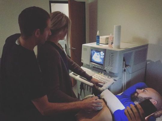
Having a rainbow baby
A rainbow baby is the name for babies born after parents have experienced the loss of another child whether due to a miscarriage, or stillbirth or in the first month after birth.
The name stems from the fact that beautiful rainbows often follow a storm and can be a symbol of hope and comfort, after a dark time.
Losing a baby is devastating, and an emotionally-draining experience, as it goes against all your expectations and you are denied the happy ending that is the norm. Often it happens before you’ve told anyone you are pregnant, so others don’t understand why you’re upset or you make a choice to keep your grief hidden.
If you have conceived your rainbow baby after such a loss, you will understandably be more anxious and worried, particularly during the first trimester when miscarriage is more common, but perhaps then all through your pregnancy.
How scans may help your anxiety
Ultrasound scans give you images of your baby made up from sound waves with no known side effects. Scans can give you a detailed snapshot of your baby’s health and be a source of reassurance and detailed information throughout your pregnancy.
Ultrasound Direct have run a network of private clinics at 80+ locations offering private baby scans near you in the UK for 20 years. The company offers 14 different types of antenatal scan that can provide the extra scans you may want to think about for your rainbow baby. The clinics are run by expert obstetric sonographers, so you are in safe hands and appointments can be arranged to suit you, at a location near you.
Reassurance in the early days
The first 12 weeks of pregnancy can be worrying, as this is when miscarriage rates are highest. One in four pregnancies end in miscarriage and 85 per cent of these occur in the first trimester, [i]but a baby scan at a key point can be immensely reassuring. Even if the news isn’t good, just knowing what is happening so that you can deal with it, is often preferable to being left to fret and worry.
The NHS offers routine booking scans (sometimes called a dating scan) at between 8 and 14 weeks of your pregnancy and earlier if you experience spotting, bleeding, or cramping.
Ultrasound Direct offers early scans to provide you with the information and - in most cases - reassurance you need. These include:
· Early scan: A 30-minute scan is offered at 6 to 11 weeks to find out if your pregnancy is viable and measuring correctly for your dates. It can also detect an ectopic pregnancy, where the baby is developing in a fallopian tube instead of the uterus.
· Dating Scan: Performed at between 12 and 16 weeks, this 20-minute scan is also referred to as a 12-week scan or booking scan. The main purpose is to date your pregnancy by measuring your baby's size and confirm how many babies you're expecting. From 12 weeks, 4DFREEVIEW™ technology is available to give you a 3D moving picture of your baby, including their heart.
· Babybond Nuchal Translucency (NT) Scan: Ultrasound Direct offer a Babybond Nuchal Translucency (NT) Scan (also called a nuchal scan)at between 11 and 13 weeks and 6 days gestation. This uses ultrasound to measure fluid at the back of your baby's neck (this area is called the nuchal fold). If a baby has extra fluid at this point, it's a marker for an extra chromosome and they may have Down's syndrome and you'll be counselled to see if you want further diagnostic tests to find out for sure.
· NIPT scan: This test combines a non-invasive pre-natal blood screening test for Down's syndrome (called Harmony) with a 30-minute diagnostic scan to check viability and dates. It's performed between 10 and 22 weeks.
Second trimester scans
Many of the detailed scans available between 12 and 24 weeks use the latest 4DFREEVIEW™ultrasoundtechnology. These include:
· Anomaly scans: This 40-minute scan is performed between 19 and 24 weeks and measures your baby's growthand checks internal organs and is probably the most important scan you'll have in pregnancy. Also available on the NHS, anomaly scans include sexing of your baby (but only if requested).
Other important second trimester scans include the Babybond Gender Scan at between 16 and 23 weeks and the JustGender™scan at 18 to 30 weeks.
Final trimester scans
The medical purpose of these baby scans is always to check on your baby's growth, wellbeing and presentation (position). If you’ve previously lost a baby at this stage in pregnancy you may appreciate the reassurance of extra monitoring scans to check on your baby’s wellbeing at this time.
Ultrasound Direct offer:
· Growth scans: Offered between 24 to 34 weeks, these can measure your baby's growth and estimate fetal weight.
· 4D bonding scans: Available between 24 and 34 weeks, these scans use 4D imaging techniques for 20 minutes, to measure your baby’s growth and estimate fetal weight.
· Presentation scans: From 37 weeks onwards, these scans can check if your baby and placenta are in the right positions for birth.
For a complete list of all scans offered pre and during pregnancy go to Ultrasound Direct.
What parents say about Ultrasound Direct
Ultrasound Direct actively obtain verified customer reviews via TrustPilot, one of the world’s leading review aggregators. The company cannot influence the reviews collected and welcome feedback from their clients. TrustPilot actively collect daily reviews from Ultrasound Direct clients and have currently in access of 5,000+ on record. Here’s a snapshot of recent feedback from their clients.
‘Was amazing experience!!’
“I loved it!! My scan visit was on Sunday 8 April!! Both ladies were very polite, doctor who took the scan was very nice and gentle.” Adelina Cani
‘Useful information’…
“The lady was lovely and gave us a lot of useful information.” Kelly Maxwell
‘Excellent from booking-in to the scan’
“Excellent from booking in to the scan, great service.” Danny Youles
‘Put my mind at rest about everything’
“Excellent experience and price. Receptionist and sonographer were lovely and welcoming and put my mind at rest about everything. Would definitely recommend/return.” Kelly-marie Robinson
‘Huge peace of mind’
“Appointment for early scan was well worth the money spent. We were seen slightly early than scheduled appointment so no waiting around, enjoyed being able to see scan on big screen whilst it was being performed and felt more time spent so we can watch, sonographer was kind and caring, lots of photos printed. Huge peace of mind having early scan, would recommend.” Kirsty Morgan
‘Explained the whole thing’
“Lovely staff very friendly and took time to get to know us and our situation. Explained the whole thing.” Samantha Fisher
References
[i]https://www.tommys.org/pregnancy-information/im-pregnant/early-pregnancy/how-common-miscarriage
0 notes
Photo
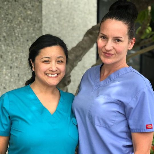
These two smiling faces are ready to welcome our new Ultrasound/Vascular Tech students starting October 7th in #OrangeCounty! Pictured left is Lani Coates RDMS, RVT, who is our #Ultrasound Program Director. She’s a Registered Diagnostic Medical #Sonographer (RDMS)—Ob/Gyn, a Registered Vascular Technologist—(RVT), Member of Society of Medical Diagnostic Sonographers (SDMS), and has 16+ yrs. scanning experience! On the RHS is Ultrasound Instructor Nicole Mattice, who is actually a graduate of our program, has over 4 years scanning experience in the field and loves working hands-on with students who are standing in the same shoes she once stood in. She knows what works, what doesn’t, and what it takes to make it in the field. Our all-inclusive & hands-on program offers students 1,000+ hours of clinical internship and prereqs are included so you can start with no prior experience! It’s open enrollment for this class so call us to get your questions answered today! 👩⚕️👨⚕️ #moderntechnologyschool . . . . . #healthcare #peopleinhealthcare #sonography #longbeach #ultrasoundtechnology #ultrasoundlife #ultrasoundstudent #medicalschool #sonographersdoitinthedark #career #anaheim #linkedin #ultrasoundtech #medicalstudent #medicalstudents #gardengrove #irvine #huntingtonbeach #ultrasound #vascular #teachersofinstagram #teacherlifestyle #sonographystudent #sonographerlife #ultrasoundschool (at Modern Technology School) https://www.instagram.com/p/B27nTF-gVoc/?igshid=24oacaatemr2
#orangecounty#ultrasound#sonographer#moderntechnologyschool#healthcare#peopleinhealthcare#sonography#longbeach#ultrasoundtechnology#ultrasoundlife#ultrasoundstudent#medicalschool#sonographersdoitinthedark#career#anaheim#linkedin#ultrasoundtech#medicalstudent#medicalstudents#gardengrove#irvine#huntingtonbeach#vascular#teachersofinstagram#teacherlifestyle#sonographystudent#sonographerlife#ultrasoundschool
0 notes
Text

"Did you know? Anomaly scans, which are essential for ruling out birth defects. To ensure a healthy start, it's vital to schedule your anomaly scan between 19th to 20th weeks of pregnancy.
#addonhealthcare#addonscanslabs#FetalAnomalyScan#PregnancyJourney#ExpectingParents#HealthyBaby#ExpertCare#PregnancyHealth#BabyDevelopment#UltrasoundScan#MaternityCare#ExpectingMoms#HealthyPregnancy#BabyOnTheWay#PrenatalCare#MomToBe#BabyCheckup#UltrasoundTechnology#FetalHealth#ParentingPrep#ScanAppointment#PregnancyWellness
0 notes