#High-Resolution 3D X-Ray Microscopy
Explore tagged Tumblr posts
Visit Tumblr Blog
Explore Tumblr blogs with no restrictions, modern design and the best experience.
Last Seen Tumblr Blogs


Fun Fact
70% of Tumblr users say the Dashboard is their favorite place to spend time online.
Text
Market Outlook for High-Resolution 3D X-Ray Microscopy Market Services
Market Overview –
The High-Resolution 3D X-Ray Microscopy Market is experiencing substantial growth due to the increasing demand for advanced imaging techniques in various scientific and industrial applications. High-resolution 3D X-ray microscopy, also known as X-ray tomography or computed tomography (CT), enables detailed three-dimensional imaging of internal structures with superior resolution and clarity.
The 3D X-ray Microscopy market is thriving as industries and research sectors demand high-resolution imaging solutions. These advanced systems offer detailed insights into the internal structures of various materials, aiding in research, quality control, and failure analysis. With continuous technological advancements, the market for 3D X-ray microscopy is poised for further growth.
Key drivers of market growth include advancements in imaging technology, such as higher spatial resolution and faster image acquisition, which allow for precise visualization of microstructures in diverse samples. Additionally, the expanding applications of high-resolution 3D X-ray microscopy across multiple industries, including materials science, life sciences, electronics, and geology, fuel market expansion.
The market offers a wide range of high-resolution 3D X-ray microscopy systems, including laboratory-based and synchrotron-based systems, tailored to specific research and industrial requirements. These systems enable researchers and scientists to analyze complex samples, such as biological tissues, composite materials, and geological specimens, with unparalleled detail and accuracy.
Furthermore, collaborations between research institutions, academia, and industry players drive innovation and technological advancements in high-resolution 3D X-ray microscopy, further stimulating market growth.
Moreover, the market benefits from increasing investment in research and development activities, supportive government initiatives, and growing awareness about the advantages of high-resolution 3D X-ray microscopy in scientific research, quality control, and product development.
Despite the market's positive outlook, challenges such as high initial costs, limited accessibility to advanced imaging facilities, and data analysis complexities may hinder market growth. Nonetheless, ongoing efforts to enhance system capabilities, improve user-friendliness, and expand application areas are expected to drive continued adoption of high-resolution 3D X-ray microscopy in the coming years.
Over the projection period of 2022-2030, the high-resolution 3D x-ray microscopy market is expected to grow at an 8.8% annual rate to reach USD 2487.83 million by 2030.
Segmentation –
The global high resolution 3D X-ray microscopy market is segmented into type, applications, end user, and region. The type is segmented into Sub-micron XRM, Nanoscale XRM and others. The applications are segmented into advanced package development, Mineralogy Discrimination, Failure analysis, Surface measurements and others. The end users are segmented into Oil & Gas, Material Science, Semiconductors, Metrology, Life Science, Healthcare and others. The market is spanned across regions including North America, Europe, Asia Pacific, and rest of the world.
Regional Analysis –
The High-Resolution 3D X-Ray Microscopy Market demonstrates distinct regional dynamics shaped by factors such as technological advancements, research and development capabilities, and industrial applications. North America leads the market, driven by a strong presence of key market players, advanced R&D infrastructure, and widespread adoption of cutting-edge imaging technologies across various industries.
The region's robust healthcare and industrial sectors contribute significantly to the demand for high-resolution 3D X-ray microscopy systems, thus holding a substantial market share. Similarly, Europe portrays a lucrative market landscape, characterized by a strong focus on technological innovation, stringent quality standards, and a well-established industrial base. Adoption of high-resolution 3D X-ray microscopy in automotive, aerospace, and materials science sectors further fuels market growth in the region. In Asia Pacific, the market is witnessing rapid expansion due to increasing investments in research and development, rising industrialization, and growing applications in fields like electronics and semiconductors.
Countries like China, Japan, and South Korea are driving market growth with their expanding manufacturing sectors and rising demand for advanced imaging solutions. Latin America and the Middle East & Africa regions present opportunities for market penetration, driven by growing industrialization and investments in scientific research. However, challenges such as limited awareness and infrastructure may impact market growth in these regions. Overall, the High-Resolution 3D X-Ray Microscopy Market displays promising growth prospects across diverse regions, driven by the increasing demand for precise imaging solutions in various industries.
Key Players –
High-resolution 3D X-ray microscopy key players include Zeiss, Rigaku Corporation, Bruker Corporation, Thermo Fisher Scientific Inc., GE Measurement & Control Solutions, National Resource for Automated Molecular Microscopy, Phenom-World BV, TESCAN, Matsusada Precision Inc., and Octopus Imaging Software.
Related Reports –
Meningitis Diagnosis and Treatment
Endodontic Devices
Fertility Drug and Surgery
Opioids
For more information visit at MarketResearchFuture
#High-Resolution 3D X-Ray Microscopy Market#High-Resolution 3D X-Ray Microscopy Market Size#High-Resolution 3D X-Ray Microscopy Market Share#High-Resolution 3D X-Ray Microscopy Market Trends
0 notes
Text
Modern technology and dinosaur fossils
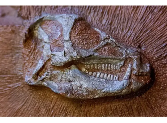
There isn't any credible scientific evidence that supports the mainstream mineralization theory but on the contrary modern technology proves that it can't happen the way the theory suggests. Modern technology, especially microscopy, has dramatically deepened our understanding of dinosaur fossils, revealing details that were unimaginable just a few decades ago. Here’s a concise overview of what microscopy and related tools have uncovered: 1. Discovery of Soft Tissues Using scanning electron microscopy (SEM) and transmission electron microscopy (TEM), scientists have found: • Blood vessel-like structures • Red blood cell-shaped objects • Bone cells (osteocytes) with preserved structures These were first reported in a T. rex femur by Dr. Mary Schweitzer in the early 2000s, challenging the idea that soft tissue couldn’t survive millions of years.
2. Protein and Molecular Structures Microscopy, combined with mass spectrometry and immunohistochemistry, has revealed: • Preserved collagen fibers, confirming a link between dinosaurs and birds. • Reactions with antibodies that only bind to certain proteins, suggesting that original molecules survived in some fossils.
3. Pigmentation and Feather Color With electron microscopes, researchers have identified melanosomes (pigment-containing structures) in fossilized feathers. These led to: • Reconstructions of actual feather colors in dinosaurs like Anchiornis and Microraptor (e.g., iridescent black, reddish tones).
4. Growth and Disease Evidence Bone cross-sections viewed under light and electron microscopes show: • Growth rings, like tree rings, indicating growth rates. • Signs of bone cancer, arthritis, and injuries helping us understand dinosaur health. 5. Cellular and Nanostructural Detail Advanced imaging reveals: • Nano-scale preservation of original tissue structure. • Differences between fossil material and possible contamination (ruling out modern biofilm or bacteria in some studies). In Summary: Microscopy and modern tech have turned dinosaur bones into biological time capsules, revealing: • Cellular structure • Protein preservation • Pigment patterns • Disease evidence This new level of insight has reshaped our view of dinosaurs not just as skeletons, but as once-living, dynamic creatures with feathers, color, disease, and growth cycles. Here’s a list of modern technologies that have been used to analyze and reveal new facts about dinosaur fossils especially those related to soft tissue, cellular structures, and ancient biology:
Scanning Electron Microscopy (SEM) • Produces high-resolution images of fossil surfaces. • Used to identify blood vessel-like structures, osteocytes, and melanosomes (pigment organelles).
Transmission Electron Microscopy (TEM) • Offers ultra-high magnification to see internal structures of cells and fibers. • Helped confirm collagen fibers and sub-cellular features in fossils.
Mass Spectrometry (LC-MS/MS) • Detects and sequences ancient proteins like collagen. • Confirmed molecular similarities between dinosaurs and birds.
Immunohistochemistry • Uses antibodies to test for specific proteins. • Proved some structures in fossils react like real biological tissue, not just mineralized look-alikes.
Synchrotron X-ray Imaging • High-energy, non-destructive imaging of fossil interiors. • Maps the chemical composition of fossils, including traces of blood, soft tissues, or pigments.
Raman Spectroscopy • Identifies molecular bonds and organic compounds. • Confirms presence of preserved proteins and pigments.
Fourier-Transform Infrared Spectroscopy (FTIR) • Detects organic molecules in fossils. • Supports claims of soft tissue preservation by matching spectral signatures of known proteins.
Confocal Laser Scanning Microscopy • Produces 3D images of soft tissue structures inside fossils. • Used to observe elasticity and fine internal details of preserved vessels.
9. Micro-CT (Computed Tomography) Scanning • Creates detailed 3D models of fossil interiors. • Reveals hidden bone structures, growth rings, and sometimes trapped soft tissue.
Stable Isotope Analysis • Measures carbon, oxygen, and other isotopes. • Reveals insights about dinosaur diets, environments, and metabolism. These technologies are revolutionizing paleontology by uncovering molecular and cellular data that were never thought possible in such ancient remains. Here’s a summary with examples of specific dinosaur species where modern technology has revealed extraordinary fossil details:
Tyrannosaurus rex • Discovery: Soft tissue structures including blood vessels, osteocytes, and collagen fragments. • Technology used: Scanning Electron Microscopy (SEM), Mass Spectrometry (LC-MS/MS), Immunohistochemistry. • Significance: First confirmed protein sequences from a dinosaur; supports a close evolutionary link to birds.
2. Anchiornis huxleyi • Discovery: Microscopic pigment structures called melanosomes in fossilized feathers. • Technology used: Transmission Electron Microscopy (TEM), Synchrotron X-ray imaging. • Significance: Revealed feather colors — likely black, gray, and reddish hues — making it one of the first dinosaurs reconstructed with accurate coloration.
Brachylophosaurus canadensis (a hadrosaur) • Discovery: Preserved blood vessels, cells, and possible nuclei in bone tissue. • Technology used: Confocal Laser Scanning Microscopy (CLSM), FTIR spectroscopy, SEM. • Significance: Demonstrated that even delicate cellular structures can persist under rare conditions.
Microraptor gui • Discovery: Feather structure and iridescent coloration patterns. • Technology used: Electron Microscopy, Melanosome analysis. • Significance: Showed it had shiny, bird-like plumage linking flight-related traits with non-avian dinosaurs. These breakthroughs have turned dinosaur bones into molecular time capsules, and they continue to reshape how we imagine these ancient creatures.
#dinosaurs#paleontology#dinosaur extinction#science#geology#cosmology#cosmos#mary schweitzer#mary schweizer#space
12 notes
·
View notes
Text
Digital imaging is a technology used across a wide range of fields, with significant demand in sectors like healthcare (medical imaging), manufacturing quality control, media and entertainment, scientific research, automotive industry, aerospace, and even consumer electronics due to its ability to capture, process, and store visual data with high quality and efficiency, enabling detailed analysis and documentation across various applications; key demands include high resolution imaging, fast data transfer, advanced image analysis capabilities, and seamless integration with other systems.
Specific areas where digital imaging is highly demanded:
Healthcare:
X-rays, CT scans, MRIs, ultrasounds - used for diagnosis and treatment planning.
Telemedicine - remote consultation with digital images.
Pathology - analyzing tissue samples with digital microscopy.
Manufacturing:
Quality control inspections - identifying defects in products during production.
Reverse engineering - creating 3D models from existing parts using digital imaging
Media and Entertainment:
Photography - high-resolution digital cameras for professional and consumer use
Filmmaking - digital cinematography and post-production editing
Graphic design - creating and manipulating digital images
Scientific Research:
Microscopy - capturing detailed images of microscopic samples
Satellite imagery - monitoring environmental changes and land use
Astronomy - capturing images of celestial objects
Automotive Industry:
Vehicle design - creating digital prototypes
Crash testing - analyzing impact forces with high-speed imaging
Key factors driving the demand for digital imaging:
Improved accuracy and precision:
Digital images provide detailed information that can be analyzed more precisely than traditional methods.
0 notes
Text
Metal X-Ray Mirror Lens Market to Hit USD152.2 Million by 2035: What’s Fueling the Growth?
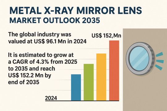
The global Metal X-Ray Mirror Lens Market is projected to grow from a valuation of USD96.1 million in 2024 to USD152.2 million by 2035, registering a steady CAGR of 4.3% over the forecast period (2025–2035). Breakthrough applications across medical imaging, semiconductor fabrication, synchrotron research and space exploration are driving demand for precision X-Ray optics. Technological advances in nanofabrication and multilayer coatings are enhancing lens performance, while high production costs and complex manufacturing processes present challenges. North America currently leads the market, but rapid growth is expected in East Asia and Europe as R&D investments and infrastructure for advanced X-Ray facilities expand globally.
Market Overview
Metal X-Ray mirror lenses are specialized optical elements crafted from highly polished metals and precision-coated multilayers, designed to collimate, focus and steer X-Ray beams with sub-micrometer accuracy. Their unique reflective properties make them indispensable in:
Medical imaging (e.g., high-resolution mammography and computed tomography)
Semiconductor inspection and lithography (EUV and X-Ray lithography)
Synchrotron and laboratory X-Ray instrumentation for materials science, biology and chemistry research
Space and defense applications, where weight, durability and precision are critical
Market Drivers & Trends
Rising Adoption in Semiconductor and Electronics Manufacturing
Advanced Lithography Needs: As chip geometries shrink below 5 nm, X-Ray and EUV lithography require ultrahigh-precision optics to pattern at the nanometer scale. Multilayer mirror lenses offer high reflectivity at EUV wavelengths, improving pattern fidelity and throughput.
Yield Enhancement: Real-time wafer inspection with X-Ray imaging identifies defects invisible to optical systems, reducing waste and boosting yields in fabs.
Growing Utilization of Synchrotron and Laboratory X-Ray Sources
Synchrotron Facilities Expansion: New synchrotron centers in Asia and the Middle East are being commissioned; each demands custom X-Ray optics for beamlines, driving lens orders.
Laboratory X-Ray Innovation: Compact, high-brightness sources integrated into university and corporate labs rely on Kirkpatrick-Baez (K-B) mirror arrays to achieve tight beam focusing for microscopy and spectroscopy.
Latest Market Trends
Multilayer Coating Breakthroughs:
Atomic-level control over film thickness is enabling reflectivities above 70% at soft X-Ray energies, boosting signal-to-noise ratios in imaging.
Additive Manufacturing for Lens Supports:
3D-printed metal mounts reduce weight and thermal distortion, critical for spaceborne telescopes and satellite payloads.
Integration with Adaptive Optics:
Piezo-driven deformable substrates correct wavefront aberrations in real time, enhancing focus precision in variable-temperature environments.
Key Players:
AXO DRESDEN GmbH
Bertin Winlight
Sigray, Inc.
Xrnanotech
Fischer Technology Inc.
JTEC Corporation
Inrad Optics Inc.
Rigaku Innovative Technologies Europe s.r.o, (RITE)
NTT Advanced Technology Corporation
X-Ray Optical Systems, Inc. (XOS)
ZEISS Group
Among Others
Recent Developments
Sigray, Inc. introduced its next-generation metal mirror lens series for synchrotron beamlines (Feb 2023), featuring a modular design that halves alignment time.
ZEISS announced a €20 million expansion of its Oberkochen facility (Oct 2023) to automate precision grinding and ion-beam figuring processes, increasing annual X-Ray lens output by 30%.
JTEC Corporation unveiled a titanium-based single-layer mirror capable of withstanding 500 °C in situ operation within plasma physics experiments (Q1 2024).
Access important conclusions and data points from our Report in this sample - https://www.transparencymarketresearch.com/sample/sample.php?flag=S&rep_id=86589
Market Opportunities
Emerging Research Hubs: Growth of synchrotron centers in South Korea, India and GCC countries opens new markets for high-precision optics.
Space Science Missions: Upcoming X-Ray astronomy satellites (e.g., Athena by ESA) require lightweight, multilayered mirror assemblies, representing multi-million-dollar contracts.
Biomedical Imaging Evolution: Shift toward phase-contrast and dark-field X-Ray modalities in preclinical research expands lens requirements beyond conventional absorption imaging.
Customized On-Site Fabrication: Fab-in-the-lab services offering ultrafast turnaround for custom mirror shapes and surface finishes.
Future Outlook
Analysts foresee sustained growth driven by:
Continuous R&D Investments: Government and private funding in advanced manufacturing to reduce production costs—from ~US $100,000 per lens to targets below US $50,000.
Technological Convergence: Integration of AI-driven metrology during manufacturing will tighten production tolerances, improving yield and reducing scrap by an estimated 15%.
Expanded End-Use Verticals: Beyond semiconductors and research, emerging applications in nondestructive testing for aerospace components and additive manufacturing inspection will diversify revenue streams.
Market Segmentation
By Metal Type
Gold: Excellent reflectivity at soft X-Ray energies.
Silver: Broad reflectance window, used in multilayer stacks.
Other Metals: Nickel, titanium and alloys for specialized applications.
By Layer Type
Single Layer: Simpler, lower-cost; suited for lab-scale sources.
Multi-Layer: High reflectivity across wider energy bands; dominate advanced applications (66.8% share in 2024).
By Surface Shape
Plane
Paraboloid
Ellipsoid
Spherical
Others: Toroidal, cylindrical
By Application
Microscopy
Inspection
Imaging
Metallurgy
Laser Processing
Others: Telescopy, metrological instruments
By Industry Vertical
Aerospace & Defense
Healthcare & Life Science
Research & Academia
Electronics & Semiconductor
Metal & Mining
Others: Pharmaceutical, telecommunication
Regions Covered
North America, Western Europe, Eastern Europe, East Asia, South Asia, Central and South America, Middle East and Africa
Why Buy This Report?
Comprehensive Data: In-depth historical analysis (2020–2024) and forecast to 2035, segmented by metal, layer type, shape, application and industry.
Strategic Insights: Detailed Porter’s Five Forces, value-chain analysis, and growth opportunity mapping.
Competitive Landscape: Profiles of >10 key players with financial overviews, product portfolios and recent strategic moves.
Actionable Recommendations: Tailored strategies for new entrants, expansion roadmap for incumbents, and risk-mitigation tactics for supply-chain challenges.
Accessible Formats: PDF report paired with Excel database for custom analyses.
About Transparency Market Research Transparency Market Research, a global market research company registered at Wilmington, Delaware, United States, provides custom research and consulting services. Our exclusive blend of quantitative forecasting and trends analysis provides forward-looking insights for thousands of decision makers. Our experienced team of Analysts, Researchers, and Consultants use proprietary data sources and various tools & techniques to gather and analyses information. Our data repository is continuously updated and revised by a team of research experts, so that it always reflects the latest trends and information. With a broad research and analysis capability, Transparency Market Research employs rigorous primary and secondary research techniques in developing distinctive data sets and research material for business reports. Contact: Transparency Market Research Inc. CORPORATE HEADQUARTER DOWNTOWN, 1000 N. West Street, Suite 1200, Wilmington, Delaware 19801 USA Tel: +1-518-618-1030 USA - Canada Toll Free: 866-552-3453 Website: https://www.transparencymarketresearch.com Email: [email protected]
0 notes
Link
0 notes
Text
The Role of Optical Lenses in Medical Devices: Enhancing Diagnosis and Treatment
Optical lenses have become indispensable components in a wide range of medical devices, playing a crucial role in improving diagnosis, treatment, and patient care. By manipulating light, these lenses enable healthcare professionals to see, measure, and interact with the human body at various scales, from microscopic to macroscopic.

Key Applications of Optical Lenses in Medical Devices
Microscopy:
Compound Microscopes: These microscopes use multiple lenses to magnify small objects, allowing scientists to examine cells, tissues, and microorganisms in detail.
Fluorescence Microscopes: These microscopes use specialized lenses to excite and detect fluorescent molecules, enabling researchers to study cellular processes and identify specific biomarkers.
Confocal Microscopes: These microscopes use pinhole apertures and lenses to eliminate out-of-focus light, providing high-resolution images of thick specimens.
Endoscopy:
Endoscopes: These flexible tubes equipped with lenses and light sources allow doctors to visualize internal organs and tissues.
Colonoscopes: These endoscopes are specifically designed to examine the colon and rectum.
Laparoscopes: These endoscopes are used for minimally invasive surgery, providing a clear view of the abdominal cavity.
Ophthalmology:
Ophthalmoscopes: These devices use lenses to examine the retina and optic nerve, allowing for early detection of eye diseases.
Refractive Surgery Lasers: These lasers use precise lenses to reshape the cornea, correcting vision problems like nearsightedness, farsightedness, and astigmatism.
Imaging:
X-ray Imaging: X-ray lenses focus X-ray radiation onto a detector, producing images of bones and internal organs.
CT Scanners: These scanners use multiple X-ray sources and detectors to create detailed 3D images of the body.
MRI Scanners: These scanners use strong magnetic fields and radio waves to generate detailed images of soft tissues.
Laser Surgery:
Laser Cutting: Laser cutting devices use lenses to focus the laser beam, enabling precise cutting of tissue.
Laser Welding: Laser welding devices use lenses to focus the laser beam, allowing for precise welding of tissue.
The Future of Optical Lenses in Medical Devices
As technology continues to advance, optical lenses are becoming increasingly sophisticated and versatile. Emerging technologies, such as adaptive optics and photonic crystal fibers, are pushing the boundaries of what is possible in medical imaging and treatment.
By understanding the fundamental role of optical lenses in medical devices, we can appreciate their impact on modern healthcare and look forward to the exciting innovations that lie ahead.
0 notes
Text
Market Dynamics and Emerging Opportunities in Cryo-Electron Microscopy
Cryo Electron Microscopy (Cryo-EM) is an advanced imaging technique that allows scientists to observe biological molecules and structures at near-atomic resolutions. Unlike traditional electron microscopy, which often involves dehydrating and chemically fixing samples, Cryo Electron Microscopy employs a rapid-freezing process to preserve biological specimens in their natural, hydrated state. This preservation minimizes artifacts and provides clear, accurate images of molecular complexes, viruses, and other biological assemblies. The rapid freezing forms a glass-like ice that stabilizes the sample and prevents structural changes, making Cryo-EM particularly valuable for structural biology.
In 2022, the market for cryo electron microscopy was projected to be worth 2.04 billion US dollars. By 2032, the global cryo electron microscopy market is projected to have grown from 2.31 billion USD in 2023 to 7.1 billion USD. CAGR (growth rate) for the cryo electron microscopy market is anticipated to be approximately 13.3% from 2024 to 2032.
Overview of Cryo Electron Microscopy
The Cryo Electron Microscopy technique has revolutionized structural biology by enabling the visualization of macromolecules that were previously difficult to study. It combines advanced cryogenic sample preparation with powerful electron microscopy to capture images of molecules in a close-to-native state. Cryo-EM has become essential for researchers working on large and complex biological assemblies like viruses, ribosomes, and membrane proteins. Unlike X-ray crystallography, which requires crystallization, Cryo-EM allows for the observation of molecules in various conformational states, providing insights into dynamic molecular processes.
Size of the Cryo Electron Microscopy Market
The global Cryo Electron Microscopy market has seen substantial growth over the past few years, driven by the expanding need for high-resolution structural data in both academic and industrial research. As of recent reports, the market is valued in the hundreds of millions and is expected to continue expanding with a high compound annual growth rate (CAGR). The growth is largely due to the increasing adoption of Cryo Electron Microscopy in pharmaceutical and biotechnological research, where it aids drug discovery and the understanding of disease mechanisms. The market size is further bolstered by technological advancements that have made Cryo-EM more accessible, improving image resolution and throughput.
Cryo Electron Microscopy Market Share
Within the Cryo Electron Microscopy market, several key players dominate, including manufacturers of Cryo-EM equipment and software developers specializing in image processing. Major companies such as Thermo Fisher Scientific and JEOL Ltd. have captured significant shares of the market, thanks to their extensive product portfolios and global presence. The market share distribution is also influenced by partnerships between academic institutions, research organizations, and industry leaders who work together to advance Cryo Electron Microscopy capabilities and applications.
Cryo Electron Microscopy Analysis
Cryo Electron Microscopy analysis is a multi-step process involving sample preparation, data collection, and image processing. Samples are flash-frozen and observed using electron beams to capture thousands of images that can then be computationally reconstructed to form a high-resolution 3D model. Advanced software tools enable researchers to analyze molecular structures in great detail, identifying features critical for understanding function and interaction. Cryo-EM analysis has proven instrumental in studying complex biological processes, such as enzyme mechanisms and membrane transport, with applications spanning drug development and biomedical research.
Cryo Electron Microscopy Trends
The Cryo Electron Microscopy field is evolving rapidly, with several notable trends. First, the development of more powerful direct electron detectors has significantly improved the quality of data collected. Second, advancements in artificial intelligence and machine learning are enhancing image processing, making it faster and more accurate. Additionally, single-particle analysis, a Cryo-EM technique for studying individual molecules, is gaining traction as it enables high-resolution imaging without the need for crystallization. Finally, Cryo-EM is being increasingly applied in drug discovery, particularly for visualizing drug-target interactions at the molecular level.
Reasons to Buy Cryo Electron Microscopy Reports
In-depth Market Analysis: Reports provide detailed information on the market size, share, and growth forecasts, helping businesses make informed investment decisions.
Competitive Landscape Insight: Understanding the market share and strategies of key players allows for better strategic planning.
Technological Advancements: Reports highlight the latest technological developments, ensuring that researchers and companies stay updated with cutting-edge techniques.
Application Insights: By examining applications of Cryo Electron Microscopy, reports reveal its potential across various industries, particularly in pharmaceuticals.
Market Trends and Future Outlook: Reports help identify emerging trends, aiding stakeholders in anticipating shifts and planning long-term strategies.
Recent Developments in Cryo Electron Microscopy
In recent years, Cryo Electron Microscopy has seen several significant developments. One key advancement is the integration of machine learning to enhance image processing, significantly reducing analysis time. Additionally, the introduction of automated Cryo-EM platforms has improved efficiency, allowing researchers to process samples and data more rapidly. New developments in direct electron detectors have also raised the achievable resolution, making Cryo-EM a more precise tool for structural biologists. Furthermore, there have been several academic-industry partnerships focused on developing cryo-tomography methods, expanding the applications of Cryo-EM beyond single-particle analysis to cellular and tissue-level studies.
Related reports:
ivus catheters market
laboratory filtration market
medical education market
Top of Form
Bottom of Form
0 notes
Text
Telecentric Lens in the Medical Field: Applications and Benefits

Telecentric lens technology has been gaining popularity in the medical field due to its unique capabilities. This specialized type of lens is designed to ensure that the light enters the lens parallel to the optical axis, resulting in reduced distortion and magnification variability.
In this blog, we will explore the various applications of telecentric lenses in the medical field and how they can benefit healthcare professionals.
What is a Telecentric Lens?
A telecentric lens is a type of lens that has the property of being telecentric. That means it renders images where all object points appear the same size, regardless of their distance from the camera sensor or film plane. Telecentric lenses also gather light in such a way that it enters the lens perpendicular to the sensor or film plane. In other words, the angle of incidence is zero, making them ideal for 3D imaging applications.
Applications of Telecentric Lenses in the Medical Field
Endoscopy
Endoscopes are medical devices that allow doctors to see inside the body without making large incisions. Telecentric lenses are used in endoscopes to provide clear and sharp images of the internal organs. The telecentric lens allows for the light rays to be captured at an angle close to zero degrees, which reduces image distortion and improves the clarity of the image.
Ophthalmology
Telecentric lenses are used in ophthalmology to capture high-resolution images of the eye. These lenses are particularly useful when examining the retina, as they provide a clear view of the entire surface of the retina. This can aid in the diagnosis of eye diseases such as macular degeneration, glaucoma, and diabetic retinopathy.
Microscopy
Microscopy is an important tool in medical research, allowing scientists to study cells and tissues at the microscopic level. Telecentric lenses are used in microscopy to improve the quality of the captured images. The telecentric lens ensures that the light enters the microscope at an angle close to zero degrees, reducing distortion and improving the resolution of the image.
Radiology
Radiologists use telecentric lenses in X-ray machines to produce clearer images of the bones and organs. Traditional lenses often produce images with varying levels of magnification, leading to distortion and errors in diagnosis. Telecentric lenses eliminate this problem by ensuring that the image remains constant in size, regardless of its position in relation to the lens.
Surgery
Telecentric lenses are also used in surgical procedures to provide a clear view of the surgical site. They are particularly useful in minimally invasive surgeries, where the surgeon must rely on a monitor to guide the procedure. The telecentric lens produces a clear and accurate image, allowing surgeons to perform delicate procedures with greater precision.
Benefits of Telecentric Lenses in the Medical Field
Improved Image Quality
One of the most significant benefits of telecentric lenses in the medical field is improved image quality. By reducing distortion and magnification variability, telecentric lenses provide clearer and more accurate images, aiding in diagnosis and treatment planning.
Enhanced Precision
Telecentric lenses offer enhanced precision in medical procedures. They provide a clear and accurate view of the surgical site, allowing surgeons to perform delicate procedures with greater accuracy and reducing the risk of complications.
Reduced Radiation Exposure
Telecentric lenses used in radiology machines can reduce radiation exposure for patients. By producing clearer images, fewer X-rays may need to be taken, reducing overall exposure.
Cost-Effective
Telecentric lenses can be cost-effective in the long run. By providing high-quality images, they can reduce the need for repeat imaging and additional procedures, saving both time and money.
Conclusion
Telecentric lenses are increasingly becoming an essential tool in the medical field due to their unique capabilities. From endoscopy to radiology, these lenses are used in various medical procedures to provide clear and accurate images, aiding in diagnosis and treatment planning. With their ability to reduce distortion and magnification variability, enhance precision, and reduce radiation exposure, telecentric lenses offer numerous benefits to healthcare professionals and patients alike.
0 notes
Text
High-Resolution 3D X-Ray Microscopy Market Key Factors for Growth and Opportunity Illuminated by New Report 2021-2028
Global High-Resolution 3D X-Ray Microscopy Market Synopsis:
With an international High-Resolution 3D X-Ray Microscopy Market report, businesses will come to know current and future of Market outlook in the developed and emerging Markets. The report provides analysis of various perspectives of the Market with the help of Porter’s five forces analysis. It highlights the segment that is expected to dominate the global High-Resolution 3D X-Ray Microscopy Market and the areas that are expected to observe the wildest growth during the predicted period. A promotional High-Resolution 3D X-Ray Microscopy Market analysis report is a comprehensive study about the Market which tells about what is the Market status in the forecast period of 2021-2028.
High-Resolution 3D X-Ray Microscopy Market report analyzes the Market status, growth rate, future trends, Market drivers, opportunities and challenges, risks and entry barriers, sales channels, distributors and Porter's Five Forces Analysis. This Market document helps identify latest growths, Market shares, and policies employed by the major Market players. In addition, this Market study affirms the leading players across the globe in the High-Resolution 3D X-Ray Microscopy Market and their key Marketing dispositions and advertising enterprise have been highlighted to offer a clear understanding of the Market. High-Resolution 3D X-Ray Microscopy Market research report gives an examination of various segments that are relied upon to witness the quickest development amid the estimate forecast frame.
Get Sample Report + All Related Graphs & Charts @ https://www.databridgemarketresearch.com/request-a-sample/?dbmr=global-high-resolution-3d-x-ray-microscopy-market .
The Global High-Resolution 3D X-Ray Microscopy Market is expected to USD 4,126.62 million by 2028 and will grow at a CAGR of 8.76% in the forecast period of 2021 to 2028.
According to the market report analysis, 3D X-ray microscopy is also termed as industrial computed tomography. 3D X-ray microscopy helps in providing effective imaging solutions for characterizing the properties and behavior of materials revealing the details of microstructures. This method provides unique details that might not be visible by any other microscopy technologies.
The most significant key factors driving the growth of the Global High-Resolution 3D X-Ray Microscopy Market are rapid rise in focus on nanotechnology and regenerative medicine and the high adoption of advanced technologies, technological advancements and increase in the demand for advanced products in image solutions.
The major players covered in the High-Resolution 3D X-Ray Microscopy Market report are Zeiss, Rigaku Corporation, Bruker, Thermo Fisher Scientific, General Electric, TESCAN ORSAY HOLDING, A.S., MATSUSADA PRECISION Inc., HORIBA, Ltd., Nikon Metrology NV, Hamamatsu Photonics K.K., PerkinElmer Inc., Leica Microsystems, OLYMPUS CORPORATION, JEOL Ltd, Hitachi High-Tech Corporation, ACCU-SCOPE, KEYENCE CORPORATION, and WITec among other domestic and global players.
Geographic analysis, North America region leads the high-resolution 3D X-ray microscopy market owing to the rapid technological advancements and high demand for advanced products in image solutions. APAC is expected to expand at a significant growth rate over the forecast period of 2021 to 2028 because of the emerging economies from developing countries and high adoption of advanced technologies.
Access Complete Report @ https://www.databridgemarketresearch.com/reports/global-high-resolution-3d-x-ray-microscopy-market .
0 notes
Link
The global high resolution 3D X-ray microscopy market is expected to reach USD ~ 3,312 million at a CAGR of over 9% by the end of the forecast period.
#High-Resolution 3D X-Ray Microscopy Market Share#High-Resolution 3D X-Ray Microscopy Market Size#High-Resolution 3D X-Ray Microscopy Market Growth#High-Resolution 3D X-Ray Microscopy Market Forecast
1 note
·
View note
Link
Based on the High Resolution 3D X-Ray Microscopy market development status, competitive landscape and development model in different regions of the world, this report is dedicated to providing niche markets, potential risks and comprehensive competitive strategy analysis in different fields. From the competitive advantages of different types of products and services, the development opportunities and consumption characteristics and structure analysis of the downstream application fields are all analyzed in detail. To Boost Growth during the epidemic era, this report analyzes in detail for the potential risks and opportunities which can be focused on.
0 notes
Text
0 notes
Photo


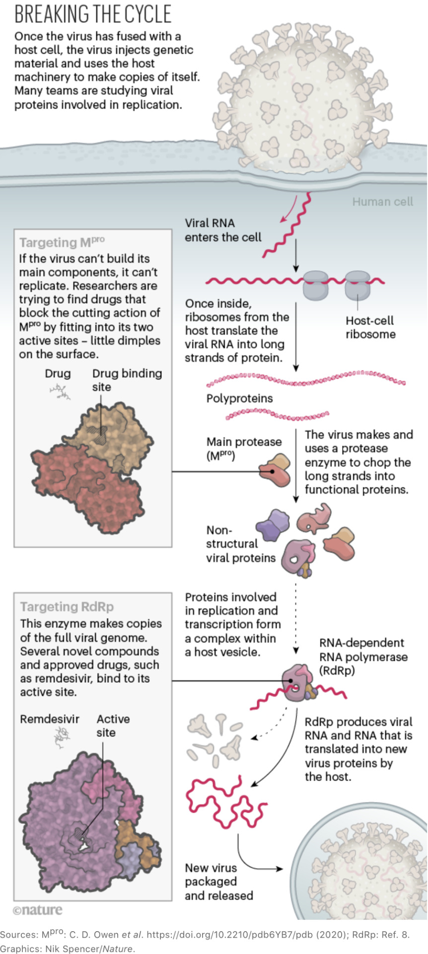
The sprint to solve coronavirus protein structures — and disarm them with drugs
by Megan Scudellari (Nature). Top Image: Cognition Studio Inc.
Lying in bed on the night of 10 January, scrolling through news on his smartphone, Andrew Mesecar got an alert. He sat up. It was here. The complete genome of a coronavirus causing a cluster of pneumonia-like cases in Wuhan, China, had just been posted online.
Around the world, similar notifications appeared on the devices of scientists who first crossed swords with coronaviruses in the 2003 outbreak of SARS (severe acute respiratory syndrome) and then again with MERS (Middle East respiratory syndrome) in 2012. Instantly, the researchers mobilized against a new adversary. “We always knew that this was going to come back,” says Mesecar, head of biochemistry at Purdue University in West Lafayette, Indiana. “It’s what history has shown us.”
In Lübeck, Germany, Rolf Hilgenfeld stopped packing boxes for his retirement and started preparing buffers for crystallography. In Minnesota, Fang Li stayed up all night analysing the new genome and drafting a manuscript. In Shanghai, China, Haitao Yang rallied a dozen graduate students to clear their schedules. In Texas, Jason McLellan instructed laboratory members to start assembling gene sequences from the viral genome.
Within 24 hours, a network of structural biologists around the world had redirected their labs towards a single goal — solving the protein structures of a deadly, rapidly spreading new contagion. To do so, they would need to sift through the 29,811 RNA bases in the virus’s genome, seeking out the instructions for each of its estimated 25–29 proteins. With those instructions in hand, the scientists could recreate the proteins in the lab, visualize them and then, hopefully, identify drug compounds to block them or develop vaccines to incite the immune system against them.
“Here we go,” thought Mesecar. “I’d better get some sleep.”
11 January: 41 confirmed cases of COVID-19 worldwide
Mesecar woke at 6 a.m. the next day, turned on the coffee pot and began blasting through the new genome looking for recognizable protein sequences. It didn’t take long. He had spent 17 years studying coronaviruses, and the new virus’s genome looked very familiar.
“Holy shit,” he thought. “This is the same thing as SARS.”
Right away, Mesecar contacted Karla Satchell, a microbiologist at Northwestern University Feinberg School of Medicine in Chicago, Illinois. Satchell is co-director of the Center for Structural Genomics of Infectious Diseases (CSGID), a consortium of eight institutions set up exactly for moments like this — to rapidly investigate the structures of emerging infectious agents.
To solve the 3D structure of a protein at high resolution, scientists first design a gene construct — a circle of DNA containing the instructions for the protein, together with regulatory sequences to control where and how it is expressed. They then insert the construct into living cells, often the bacterium Escherichia coli, using the cells’ own machinery to churn out the desired protein. Next, they purify the protein so that they can visualize its structure using either of two methods. One is X-ray crystallography, which involves growing tiny crystals of pure protein and revealing their internal structure by bombarding them with X-rays from a high-energy electron beam. The other is cryo-electron microscopy (cryo-EM), a process of scanning flash-frozen proteins using a high-powered electron microscope.
Either process can take months, even years, for an unfamiliar protein. Luckily, many of the new coronavirus proteins were familiar, with 70–80% sequence similarity to SARS-CoV, the virus that caused the 2003 SARS outbreak. By 7:30 a.m., Mesecar and his team had begun designing gene constructs for the new viral proteins, and even predicted which of their existing coronavirus inhibitors might block these proteins.
Satchell, who had been following early news reports about the virus, organized a virtual meeting of consortium members to start solving the virus’s proteins. “We’ve thrown the weight of every investigator at every site behind COVID,” says Satchell. Mesecar, a CSGID investigator, started with Mpro, the virus’s main protease, an enzyme that cuts out proteins from a long strand that the virus produces when it invades a cell, like a tailor cutting out pattern pieces. Without Mpro, there is no viral replication. Humans do not have a similar protease, so drugs targeting this protein are less likely to cause side effects.
13 January: 42 confirmed cases
In McLellan’s molecular biosciences lab at the University of Texas at Austin, graduate student Daniel Wrapp spent the weekend designing a gene construct for another key protein — the outer, three-pronged spike that gives the coronavirus its crown-like appearance and name (see ‘The key coronavirus proteins’). Wrapp placed an order for the constructs with a commercial firm that Monday, 13 January.
McLellan had been involved in determining the structures of two other coronavirus spikes — from HKU1, a cause of common colds1, and from the MERS virus2. The work was done in collaboration with structural biologist Andrew Ward at the Scripps Research Institute in La Jolla, California, and virologist Barney Graham at the US National Institute of Allergy and Infectious Diseases’ Vaccine Research Center in Bethesda, Maryland. So, the group knew how to tweak the spike protein’s genetic sequence so that it would stabilize in a pre-fusion shape — the form it adopts before it docks onto a host cell. “Our ability to get this particular structure was based upon all our prior knowledge from working on HKU1 and MERS and SARS,” says McLellan.
While McLellan’s team waited for the construct to arrive, Graham called Moderna Therapeutics, a drug-discovery company in Cambridge, Massachusetts, with which the Vaccine Research Center had been working on a pandemic-preparedness project. On 13 January — before any spike protein had been made — Moderna began preparing its manufacturing facilities to make a coronavirus vaccine based on that protein.
26 January: 2,014 confirmed cases
At ShanghaiTech University in China, Zihe Rao, Haitao Yang and their colleagues worked day and night, sacrificing their week-long Chinese Lunar New Year holiday, to solve the Mpro structure and those of another trio of proteins that the coronavirus uses to replicate.
Using X-ray data acquired at the Shanghai Synchrotron Radiation Facility and the National Center for Protein Science Shanghai — which both allocated special beam time for the project — the team solved the crystal structure of Mpro bound to an inhibitor3. In 2003, it had taken them two months to solve the structure of the SARS-CoV main protease. This time, it took one week.
Mpro in coronaviruses is made up of two identical subunits and looks like a moth-eaten heart, with an active enzyme site on each side of the structure. On 26 January, Rao and Yang submitted the Mpro structural data to the Protein Data Bank (PDB), an open-access digital resource for 3D structures of biological molecules. By 5 February, the data had been processed and the final structure was released online — not a moment too soon, says Yang. The laboratory had already received an overwhelming 300 requests for the structure.
While working on Mpro, Rao contacted a former co-worker, David Stuart, a structural biologist at the University of Oxford, UK, who is life-sciences director at Diamond Light Source, the United Kingdom’s synchrotron facility. The UK and Shanghai groups began collaborating closely to share advice and avoid overlap, says Martin Walsh, deputy life-sciences director at Diamond. “We keep each other up to date on things, and try to benefit from the different approaches they’re using and we’re using.”
Because the Shanghai team solved Mpro in complex with an inhibitor, the Diamond team decided to focus on crystallizing the protein with no molecule attached, hoping to identify active sites to which potential drug compounds might bind. Over two weeks, Walsh’s group ran 17,000 experiments to hit on the best recipe for precipitating the unbound protein into a crystal.
1 February: 11,953 confirmed cases
In Hilgenfeld’s lab at the University of Lübeck, researcher Linlin Zhang had taken to phoning the company making the Mpro gene construct daily until it finally arrived. Thanks to the lab’s experience crystallizing other coronavirus proteases, Zhang grew Mpro crystals in 10 days, and on 1 February, she took the precious samples to the BESSY II synchrotron in Berlin, which opened up a beamline especially for the project.
In addition to focusing on the unbound Mpro structure, Hilgenfeld docked a small-molecule inhibitor called 13a, which he had designed to inhibit the MERS virus, into the protein’s active site. It wasn’t a perfect fit, so the team altered a residue on the compound and named it 13b. This one “fit nicely”, says Hilgenfeld, and in ten more days his team had solved the structure of Mpro bound to the inhibitor4.
McLellan’s group in Texas was solving the spike protein structure at similar speed. As soon as the group had finished gathering high-resolution electron-microscopy data of the stabilized spike, thanks to a multimillion-dollar cryo-EM facility at the university, McLellan sent the data to Graham at the Vaccine Research Center.
Vaccines are often based on presenting parts of a virus to the human immune system to provoke a response, and the spike protein is an obvious candidate because it has a crucial role in infection.
The spike is formed of three identical molecules stuck together in the shape of a pyramid, with a hinge-like trapdoor. This opens to expose a portion that grabs onto a receptor on a human cell (see ‘The spike locks on’). Graham and McLellan’s past work on a similar protein5 suggested that presenting the spike protein in its pre-grab state would provoke the human immune system. From the complete structure, Graham could see that McLellan’s gene construct made a high-quality protein arranged in the right conformation. “It was really, really important to have that electron-microscopy information,” says Graham.
Graham tested the spike protein in mice, working to improve its expression levels and the strength of its effect on the immune system, and sent the sequence to Moderna, where the production line was ready and waiting. On 7 February, Moderna completed its first batch of the vaccine based on that protein.
Meanwhile, on 10 February, just 12 days after harvesting the protein, McLellan and his group submitted its cryo-EM structure6 to the PDB. By studying the spike in detail, they found that it binds to its human cell receptor, a protein called ACE2, at least ten times more tightly than SARS-CoV does.
At the University of Minnesota in Saint Paul, Li’s team was on its way to working out why. On 11 February, Li and his colleagues began collecting X-ray data from the spike protein using the Advanced Photon Source (APS), the synchrotron facility at the US Department of Energy’s Argonne National Laboratory near Chicago, Illinois. By 13 February, the researchers had defined the small, important spot where the spike protein locks on to the ACE2 receptor7. They found that the new coronavirus spike protein has small molecular differences in its binding region compared with that of SARS-CoV, which might be why the new virus attaches to ACE2 more strongly. These changes could also explain why it seems to infect cells better and spreads faster than the SARS virus. That same week, the virus also got a name: SARS-CoV-2.
18 February: 73,332 confirmed cases
By mid-February, protein structures were pouring out (see ‘Breaking the cycle’). On 18 February, Hilgenfeld, Zhang and their colleagues submitted a paper4 on the Mpro structure alone and bound to 13b, and posted the preprint on the bioRxiv server on 20 February. “It was pretty fast,” Hilgenfeld admits. “The longest time period was just getting it published.” That same day, the Diamond team released the high-resolution crystal structure of unbound Mproon its website.
To support US teams, the APS and other national synchrotrons coordinated their schedules to ensure there would be no interruption in beamtime if one facility had to close for maintenance or because of a local outbreak. “Our goal is just to keep the research going,” says Stephen Streiffer, director of the APS. “The rate at which people are working at this is an order of magnitude faster than they’ve been able to work on other problems.”
So far, the CSGID consortium has solved 12 unique SARS-CoV-2 protein structures, which are kept in a new online database with their accompanying genomic information. “We’ve been part of projects like this on cancer, but it took five years to set that all up,” says Adam Godzik, a bioinformatician at the University of California, Riverside, and a CSGID investigator. “This happened spontaneously in the course of months.”
16 March: 167,515 confirmed cases
With 3D structures in hand, structural-biology teams moved straight to next steps. “Structures aren’t everything,” says Mesecar. “You want to get to compounds — to antivirals and vaccines.”
On 16 March, just 65 days after the viral genome was released, clinicians gave the first dose of Moderna’s vaccine candidate to a patient in a clinical trial funded by the US National Institutes of Health.
“It was a lot faster than even the fastest one we’d previously done,” says Graham. Because of research on SARS and MERS, coronaviruses were probably the only viral family for which that was possible, he adds. “If it was a bunyavirus or an arenavirus, we would have been lost for two to three years.”
But even a vaccine developed at record-breaking speed is likely to be a slower solution than repurposing an approved drug, or at least finding one for which safety testing has begun. “That’s absolutely going to be the fastest way to help patients sick in the hospital today,” says Satchell.
That was exactly what Andrew Hopkins was planning. On 19 March, Hopkins, the chief executive of Exscientia, an artificial-intelligence drug-discovery company in Oxford, UK, took delivery of a large styrofoam cooler packed with dry ice. Inside was a library of 12,000 drug compounds known to be safe and ready for human use, sent from Scripps Research in California. The Exscientia team, working closely with Diamond, immediately began screening the collection against four of Diamond’s structures: Mpro, the spike protein, a second protease and the replication-machinery complex. Exscientia is currently preparing to test compounds that bind to the first two proteins for antiviral activity, says Hopkins.
Similarly, the ShanghaiTech team conducted virtual and high-throughput screening of a library of more than 10,000 approved drugs and compounds already in clinical trials, to see whether any would disable Mpro (see ‘Breaking the cycle’). They identified six promising candidates3. One of them, ebselen, is already in clinical trials for the treatment of bipolar disorder and hearing loss, and the group is preparing animal tests to study its activity in vivo, says Yang.
On 10 April, Rao, Yang and their collaborators published8 the structure of the virus’s replication complex — a large protein called RNA-dependent RNA polymerase (RdRp, or nsp12) that forms a complex with two others, nsp7 and nsp8. They also modelled how it binds to the antiviral drug remdesivir, originally developed to treat Ebola and now in phase III trials for coronavirus. Another recently completed structure of the protein in complex with the drug9 could provide a template to help model and modify other existing antivirals.
22 April: 2,471,136 confirmed cases
The hard-core biochemistry of designing brand-new, custom drugs to inhibit SARS-CoV-2 proteins will take months, even years, but could eventually lead to the best-performing drugs against the infection.
The ShanghaiTech team and collaborators have designed and synthesized a series of compounds targeting the active site of Mpro. On 22 April, after much chemical tweaking, they published details of one that inhibits viral replication in cells and was not toxic when tested in rats and dogs10. The team will continue developing that compound as a drug candidate, says Yang.
The Diamond team has identified 91 chemical fragments — bits of molecules that are less than one-third the size of a normal drug — that bind to Mpro. Those fragments inspired the launch of a non-profit crowdsourced initiative, the COVID Moonshot, to engage chemists around the world to use the fragments to design antiviral drug candidates. The initiative has received more than 4,600 design submissions, and several therapeutic possibilities are already emerging.
In Germany, researcher Katharina Rox at the Helmholtz Centre for Infection Research in Braunschweig tested Hilgenfeld’s 13b compound in mice, showing that it was safe and accumulated well in the lungs4, a key infection site. Meanwhile, a compound that Mesecar developed to inhibit SARS-CoV, compound 77, has been shown in unpublished work to have antiviral activity against SARS-CoV-2 in cells, and he hopes to complete animal studies by the end of the summer.
14 May: 4,248,389 confirmed cases
Structural biologists continue to plug away at the remaining unsolved proteins in the coronavirus genome. These include ORF8, a protein whose function remains mysterious. “We predict it should be crystallizable, but nobody has done it, so we’re trying,” says Godzik.
In the United Kingdom, the Diamond team is screening various compounds against a second coronavirus protease. In Texas, McLellan has shipped spike constructs to more than 100 labs worldwide. Many are looking for treatments, using the protein to fish antibodies out of the blood of people who have had COVID-19, and McLellan’s team is now characterizing the first of these potentially therapeutic antibodies.
Hilgenfeld, who was officially scheduled to retire on 1 April as a result of a mandatory retirement policy, has packed up his office but continues to work. “I’ve been working on coronaviruses for 20 years, and most of the time it was neglected and not taken seriously,” he says. “Now that it’s happened, how can I leave?” His team is investigating other SARS-CoV-2 structures, including nsp3, a large protein that the virus uses to shut down host-cell defences.
The race against the virus can’t afford to slow down anytime soon. As soon as countries start lifting restrictions on people’s movement, the virus will return and “flip around the world again”, says Satchell. “When that happens, it would be really great to have beautiful drugs that were designed specifically to target this coronavirus,” she says. “But we need to do it fast.”
Nature 581, 252-255 (2020) doi: 10.1038/d41586-020-01444-z
References
Kirchdoerfer, R. N. et al. Nature 531, 118–121 (2016).
Pallesen, J. et al. Proc. Natl Acad. Sci. USA 114, E7348–E7357 (2017).
Jin, Z. et al. Nature https://doi.org/10.1038/s41586-020-2223-y (2020).
Zhang, L. et al. Science 368, 409–412 (2020).
McLellan, J. S. et al. Science 342, 592–598 (2013).
Wrapp, D. et al. Science 367, 1260–1263 (2020).
Shang, J. et al. Nature 581, 221–224 (2020).
Gao, Y. et al. Science 368, 779–782 (2020).
Yin, W. et al. Science https://doi.org/10.1126/science.abc1560 (2020).
Dai, W. et al. Science https://doi.org/10.1126/science.abb4489 (2020).
118 notes
·
View notes
Link
The global high resolution 3D X-ray microscopy market is eyeing for a promising CAGR of 9% over the forecast period of 2016-2027, proclaims Market Research Future (MRFR) in a minutely analyzed research report. The high resolution 3D X-ray microscopy market has observed rapid development in the recent years. High-resolution 3D X-ray microscopes (XRM) have gained immense popularity as they solve the inherent challenges associated with 2D methods. In response to the limitations of 2D methods such as inaccuracy and sample consumption during the imaging process, new techniques were developed for 3D characterization. X-ray microscopy (XRM) of additive manufacturing parts is essential in ensuring efficient and effective process development of such components. Defect detection and characterization within metal parts with the help of high resolution 3D x-ray microscopy results in better understanding of interior structures of complex parts. The development of metal additive manufacturing is progressively moving to the mainstream which necessitates for improved understanding of the process and is a key driver of the growth of the global High Resolution 3D X-Ray Microscopy Market. Rigorous research and development activities in the field of image solutions and microscopy has spurred the growth of the market. High resolution 3D x-ray microscopes find their application in diverse industries such as oil & gas, material science, semiconductors, metrology, life science, healthcare, and others and growth in end-use industries augments the growth of the global high resolution 3D x-ray microscopy market.
0 notes
Text
Facelift surgical Treatment, Eye & Neck Lifts
Skin geek
Content
exists any Kind Of Downtime following treatment?
Fat Freeze Is remarkable
treatment
Cryopen Cryotherapy.
is There An Age limit For This type Of surgery?
Fat Freeze Is really good.
The proposition must consist of photos and also evidence that appropriate cryo-EM samples have currently been generated. Our complimentary software program as well as web site for remote control, tracking as well as logging of your Oxford Cryosystems device.
How many fingers means you're loose?
Then, removed it and inserted two fingers, followed by three fingersto assess tightness as compared to a single finger?” The rule of the thumb is that if you can insert your ring, middle, and index finger together into your vagina and cannot feel anything, then it is most likely that you're loose.
The example is applied to a grid, which is then frozen in liquid ethane and also kept in fluid nitrogen. Freezing should be fast sufficient to prevent the water existing developing ice crystals. Ice lattices will not only take in the electron beam of light and also obscure the image, but are likely to harm the sample's structures. If the example is frozen quickly sufficient, water solidifies as an amorphous solid and does not crystallise.
Does insurance pay for ThermiVa?
Please note: ThermiVa is not covered by insurance and pricing is subject to change. For your convenience, we offer United Medical Credit and Care Credit to help patients secure the funding they need for their healthcare procedures.
All of our products stay true to the goals of our creators; to understand the demands of our client, and to develop the maximum service. The GW4 allianceopened a ₤ 3.7 M center for high resolution electron cryo-microscopy at the College of Bristol. Photo evaluation is made it possible for by a high performance computer cluster moneyed by a BBSRC Alert 17 grant.
exists any Kind Of Downtime following therapy?
Every one of our therapies are strongly evaluated internal before we provide them to our clients to guarantee they are secure, effective and will certainly offer you, our fantastic clients, the outcomes you want.
We have extensive understanding and experience in the medical and aesthetic appeals sectors, but we understand that above all, the outcomes matter!
Making use of ultra-sound waves, the superficial muscular aponeurotic system is targeted.
Your happiness, health and also complete satisfaction are at the very core of every little thing we do right here at Elite Aesthetic appeal, and also as a result we will certainly never over-treat or perform a treatment that Dr. Shirin, considers unacceptable or hazardous.
This guarantees you have chosen the right therapy for your skin appearance objectives.
The SF HIFU Medication is unmatched by other non-invasive cosmetic devices, with its capacity to deal with the cells generally dealt with throughout medical renovations.
This implies you can constantly feel great that you remain in risk-free hands when you pick Elite Aesthetic appeals.
In time, collagen fibers start to shed flexibility, which consequently creates the skin to lose its suppleness and also start to sag.
The cells are heated up to a range in between 60-70 ˚C, hence the natural injury healing procedure is promoted to produce brand-new collagen and elastin.
They symbolize Air Liquide's scientific region and also have gone to the core of the business's activities since its creation in 1902. At PSS we pride ourselves on our customer service and are happy to provide as much help as required no matter the inquiry size. Improvements to skin disease such as psoriasis, dermatitis, acnes, and also acne. An all natural wellness brand like nothing else, the UK's very first electrically cooled down Cryotherapy chamber established by cosmetic surgeon Dr. Yannis Alexandrides.
How long does a vaginal rejuvenation last?
By the 6 week clearance appointment, the stitches should have dissolved and patients are cleared to resume regular activities. How long will results last after vaginal rejuvenation? The results of vaginal rejuvenation typically last for a lifetime, unless a new vaginal delivery reverses its affects.
This is a fantastic handheld device that enables you to proceed treatment in the house, in between sessions with ourselves. Swedish massage therapy is the most usual type as well as form of massage carried out in the West, and also it is where lots of people have their very first experience of enjoying the advantages of massage therapy. This type concentrates on the surface layers of muscle rather than deep-tissue massage therapy. Dermapen is a little tool that resembles a pen as well as is usually used for microneedling on people with skin issues as well as the end product is a tightened up, invigorated as well as beautiful skin. The ZO Red rug Peel is developed to exfoliate the skin by stimulating mobile turnover to improve complexion, structure and also quality. This treatment is best matched to customers that are experiencing discomfort, swelling inflammation in one certain component of the body.
Additionally, if you've lately had fillers, we recommend waiting 2 weeks before you have an ACTIVE CryoFacial. Unlike a conventional face, it's a quick, simple therapy that will certainly leave your face looking revitalized as well as glowing.
Fat Freeze Is remarkable
Occasionally healthy proteins embrace favored positionings that make 3D reconstruction difficult. Because of an absence of tarnish and for that reason an absence of contrast, pictures typically have a really reduced signal-to-noise ratio, calling for highly advanced discovery equipment and also photo handling. A major advantage of cryo-EM over x-ray crystallography is that the particle of rate of interest does not have to be crystallised.
treatment
Our tangential product is our range of single-stage and also two-stage GM coolers, which are finding uses in a series of applications, from our own XRD cryostats to use in radioastronomy and HTS magnets. Oxford Cryosystems specialises in the layout and manufacture of world-class cryogenic tools. Adipose tissue, or fat, is an anatomical term for loose connective tissue composed of adipocytes. Its major function is to store energy in the form of fat, although it likewise pillows and also protects the body. Excessive of this is not an advantage for consumers wishing to get rid of excess fat as well as attain that tone as well as interpretation they desire. The most sophisticated cryo-EM equipment remains incredibly costly. The growth of centralised facilities might have the ability to assist increase accessibility to cryo-EM devices.

Cryo-electron microscopy, or cryo-EM, uses electrons to research samples frozen at cryogenic temperatures. The Cryo Cabinet is a totally bonded, stainless-steel cryogenic cupboard freezer developed by Air Liquide based upon the current health standards from the European Hygienic Engineering & Design Team and also the United States Department of Farming. By utilizing Trimbio for all your medical and also non medical device testing, we can decrease the overall expenses to your company. We have proficiency with logistics, regulated temperature states, and also recognize the crucial value of preserving top quality and also standards at all times.
Yet opting out of a few of these cookies may have a result on your surfing experience. We make use of cookies on our site to give you one of the most relevant experience by remembering your choices and repeat gos to. By clicking "Accept", you consent to using ALL the cookies. Wonderfully crafted devices, providing the most recent developments in the elegance and also wellbeing area. Specimen prep work can be tricky-- not only to optimise ice thickness, however also to optimise bit distribution.
Cryopen Cryotherapy.
https://csgrid.org/csg/team_display.php?teamid=655120 ='text-align:center'>
youtube
exists An Age restriction For This sort Of surgical Treatment?
You might call for short term storage for stem cells, or require to keep certain samples or reagents offsite. We make use of cookies to guarantee we give you the most effective experience on our site. By continuing to use this site you are agreeing to our cookie policy. Photon Surgical Equipment sells new and pre-owned tools, as well as supplying maintenance and also repair services to the Veterinary as well as Human Medical Occupations at reasonable costs. We have a values of offering the most effective top quality products at cost effective rates and completely supporting them with our very own group of extremely competent as well as seasoned professionals as well as engineers. Please do not hesitate ahead with a clean face or we can offer a wipe to cleanse your face before the therapy; maintaining your makeup on is great too. In either case, you get the terrific advantages of an ACTIVE CryoFacial.
For a complexion that needs restoring opt for our Cryotherapy face therapy. The controlled system of -30 ° C cooled down air as well as pure CO2 assists to enhance oxygen degrees, aid the improvement of microcirculation, and offer optimum conditions for collagen and elastin excitement. These icy liquid-filled globes will assist move toxins that linger under the eyes, and increase oxygen circulation at the skin surface area. These borosilicate glass worlds are loaded with a non-freezing liquid, which indicates they can be rolled under the eyes as well as along the jawline, straight from the freezer. They're likewise especially great for holding over cystic outbreaks-- the amazing temperature will rapidly bring down upset inflammation.
youtube
This website makes use of cookies to improve your experience while you browse through the site. Out of these cookies, the cookies that are classified as needed are kept on your internet browser as they are crucial for the working of standard capabilities of the internet site. We likewise use third-party cookies that assist us assess and also recognize exactly how you use this website. These cookies will certainly be kept in your web browser just with your permission.
The air is supplied with premium fan aided air flow control, utilizing specialized followers able to stand up to extreme temperatures. Consequently, the air within is regularly circulated and also cooled down as well as gives an even temperature.
For the very best experience on our site, make certain to switch on Javascript in your browser. A globe leader in gases, innovations as well as services for Market and also Wellness, Air Liquide exists in 80 countries with about 66,000 workers as well as serves more than 3.6 million customers as well as people. https://turkeymist40.tumblr.com/post/633666945762328576/hifu-treatment-uk , nitrogen as well as hydrogen are essential little molecules forever, matter and also power.

What Are Some usual Facelift Faqs?
A Cryotherapy + Health center with a simple approach as well as a bespoke method to your details skin as well as body requirements. Blood retreats from the extremities and also is diverted to your core to protect your crucial organs. Blood begins to flow much faster, as well as your blood temperature may rise up to 40 ° C. For full Thermic shock to happen the head should be consisted of in the treatment.
How long does cryotherapy take to work?
How Does Cryotherapy Work? Essentially, cryotherapy delivers a short, sharp temperature shock, typically for a period between two and five minutes. Physiologically, the process leads the human brain, subconsciously, to believe that it faces a fight or flight situation.
set up an account, or crucial macromolecules simply can not be crystallised; others have their structures irreversibly transformed by crystallisation. Images of multiple copies of the molecule suspended in arbitrary orientations in vitrified ice are tape-recorded, based on the interactions of the matter with a beam of light of electrons. Newly established 'direct electron detectors' record better-quality images than older digital camera-style imaging. Large macromolecules and also subcellular structures have actually confirmed specifically excellent targets for imaging with cryo-EM. A few of the most amazing advances in the area have actually remained in figuring out the 3D framework of ribosomes, healthy proteins and infections, practically to the atomic scale.
youtube
Ada Ooi's tool sustains a temperature of -8 to -2 degrees, indicating it's perfectly topped to depuff as well as awaken the skin. It's the easiest one to make use of, as well-- simply roll the big, dimpled surface area over just-cleansed skin,, then adhere to with a lotion.
#hifu treatment#facelift#fat freezing#femiwand#Cryo#Cryopen#cellulite#Femiwand treatment#skin tag removal#Bucks vaginal tightening#hifu facial#fat freezing service#cryolipolysis#Bodybuilding for Weight Loss#lose stomach weight#fat legs treatment#Anti aging hifu#Mens facelift treatment#Double chin removal#coolsculpting#wart removal#cellulite treatments#Non surgical facelift#body and face toning#Lipo freeze
1 note
·
View note