#Microscope Slides
Explore tagged Tumblr posts
Visit Tumblr Blog
Explore Tumblr blogs with no restrictions, modern design and the best experience.
Last Seen Tumblr Blogs


Fun Fact
Post activity is at the highest at 4:00 pm EDT; notes peak at 10:00 pm EDT.
Photo

Plate I. Principles of microscopy, being a handbook to the microscope. 1906. Frontispiece.
575 notes
·
View notes
Photo

Victorian microscope slides
419 notes
·
View notes
Photo


Arranged slide, Watson & Sons “Eggs of Butterflies, Etc.” shown in detail, and three Victorian arranged slides, shown on opaque background
#victorian#arranged slides#butterfly eggs#diatoms#microscope slides#microscope#insect eggs#eggs#insects#vintage#vintage science#victoriana#19th century#entomology
104 notes
·
View notes
Photo

Antique microscope slides.
30 notes
·
View notes
Photo

(GIF) What are all those tiny moving dots?
Every single one of those tiny little dots is a bacteria. What you are looking at is only a small view of a microscope slide of just a few drops of activated compost water.
200x magnification
26 notes
·
View notes
Photo

William Allott Firth, 1853-1923
W.A. Firth is widely recognized as one of the premier diatom slide preparers of all-time. He mounted slides with single species (consisting of several specimens that illustrate different angles of view), test slides with lines of several species that have differently-sized microscopic structures, slides with arrangements of 10-12 species from a certain location, or geometric arrangements of numerous different species. From the late 1890s onward, Firth sold his preparations, either with his own labels or through professional retailers (Figures 1 and 2). In addition to diatoms, Firth also prepared slides of foraminifera, spicules from Synapta, butterfly wings and scales, and various other small objects (Figures 3 and 4).
22 notes
·
View notes
Text
Wanna see what I do all day?
Besides create hilariously witty posts *cough*
Well, I'm a contract lab tech (damn you, shit economy! I might be a FULL TIME lab tech if not for you!) I actually love the work though, and most of it involves histological prep. I've been doing histology in some form or another for 7 years (can you believe it?) and I thought it might be cool to take pictures of the work I'm doing right now.

This is a microtome. See that wax block inside the small blue plastic frame, above the blade? Here, have a blurry closeup:

That particular block contains a fixed piece of snail oviduct tissue. This machine acts like a very small cheese slicer-- pound that crank on the side and you get little sections that come off the wax in a continuous ribbon, like so:

Or at least, that's the idea. It's a beautiful thing when it actually works. I won't bore you with all the things that can go wrong at any given stage in this process, but trust me, lots of fuckups are inevitable.
Okay so I got a ribbon of wax and snail tissue sliced up like so much sausage meat. Now what, genius?

Well, I poke you for a while with this rusty metal probe, is what.
Nah seriously, now you float the ribbons on this heated water bath. This smooths out all the wrinkles and makes it possible to transfer them in rows onto a glass slide. Y'know, as long as you dON'T SCREW IT UP

Ah, lovely. Here's some I prepared earlier...

Congrats, you have some unstained slides! Which are nice, but not very useful. Once they dry thoroughly, they can be stained so that cellular details will show up under the microscope. We use a real bog-standard two-stain dealie called H&E, hematoxylin and eosin. Nothin' fancy.

Behold, my messy station inside the fume hood. I didn't take very good pictures of the staining trays, but each one of those little plastic containers holds a different chemical (xylene, alcohols, stains, acid, water, but mostly those first two). The slides are put into racks and dunked in the various toxins for a while until they take up the right amount of stain and the excess wax is dissolved. All VERY complex I assure you (except not really). :P

Here's what finished slides look like right after staining and coverslipping (I probably should have chosen some nicer samples because this was the batch that sectioned poorly and they look like shit, but shhhhh, you didn't know that). ;) Now they just need to dry to let the Permount resin harden, and you got yourself some microscope slides for science. Whoot!
Now for a bonus picture of the microtome station...

...my collection of razor blades...

...and other instruments of torture.

Don't even ask me what the spoon is for. Seriously, I came in one day to find a metal spoon in the tray, and I have no idea why it's there. I have 7 fucking years of histo experience and not once have I ever employed a spoon. WTF, co-workers.
(Not pictured: me singing and dancing to Rush and/or Genesis albums when no one is looking. Hey I gotta do something while waiting for slides to stain).
Oh, and as a sidenote, this isn't even the really difficult form of histology. Wax histo is a piece of cake. I was actually first trained to do plastic embedding for transmission electron microscopy. It's the same basic procedure, but imagine a sample ten times smaller, sections a whole order of magnitude thinner (you need an ULTRAmicrotome for that, no lie), and then dialing up the difficulty by 50. When I tell new students this, I love watching their eyes glaze over in shock. Heh heh heh. I'm terrible.
10 notes
·
View notes
Text
I made my first (slightly) successful and properly-made microscope slide this weekend!!! :D

It's tomato skin! I'm unsure if it's called tomato peel or tomato skin, so I'll just call it both. Look, what can I say? English isn't my native language, y'all can't judge me too much, lol.
It was relatively easy to prepare the slide, since I didn't need any dyes to view the cells. I cut a tiny piece of a tomato with my scalpel, and with the help of tweezers I slowly peeled the piece (if I tried to remove it fast, the skin would break apart). I also scraped the inside of the skin with the scalpel to make sure no tomato bits were stuck to it.
After making sure the slide was clean (I personally don't think I need to worry too much about it being perfect, because this is a hobby! It's not meant to be an actual professional slide. Especially since I have never taken classes on any of this). I put the skin on top of it with the tweezers, and then I put a single droplet of water on top of the skin.
The hardest part for me was to place the coverslip. I know I need to place it at a 45⁰ degree angle on top of the slide, and then slowly lower it until it's covering the specimen (or else air gets trapped inside and makes it hard to view the image.)
Doing that was waaaaay harder than I thought it was going to be.
After endless suffering and having to restart because I messed up, I finally got it right.
This is the result!

This is the tomato skin on 100x magnification on an optic microscope.
Unfortunately, my phone is really low quality, so the photo is also very low quality. Seeing it on the microscope was way better, trust me.
I'm thinking about what to try next. :)
#microscope#microscope slides#send help#i promise I'm trying#cells are really cool to look at#yes I'm neurodivergent
7 notes
·
View notes
Text


Stem tissue under light microscope
#studyblr#study hard#microscopy#microscope slides#light microscope#microscopic#microscope#biology studyblr#biology lab#a level biology#study biology#biology#biological#cells#plant cell#plant tissue
7 notes
·
View notes
Photo


Forgive my poor photography skill, these microscope slides actually look perfect and very delicate.
#biological microscope slides#microscope slides#prepared microscope slides#tissue slide#glass slide#amoeba#lab
3 notes
·
View notes
Text

Hello there
#microscopy#microscope#microscopic#light microscope#microscope slides#a level biology#biology studyblr#alevelblr#alevel#a levels#plants#plant cell
3 notes
·
View notes
Photo
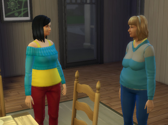
Amaya has her mother’s nose and her mother’s eyes, but something about her makes me see her as a younger version or Grandma Cora.

Amaya does her homework while Grandma Cora watches. Shane’s microscope slide photos are on the wall behind them. For some reason, when he worked from home, he was asked to collect a slide for the microscope. They make nice pictures.

Grandma left the table and good little Amaya is still doing her homework. Seeing those pictures on the wall make Amaya want to go into science like her dad.
#sims 4#simblr#homework#microscope slides#botanist#amaya donahue#coraline donahue#inspiring artwork#the donahues
2 notes
·
View notes
Text
THERE IS NEVER AN AGE AT WHICH YOU OUTGROW GIGGLING AT HANDLING SLIDES OF BULL SPERM.
2 notes
·
View notes