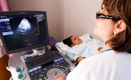
#cardiac sonography
Text

studying for an exam :)
#cardiac sonography#cardiac sonography student#cardiachealth#sonography#sonography school#ultrasound#echocardiography#health care student#healthcare#health#healthcareworkers
9 notes
·
View notes
Text
i wish i didnt need like, food and housing otherwise culinary school would be crazy cool
#i'll live cardiac sonography makes money#but grrrrrrr i wanna cook#im like that rat#you know#skyler posting
7 notes
·
View notes
Text

Made a lil something today for the sonography girlies ✨🐶🔊
#silence of the lambs#sonography#ultrasound#echocardiography#sonographer#cardiac sonographer#diagnostic medical sonography
3 notes
·
View notes
Text
Cardiac Sonography Diploma Program at Pharma-Medical College
Explore our comprehensive Cardiac Sonography Diploma program at Pharma-Medical College. Prepare for a rewarding career in diagnostic cardiac sonography with hands-on training and expert faculty. Visit our website to learn more and kickstart your journey in the world of cardiovascular healthcare.
Join us today - https://pharmamedical.ca/programs/medical-health/diagnostic-cardiac-sonography/
0 notes
Text
Sound Waves Unveiling Health: Exploring the Benefits of Sonography
Introduction:
Sonography, also known as ultrasound imaging, has revolutionized the field of medical diagnostics, offering a safe, non-invasive, and versatile method of visualizing internal structures within the human body. By utilizing high-frequency sound waves, sonography provides valuable diagnostic information across various medical specialties. In this blog post, we will explore the numerous benefits of sonography and how it continues to improve patient care and outcomes.
1. Non-Invasive and Radiation-Free Imaging:
One of the most significant advantages of sonography is its non-invasive nature. Unlike imaging techniques such as X-rays or CT scans, sonography does not involve ionizing radiation. Instead, it uses harmless sound waves to create images, making it a safe and preferred option for patients, including pregnant women and children. The absence of radiation exposure allows for repeated examinations without health risks, ensuring ongoing monitoring and effective treatment planning.
2. Real-Time Imaging and Dynamic Evaluation:
Sonography provides real-time imaging, allowing healthcare professionals to visualize the movement and functioning of internal structures. This dynamic evaluation is particularly beneficial for assessing organs, blood flow, and fetal development. Real-time sonography enables immediate feedback, aiding in the diagnosis and monitoring of conditions, such as cardiac abnormalities, gastrointestinal disorders, and musculoskeletal injuries. It also helps guide interventions, such as needle biopsies or aspirations, with precision and accuracy.
3. Versatility and Wide Range of Applications:
Sonography is a versatile imaging modality used across various medical specialties. It can examine multiple systems in the body, including the abdomen, pelvis, cardiovascular system, musculoskeletal system, and reproductive organs. Obstetric sonography allows monitoring of fetal growth and development during pregnancy. Additionally, specialized forms of sonography, such as Doppler ultrasound, can assess blood flow, detect vascular abnormalities, and aid in the diagnosis of deep vein thrombosis, arterial stenosis, or organ perfusion.
4. Safe Imaging Method for Pediatric Patients:
Sonography plays a crucial role in pediatric healthcare due to its safety and non-invasiveness. It is often the preferred imaging method for infants and children, as it does not require sedation or expose them to radiation. Sonography can be used to evaluate congenital anomalies, monitor growth and development, assess abdominal or urinary tract issues, and guide procedures like drainage or biopsy. Its child-friendly nature helps alleviate anxiety and discomfort for young patients and their families.
5. Cost-Effective and Widely Accessible:
Compared to other imaging techniques, sonography is generally more cost-effective, making it accessible to a wider range of patients. It utilizes readily available equipment and does not require expensive contrast agents or specialized facilities. Sonography can be performed at various healthcare settings, including clinics, hospitals, and even mobile units, facilitating timely and convenient access to imaging services. This accessibility contributes to early detection, accurate diagnosis, and improved patient outcomes.
6. Collaboration with Other Imaging Modalities:
Sonography often complements other imaging modalities, such as CT scans or MRI, by providing real-time guidance for procedures or confirming initial findings. It can help determine the need for further investigations or guide interventions, such as needle aspirations or biopsies. The combination of sonography with other imaging techniques enhances diagnostic accuracy, streamlines patient management, and improves treatment planning.
Conclusion:
Sonography has transformed medical diagnostics, offering a safe, non-invasive, and versatile imaging modality with numerous benefits. Its real-time imaging, non-ionizing radiation, versatility, and accessibility make it an indispensable tool in healthcare. Sonography aids in the diagnosis, monitoring, and treatment of various conditions, ensuring better patient outcomes and improved quality of care. As technology continues to advance, the future of sonography holds even
It is all about the benefits of sonography also known as ultrasound imaging. Searching for the best sonography center in Indore is one of the best options for you because ultrasound imaging has many benefits over other diagnostic methods. It is noninvasive and non-radiating technology that provides real time imaging for the inner body part that is expected to be location of a disease, thus it is one of the best diagnostic tools.
#sonography services in indore#ultrasound imaging in indore#diagnostic sonography in indore#sonography centers in indore#ultrasound technology in indore#sonogram facilities in indore#obstetric ultrasound in indore#abdominal ultrasound in indore#musculoskeletal ultrasound in indore#cardiac ultrasound in indore
0 notes
Note
What are you taking in school?
I'm in a program for cardiac sonography :)
4 notes
·
View notes
Text
Hepatic Arterial Infusion Radiation of 5-gelatin with regard to Individuals along with Unresectable Hard working liver Metastases through Intestinal tract Most cancers Refractory to Standard Systemic Radiation: A Multicenter Retrospective Research
The end result demonstrated that the growth regarding bacterial cells along with the creation of alpha-amylase on a wheat or grain bran substrate could possibly be portrayed by simple versions including the actual mathematical concept of every single cycle as well as the alternative in dried up substrate excess weight over the incubation time. Fresh information obtained from the series of set fermentations were utilized for you to verify the actual types offered. In both cases, the particular design simulator harmonized nicely together with the trial and error studies, making it very easy to conclude the types could be successfully used for the introduction of development as well as alpha-amylase creation throughout solid-state fermentation techniques. (C) The year 2010 Elsevier B.Sixth is v. All rights earmarked.Aspires: The actual INSPIRON-I tryout can be a first-in-man look at the security as well as effectiveness from the Inspiron drug-eluting stent, a new sirolimus-eluting stent together with abluminal biodegradable polymer bonded coating as well as thin cobalt-chromium blend. Approaches along with benefits: This is the randomised, multicentre comparability among Inspiron as well as a stent sticking with the same steel structure however with out polymer layer or medicine elution (Cronus). The key objective was to measure the in-segment overdue decline (LLL) at 6 months. Secondary endpoints incorporated per cent in-stent impediment since assessed by simply intravascular sonography (IVUS) with #Link# half a year as well as significant unfavorable cardiac activities (MACE). Fifty-eight individuals have been enrollment (60 lesions), 22 with regard to Inspiron as well as 19 pertaining to Cronus. Standard specialized medical as well as angiographic qualities regarding equally organizations were similar. In half a year, the particular in-segment LLL has been lowered in the Inspiron group when compared to handle party (3.Nineteen +/- 3.16 millimeters as opposed to. 0 #Link# .Fifty eight +/- 0.Some mm, correspondingly; s less space-consuming than 0.001), and also the pct neointimal impediment (Seven.8-10 +/- Seven.1% versus. Twenty six.5 +/- Eleven.4%; r smaller compared to 3.001). In two-year follow-up, likelihood of MACE ended up being comparable in between groups (Seven.In search of compared to. 21 years of age.1%, correspondingly; p=0.Something like 20), using reduced target patch revascularisation regarding Inspiron (2 as opposed to. Twenty one.1%, respectively; p=0.01) no stent thrombosis. A conclusion: Sirolimus eluted from a great abluminal naturally degradable polymer bonded over a cobalt-chromium alloy proven good at lowering restenosis with few months.Although peroxisome proliferator-activated receptor agonists are generally approved to boost cardio risk factors, their particular cardiovascular safety factors are dubious. We all therefore reviewed your materials to distinguish milestone randomized manipulated studies considering the effects of peroxisome proliferator-activated receptor gamma agonists (pioglitazone along with rosiglitazone), alpha agonists (fenofibrate along with gemfibrozil), and also griddle agonists (bezafibrate, muraglitazar, ragaglitazar, tesaglitazar, and aleglitazar) on aerobic outcomes. Pioglitazone may well slightly reduce heart activities but additionally might increase the probability of kidney cancer malignancy. Rosiglitazone boosts the #Link# risk of myocardial infarction and has been recently pulled in Eu along with constrained in america. Fibrates boost cardio results simply throughout choose subgroups: fenofibrate within diabetic patients along with metabolism syndrome, gemfibrozil in sufferers with dyslipidemia, as well as bezafibrate within patients using diabetes or perhaps metabolic affliction.
1 note
·
View note
Text
"Sonography: Understanding the Procedure and Pricing"

Introduction:
In the realm of diagnostic medicine, sonography stands out as a versatile and invaluable tool, offering a non-invasive glimpse into the intricate structures within our bodies. Whether it's monitoring fetal development during pregnancy or investigating potential health concerns in various organs, sonography has become an integral part of modern healthcare. This blog aims to delve into the world of sonography, exploring its procedures and shedding light on the crucial aspect of price considerations.
Understanding Sonography:
**Sonography, or ultrasound imaging, utilizes high-frequency sound waves to generate real-time images of the inside of the body.** The procedure involves a transducer, a small hand-held device, which emits sound waves and captures the echoes as they bounce back. These echoes are then translated into visual images, providing a detailed view of organs, tissues, and blood flow. One of the key advantages of sonography is its non-invasive nature, making it a safe and widely used diagnostic technique.
Common Types of Sonography Procedures:
1. Obstetric Sonography:
- This type of sonography is well-known for monitoring fetal development during pregnancy. It helps assess the health and growth of the fetus, detect any abnormalities, and determine the baby's gender.
2. Abdominal Sonography:
- Abdominal sonography is used to examine the organs in the abdominal cavity, such as the liver, gallbladder, kidneys, and pancreas. It aids in identifying abnormalities, including tumors or cysts.
3. Pelvic Sonography:
- Pelvic sonography is commonly employed to assess the health of the reproductive organs in both males and females. It can detect issues like ovarian cysts, fibroids, or prostate abnormalities.
4. Vascular Sonography:
- Focusing on blood vessels, vascular sonography helps in diagnosing conditions such as blood clots, aneurysms, or arterial blockages.
5. Cardiac Sonography:
- Also known as echocardiography, this type of sonography evaluates the structure and function of the heart. It aids in detecting heart conditions and assessing overall cardiac health.
**Factors Influencing Sonography Price:**
The cost of a sonography procedure can vary significantly, and understanding the factors influencing these prices is crucial for making informed decisions about healthcare expenditures.
1. Type of Sonography:
- Different types of sonography procedures come with varying levels of complexity. Obstetric sonography, for instance, may be more routine compared to a detailed vascular or cardiac sonogram, influencing the overall cost.
2. Geographical Location:
- The cost of healthcare services often varies based on the region. Urban areas and well-established medical facilities might have higher prices due to increased operational costs.
3. Facility Reputation:
- Renowned hospitals and diagnostic centers with a reputation for accuracy and reliability may charge higher prices. The assurance of quality service can be a significant factor for many individuals.
4. Technology and Equipment:
- Facilities equipped with state-of-the-art ultrasound machines and advanced technology may charge more for their services. However, the higher cost may be justified by the improved precision of the imaging.
5. Additional Services:
- Some sonography centers offer additional services, such as consultations with specialists, 3D imaging, or extended reports. These extras can contribute to an increase in overall test prices.
Navigating Sonography Prices:
1. Government Healthcare Facilities:
- Government hospitals and clinics often provide sonography services at subsidized rates or even for free. This makes them an accessible option for those on a tight budget.
2. Private Diagnostic Centers:
- Private diagnostic centers offer a range of sonography services, and their prices can vary. It's advisable to research multiple centers to find a balance between affordability and quality.
3. Hospital Sonography Departments:
- Many hospitals have dedicated sonography departments, providing integrated services with other medical specialties. While prices may be slightly higher, the convenience and comprehensive healthcare approach can be beneficial.
4. Specialized Sonography Clinics:
- Specialized clinics focusing solely on sonography may offer competitive prices, especially if they operate efficiently with lower overhead costs.
Srishti Hospital and Dr. Mamta Gupta for IVF Treatment
Srishti Hospital in Jaipur, Rajasthan is a leading fertility and IVF center under the direction of Gynecologist, Obstetrician and IVF Specialist Dr. Mamta Gupta. With over 21 years of experience helping couples conceive, Dr. Mamta Gupta offers comprehensive fertility treatments including IVF, IUI, ICSI, egg/embryo freezing, egg donation, and gestational surrogacy.
As both a gynecologist and infertility specialist, Dr. Gupta provides science-based care paired with emotional support throughout the challenges of infertility treatment. Srishti Hospital has state-of-the-art IVF labs and nursing staff to monitor progress every step of the way. From your first injection to your embryo transfer, you can trust Dr. Mamta Gupta and her team at Srishti Hospital for your best chance at successful conception through IVF.
0 notes
Text
THE ROLE OF ULTRASOUND IN MODERN MEDICINE: BEYOND PREGNANCY
Ultrasound technology has revolutionized modern medicine, offering non-invasive imaging capabilities that have transcended its traditional association with pregnancy. While it remains an indispensable tool in obstetrics, allowing expectant parents to catch a glimpse of their developing baby, the utility of ultrasound extends far beyond the realm of prenatal care. In this blog post, we explore the multifaceted role of ultrasound in modern medicine and its diverse applications across various medical specialties.
Ultrasound, also known as sonography, operates on the principle of sound waves to generate real-time images of internal body structures. Unlike other imaging modalities such as X-rays or CT scans, ultrasound uses harmless sound waves, making it safe for both patients and healthcare providers. This safety profile, coupled with its portability and cost-effectiveness, has contributed to its widespread adoption across medical disciplines.
In cardiology, ultrasound plays a critical role in the diagnosis and management of heart disease. Echocardiography, a specialized form of ultrasound, allows cardiologists to visualize the heart's structure and function in exquisite detail. By assessing parameters such as ejection fraction, valve function, and chamber dimensions, echocardiography aids in the diagnosis of conditions such as heart failure, valvular disorders, and congenital heart defects. Additionally, stress echocardiography enables clinicians to evaluate cardiac function during exercise, providing valuable insights into coronary artery disease.
Beyond cardiology, ultrasound finds applications in numerous other specialties, including gastroenterology, urology, and musculoskeletal medicine. In gastroenterology, ultrasound is used to assess liver and gallbladder pathology, guide procedures such as liver biopsies, and diagnose conditions such as pancreatitis and inflammatory bowel disease. In urology, ultrasound facilitates the evaluation of the kidneys, bladder, and prostate, aiding in the diagnosis of urinary tract infections, kidney stones, and prostate cancer. Moreover, musculoskeletal ultrasound enables precise localization of soft tissue injuries, tendonitis, and joint effusions, guiding interventions such as injections and aspirations.
One of the most exciting developments in ultrasound technology is its integration with artificial intelligence (AI) algorithms. Machine learning algorithms trained on vast datasets of ultrasound images have shown remarkable accuracy in diagnosing various conditions, often outperforming human clinicians. These AI-powered ultrasound systems have the potential to enhance diagnostic accuracy, reduce interpretation times, and improve patient outcomes across a wide range of medical specialties. From detecting breast cancer to assessing fetal well-being, AI-enhanced ultrasound holds promise for revolutionizing healthcare delivery in the 21st century.
In addition to its diagnostic capabilities, ultrasound serves as a valuable tool in interventional procedures. Ultrasound-guided interventions offer real-time visualization of anatomical structures, enhancing precision and safety. Whether performing nerve blocks, joint injections, or central line placements, clinicians rely on ultrasound to ensure accurate needle placement and minimize complications. Furthermore, ultrasound-guided biopsies enable targeted sampling of suspicious lesions, facilitating the diagnosis of cancer and other pathological conditions.
The versatility of ultrasound extends beyond clinical settings, finding applications in disaster relief efforts, remote healthcare delivery, and resource-limited settings. Portable ultrasound devices enable healthcare providers to perform rapid assessments in field hospitals, ambulances, and remote clinics, facilitating timely triage and treatment of injured individuals. Moreover, telemedicine platforms leverage ultrasound technology to connect patients with healthcare providers across geographic distances, enabling real-time consultation and diagnosis.
In conclusion, ultrasound represents a cornerstone of modern medicine, offering unparalleled versatility and utility across a diverse array of medical specialties. From its origins in obstetrics to its expanding role in cardiology, gastroenterology, urology, and beyond, ultrasound continues to redefine the practice of medicine in the 21st century. With ongoing advancements in technology and the integration of AI algorithms, the future holds tremendous promise for ultrasound as a transformative tool in healthcare delivery, enhancing diagnostic accuracy, improving patient outcomes, and expanding access to quality medical care worldwide.
Connect with us to learn more about how the AV Imaging team can help!
Orignal Source: https://av-imaging.com/the-role-of-ultrasound-in-modern-medicine-beyond-pregnancy.html
0 notes
Text
CAREER OPTIONS IN ULTRASOUND
A career in ultrasound, also known as diagnostic medical sonography, offers various opportunities for individuals interested in medical imaging and healthcare. Here are some career options in ultrasound:
Diagnostic Medical Sonographer: These professionals operate ultrasound equipment to produce images of the body’s internal structures. They work closely with physicians to assist in the diagnosis and treatment of medical conditions. There are specialties within diagnostic medical sonography, such as abdominal, obstetric and gynecological, vascular, and cardiac sonography.
Cardiac Sonographer (Echocardiographer): Specializing in imaging the heart, cardiac sonographers use ultrasound to create detailed pictures of the heart’s chambers, valves, and blood vessels. This helps in assessing heart function and identifying cardiac issues.
Vascular Technologist: Vascular technologists specialize in imaging blood vessels and blood flow to detect issues such as blood clots, arterial blockages, or venous disorders. They may work in hospitals, clinics, or vascular laboratories.
Obstetric and Gynecologic Sonographer: These sonographers focus on imaging the female reproductive system, including monitoring fetal development during pregnancy. They play a crucial role in providing information about the health of the fetus and supporting obstetricians in managing pregnancies.
Musculoskeletal Sonographer: Specializing in imaging muscles, tendons, ligaments, and joints, musculoskeletal sonographers assist in diagnosing conditions such as injuries, arthritis, or tumors affecting the musculoskeletal system.
Pediatric Sonographer: Working with children and infants, pediatric sonographers specialize in imaging pediatric organs and tissues. They collaborate with pediatricians to diagnose and monitor various medical conditions in young patients.
Research and Development: Some professionals in ultrasound pursue careers in research and development, working for companies that develop and improve ultrasound technology. They may be involved in designing new equipment, enhancing imaging techniques, or conducting clinical trials.
Education and Training: Experienced sonographers may choose to become educators, teaching ultrasound techniques and principles to students in educational institutions, hospitals, or training programs.
Management and Administration: Sonographers with experience and leadership skills may transition into managerial or administrative roles within healthcare settings, overseeing ultrasound departments or clinics.
Mobile Sonography Services: Some sonographers work for mobile ultrasound services, providing imaging services at various locations, including hospitals, clinics, and patients’ homes.
To pursue a career in ultrasound, individuals typically need formal education and training in diagnostic medical sonography, usually through an accredited program. Certification by professional organizations, such as the American Registry for Diagnostic Medical Sonography (ARDMS) or Cardiovascular Credentialing International (CCI), is often required or preferred for employment. Additionally, staying updated on advancements in technology and techniques through continuing education is essential in this field. If you really interested to build a career in ultrasound. join ultrasound certification courses in StudyUltrasound is the best option.
0 notes
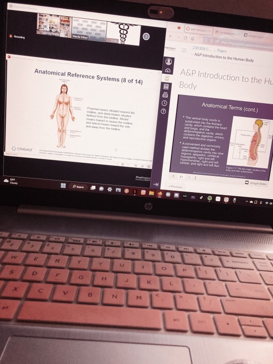
Text
this is what every morning of my life is now :)
#anatomy and physiology#anatomy#anotomical#sonography#cardiac sonography#cardiachealth#health#healthcare#healthcareworkers#ultrasound#sonographer#sonography school#cardiac sonography student#sonography student
3 notes
·
View notes
Text
Sonography: Understanding the Procedure and Pricing
Sonography: Understanding the Procedure and Pricing

Introduction:
In the realm of diagnostic medicine, sonography stands out as a versatile and invaluable tool, offering a non-invasive glimpse into the intricate structures within our bodies. Whether it's monitoring fetal development during pregnancy or investigating potential health concerns in various organs, sonography has become an integral part of modern healthcare. This blog aims to delve into the world of sonography, exploring its procedures and shedding light on the crucial aspect of price considerations.
Understanding Sonography:
**Sonography, or ultrasound imaging, utilizes high-frequency sound waves to generate real-time images of the inside of the body.** The procedure involves a transducer, a small hand-held device, which emits sound waves and captures the echoes as they bounce back. These echoes are then translated into visual images, providing a detailed view of organs, tissues, and blood flow. One of the key advantages of sonography is its non-invasive nature, making it a safe and widely used diagnostic technique.
Common Types of Sonography Procedures:
1. Obstetric Sonography:
- This type of sonography is well-known for monitoring fetal development during pregnancy. It helps assess the health and growth of the fetus, detect any abnormalities, and determine the baby's gender.
2. Abdominal Sonography:
- Abdominal sonography is used to examine the organs in the abdominal cavity, such as the liver, gallbladder, kidneys, and pancreas. It aids in identifying abnormalities, including tumors or cysts.
3. Pelvic Sonography:
- Pelvic sonography is commonly employed to assess the health of the reproductive organs in both males and females. It can detect issues like ovarian cysts, fibroids, or prostate abnormalities.
4. Vascular Sonography:
- Focusing on blood vessels, vascular sonography helps in diagnosing conditions such as blood clots, aneurysms, or arterial blockages.
5. Cardiac Sonography:
- Also known as echocardiography, this type of sonography evaluates the structure and function of the heart. It aids in detecting heart conditions and assessing overall cardiac health.
**Factors Influencing Sonography Price:**
The cost of a sonography procedure can vary significantly, and understanding the factors influencing these prices is crucial for making informed decisions about healthcare expenditures.
1. Type of Sonography:
- Different types of sonography procedures come with varying levels of complexity. Obstetric sonography, for instance, may be more routine compared to a detailed vascular or cardiac sonogram, influencing the overall cost.
2. Geographical Location:
- The cost of healthcare services often varies based on the region. Urban areas and well-established medical facilities might have higher prices due to increased operational costs.
3. Facility Reputation:
- Renowned hospitals and diagnostic centers with a reputation for accuracy and reliability may charge higher prices. The assurance of quality service can be a significant factor for many individuals.
4. Technology and Equipment:
- Facilities equipped with state-of-the-art ultrasound machines and advanced technology may charge more for their services. However, the higher cost may be justified by the improved precision of the imaging.
5. Additional Services:
- Some sonography centers offer additional services, such as consultations with specialists, 3D imaging, or extended reports. These extras can contribute to an increase in overall test prices.
Navigating Sonography Prices:
1. Government Healthcare Facilities:
- Government hospitals and clinics often provide sonography services at subsidized rates or even for free. This makes them an accessible option for those on a tight budget.
2. Private Diagnostic Centers:
- Private diagnostic centers offer a range of sonography services, and their prices can vary. It's advisable to research multiple centers to find a balance between affordability and quality.
3. Hospital Sonography Departments:
- Many hospitals have dedicated sonography departments, providing integrated services with other medical specialties. While prices may be slightly higher, the convenience and comprehensive healthcare approach can be beneficial.
4. Specialized Sonography Clinics:
- Specialized clinics focusing solely on sonography may offer competitive prices, especially if they operate efficiently with lower overhead costs.
Srishti Hospital and Dr. Mamta Gupta for IVF Treatment
Srishti Hospital in Jaipur, Rajasthan is a leading fertility and IVF center under the direction of Gynecologist, Obstetrician and IVF Specialist Dr. Mamta Gupta. With over 21 years of experience helping couples conceive, Dr. Mamta Gupta offers comprehensive fertility treatments including IVF, IUI, ICSI, egg/embryo freezing, egg donation, and gestational surrogacy.
As both a gynecologist and infertility specialist, Dr. Gupta provides science-based care paired with emotional support throughout the challenges of infertility treatment. Srishti Hospital has state-of-the-art IVF labs and nursing staff to monitor progress every step of the way. From your first injection to your embryo transfer, you can trust Dr. Mamta Gupta and her team at Srishti Hospital for your best chance at successful conception through IVF.
0 notes
Text
Paramedical Courses: A Stepping Stone to a Successful Healthcare Profession

Paramedical courses are increasingly becoming a popular choice for those interested in a career in healthcare. These courses offer a stepping stone to a successful healthcare profession, providing students with the necessary skills and knowledge to excel in various medical fields. This blog will explore some of the top paramedical courses, including medical diploma, medical lab technology, radiology & medical imaging technology, operation theatre technology, and cardiac care technology.
Medical Diploma
A medical diploma is a foundational course that introduces students to the healthcare sector. It covers a broad range of topics, including basic health facilities, emergency handling, and community healthcare. These courses are designed to equip students with the necessary skills to deal with medical emergencies and provide healthcare services to communities.
Medical Lab Technology
Medical lab technology is another crucial paramedical course that trains students to perform clinical laboratory testing in various fields such as hematology, clinical chemistry, immunohematology, microbiology, and serology.Students learn to operate state-of-the-art instruments, make specimen-oriented decisions, and report results on Laboratory Information Systems. This course provides the hands-on skills needed to become a medical laboratory technician, a role that is integral to the diagnosis, treatment, and prevention of diseases.
Radiology & Medical Imaging Technology
Radiology and medical imaging technology is a rapidly growing field in healthcare. This course develops critical thinking and problem-solving skills, knowledge of human anatomy and physiology, and the ability to use various imaging technologies. Medical imaging technologists work with patients undergoing interventional procedures, computed tomography, sonography, and magnetic resonance imaging. These professionals play a vital role in diagnosing and monitoring various health conditions, making this course a valuable stepping stone for those interested in a career in medical imaging.
Operation Theatre Technology
Operation theatre technology is a specialized course that prepares students for a career as an operation theatre technician. These professionals play a crucial role in managing the operation theatre, assisting doctors during surgeries, and ensuring the smooth functioning of all procedures.This course provides practical knowledge and skills, making it an excellent choice for those interested in a hands-on healthcare profession.
Cardiac Care Technology
Cardiac care technology is a specialized paramedical course that focuses on the diagnosis and treatment of heart diseases. This course provides students with the necessary skills to operate various cardiac diagnostic equipment, perform cardiac procedures, and provide patient care. With the increasing prevalence of heart diseases, there is a growing demand for professionals trained in cardiac care technology.
Conclusion
Lumeen Paramedical stands as a beacon of excellence in the field of paramedical education in Delhi. Offering a diverse range of top-notch courses, including Medical Diploma, Medical Lab Technology, Radiology & Medical Imaging Technology, Operation Theatre Technology, and Cardiac Care Technology, Lumeen Paramedical is committed to equipping students with the necessary skills and knowledge to excel in the healthcare sector. Recognized for their excellence, these courses have positioned Lumeen Paramedical as the provider of the best paramedical courses in Delhi, making it a top choice for aspiring healthcare professionals.
#best paramedical courses in noida#medical diploma in noida#radiology medical lab in noida#medical lab technology#b. voc in radiology medical lab#bsc.voc in medical lab technology
0 notes
Text
Exploring the World of Diagnostic Cardiac Sonography in Canada
In the ever-evolving field of healthcare, the role of diagnostic imaging is crucial for accurate diagnosis and effective patient care. One such specialized area is Diagnostic Cardiac Sonography, a discipline that combines cutting-edge technology with medical expertise to visualize the heart’s structure and function. If you’re intrigued by the prospect of a career in healthcare and have a fascination for technology and the heart, a Diagnostic Cardiac Sonography program in Canada might be your perfect choice.
To read full blog post, visit: https://pharmamedical.ca/exploring-the-world-of-diagnostic-cardiac-sonography-in-canada/
#Diagnostic Cardiac Sonography in Canada#Cardiac Sonography programs in Canada#Diagnostic Cardiac Sonography
0 notes
Text
The Benefits of Hands-On Ultrasound Training for Medical Professionals.
Introduction
The medical field is constantly evolving with advances in technology and methods. A notable advancement is the significantly increased use of ultrasounds in diagnostics and treatment planning, highlighting the importance of comprehensive ultrasound training for medical professionals.
Definition of hands-on ultrasound training
Hands-on ultrasound training is a clinically oriented training approach where medical professionals interact directly with patients under expert supervision. This method not only involves a theoretical understanding of ultrasounds but also practical application, allowing trainees to:
- Gain familiarity with ultrasound equipment.
- Enhance image acquisition and interpretation skills.
- Develop appropriate bedside manners.
Importance of ultrasound training for medical professionals
For medical professionals, undergoing an effective ultrasound training program is crucial. This comprehensive training provides them with an understanding of the topography, imaging optimization techniques, and interpretation of normal and pathological conditions which are essential in guiding diagnosis and facilitating treatment. It's a significant skillset that highly improves patient care.
Benefits of Hands-On Ultrasound Training
The importance of hands-on ultrasound training cannot be overstated; it offers clinicians a unique opportunity to refine their skills and optimize patient care. These benefits range from enhanced diagnostic abilities, improved patient care, increased confidence and proficiency, to cost-effectiveness and expanded career opportunities.
Enhanced Diagnostic Skills
Attending a hands-on ultrasound course offers a practical approach to learning. This format allows medical professionals to directly apply theoretical knowledge to real clinical scenarios, significantly enhancing their diagnostic skills.
Improved Patient Care
Ultrasound is non-invasive and risk-free, offering immediate results. Its versatility makes it a valuable tool for assessing a variety of conditions, resulting in early diagnosis and treatment, hence, improved patient care.
Increased Confidence and Proficiency
Through hands-on ultrasound training, clinicians increase their comfort and proficiency with sonography equipment. This confidence translates into improved patient care, as well-prepared professionals are less likely to make diagnostic errors.
Cost-effective Imaging Option
Ultrasound is a cost-effective imaging modality when compared to other imaging techniques like MRI and CT scans. The skills acquired during sonographer training help doctors provide quality care while simultaneously reducing healthcare costs.
Expanded Career Opportunities
An in-depth understanding and practical experience in ultrasound offer doctors an edge in the competitive medical field. Receiving sonography training can open doors to various specialties including obstetrics, musculoskeletal, cardiac, and vascular ultrasound.
Types of Hands-On Ultrasound Training Programs
Different endeavors have been implemented to address the escalating demand for skilled sonographers, including a variety of hands-on ultrasound training programs. These programs vary significantly, harmonizing with the diversity of medical practitioners.
Sonography Training Programs
These are comprehensive courses that typically entail both classroom learning and hands-on practice. Professionals gain in-depth knowledge of the principles of ultrasound and receive training on state-of-the-art equipment guided by seasoned sonographers. These programs often include:
• Training on both basic and advanced principles of sonography
• Clinical experience in various ultrasound specializations
Ultrasound Training Centers
These institutes provide a robust, hands-on training environment where medical professionals can without delay implement the theories they learn. These centers offer:
• A chance to work with a variety of patients
• Opportunities to learn the latest ultrasound techniques
Online Ultrasound Courses
With increasing technological advancements, learning has transitioned into digital platforms. These online sonography programs offer:
• Flexible scheduling for busy medical professionals
• Access to professional sonographers for virtual hands-on training.
Choosing the Right Hands-On Ultrasound Training Program
Choosing the right hands-on ultrasound training program is crucial for the enhancement of one's medical career. Key factors to consider include the training center’s accreditation, its faculty's experience, practical training opportunities, current course structure, and feedback from previous students.
Accreditation and Certification
Ensure the ultrasound training center you select is accredited and that the course leads to certification. Accreditation guarantees that the program meets established standards for curriculum, facilities, staff qualifications, and ethics. Certification indicates that a professional body recognizes the course and that it equips you with the vital skills you need in the profession.
Experienced Faculty and Staff
A high-quality ultrasound training program is characterized by knowledgeable and experienced faculty and staff. The instructors should have a good grasp of ultrasound technology and should be competent in teaching and demonstrating hands-on procedures.
Practical Training Opportunities
The ideal ultrasound training program should have a strong emphasis on practical training opportunities. This could include clinical rotations, lab work, or supervised practice sessions. Hands-on practice allows you to apply the theory you learn in class and to become competent in using ultrasound technology.
Curriculum and Course Structure
Look for a comprehensive curriculum that covers all aspects of ultrasound technology and a well-structured course that progresses logically. The course should ideally balance theory with practical sessions and provide a thorough grounding in all areas of sonography.
Feedback and Testimonials from Previous Students
Lastly, consider feedback and testimonials from previous learners to ensure the course provides positive outcomes. Past students' experiences could give valuable insights into the course's quality and effectiveness.
Real-Life Case Studies
The real-world impact of hands-on ultrasound training is evidenced in countless success stories across the global healthcare community.
Success stories of medical professionals who underwent hands-on ultrasound training
From seasoned physicians to fresh graduates, many medical professionals have found hands-on ultrasound training instrumental in enhancing their clinical skills. Some of the benefits reported include:
- Improved procedural confidence
- Increased precision in diagnosis
- Enhanced patient interaction and satisfaction
How ultrasound training improved their diagnostic abilities and patient care
The direct application of hands-on ultrasound training in medical practice is vast. Doctors and other medical professionals have remarked on how the following benefits significantly improved their patient care:
- Faster, more accurate results leading to quicker treatment.
- Reduction in the use of invasive diagnostic procedures
- Better ongoing monitoring of patient conditions.
The Future of Hands-On Ultrasound Training
The future landscape of ultrasound training holds promising prospects for medical professionals. Rapidly evolving technological advancements and growing demand are catalyzing new directions in sonographer training, enhancing both the intricacy and the efficacy of these vital services.
Technological Advancements in Ultrasound Training
In recent years, emerging innovations in technology have revolutionized the ultrasound landscape. These advancements include, but are not limited to:
- High-definition imagery: Offers superior image quality, improving diagnostic accuracy and contributing to better patient outcomes.
- 3D and 4D ultrasound: These technologies facilitate real-time visualization and examination of anatomical structures, enhancing the depth and scope of ultrasound training.
Integration of Ultrasound in Medical Specialties
Ultrasound is increasingly integrated into various medical disciplines, offering unique investigative insights for specialties like cardiology, obstetrics, and musculoskeletal medicine. As a result, flexible hands-on ultrasound courses tailored for these respective specialties are becoming increasingly crucial.
Continuing Education and Professional Development Opportunities
Many ultrasound training centers are recognizing the necessity for continual skills development in the face of advancing technology and integrating ultrasound. As such, they are offering ongoing education programs and professional development opportunities for practitioners. This approach ensures that doctors and medical professionals remain competent and updated, enhancing their ability to deliver outstanding patient care.
Conclusion
Recap of the benefits of hands-on ultrasound training for medical professionals
In summary, hands-on ultrasound training offers numerous advantages for doctors and all medical professionals. The most notable benefits include:
- Direct, experiential learning opportunities, fostering comprehensive knowledge and skill-building.
- Real-time feedback and guidance from experienced trainers.
- Enhancing diagnostic accuracy and patient care.
- Promoting confidence and proficiency in ultrasound usage.
#"ultrasound training#hands on ultrasound course#sonography training program#sonographer training#best ultrasound course#ultrasound training centre#ultrasound training for doctors#best ultrasound training centre#Hands on ultrasound centre in India
0 notes
Text
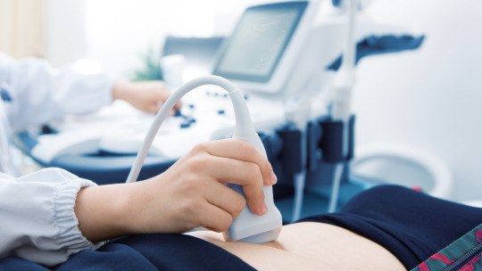
Sonography is a medical diagnostic procedure that uses sound waves to acquire real-time images inside our body without incisions. It helps physicians to look at the organs and the blood vessels and see the action of the internal organs such as cardiac movements. To get the best doctor for pregnant female sonography for better guidance and medical care. Call Now: 9399601809 Email at [email protected].
0 notes
Last Seen Blogs

naruto-ol-protags
Protags’ content archive

muitindia
Maharishi University of Information Technology

no--thx
jaclyn

thingwithasoul
Say you love me too

jemscorner
JemsCorner