#congenital hydrocephalus
Photo
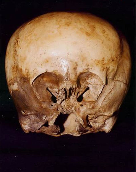
The Starchild skull
#The Starchild skull#skull#congenital hydrocephalus.#died#human skull#malformed#human skull of a child who likely died as a result of congenital hydrocephalus#It received widespread publicity after paranormalist Lloyd Pye claimed it was of extraterrestrial origin.#extraterrestrial origin#photo#photography#anatomy
35 notes
·
View notes
Text
Find Me Friday: Theo, Phil, Vincent, & Eddie!
Logo that says Reece’s Rainbow Special Needs Adoption Support in blue, below a blue & yellow paint stroke rainbow graphic with a yellow Ukrainian trident symbol on the right half.
In this series, each Friday I’m able, I want to share a different child or group of children who are available for adoption and listed through the adoption advocacy website Reece’s Rainbow. Please note, names used on…

View On WordPress
#accessible post#adoption advocacy#asthma#available for adoption#available to large families#available to married couples#available to older parents#available to single parents#bronchopulmonary dysplasia#cognitive delay#congenital STD#cortical blindness#Eastern Europe adoption#education#Edwards syndrome#esophogeal atresia#Find Me Friday#FSP#g-tube#gastric reflux#heart defect#hydrocephalus#low birth weight#medical history#Phil#photo descriptions#polymalformative syndrome#premature birth#psychomotor delay#Reece&039;s Rainbow
0 notes
Text
I have personally witnessed so many instances of pet owners forcing beloved animals to continue living far beyond the point of their quality of life hitting zero that the whole "Deux Face" situation was absolutely nothing new to me, except for the wave of genuine nausea I experienced at the social media reaction to her existence.
I can honestly say that I feel a palpable sense of relief, knowing that that calf is no longer being forced to live in that condition, and there's no amount of "well, she had vet care!" or "it doesn't seem like she's in pain!" that would have mollified me. I have been in hundreds of QoL consults with clients who parrot the exact same sentiments while their animal lies, completely recumbent and unresponsive, on the examination table. It's the exact same thing, except compounded by the ghoulish addition of the calf's value as a sideshow act.
Any normal presenting newborn calf that failed to thrive as she did would have been euthanized on welfare grounds long before the 26 day mark. A dairy calf is literally supposed to be able to walk away from its birthing site, a calf whose only developmental milestone was that it "can kinda lift its head" is a calf that has something inherently wrong with it. If Deux Face wasn't deformed in a way that makes well-meaning assholes spout that stupid poem, then she probably would have been put out of her misery ages ago. I keep thinking of the grotesque congenital deformities that domestic cattle can present with, twisted spines and fused joints and extreme hydrocephalus and cleft palates, and how, if Deux Face had presented with QoL reducing examples of any of those conditions, people wouldn't have even batted an eye at her euthanasia. She was literally forced to keep on living, just so people could keep "consuming" her continued existence. She was forced to live, not because it was to her benefit, but for the benefit of the farm, who romanticised and profited from her, and the benefit of idiots who think a goddamn newborn calf should be a source of inspiration in their own lives.
The people on this site who mourned that we didn't get to gawk at this animal a little longer disgust me. The only difference between you and the woman who keeps her 19 year old constant DKA, cushingoid, and severely arthritic Shih Tzu alive is that she, at least, has the excuse that this is an animal she has loved and cared for for decades. She's blinded by love, and needs to be counselled towards the realisation that the dog's existence is now more for her benefit than his. You're just upset that there are no more juicy pics of a recumbent, half-dead calf that you can caption with "TWice aS MaNY STArs As UsuaL!!!!!" in goofy ass fonts.
The only sad thing about Deux Face's death is that it took so long. Fight me on it, I don't care. Your gross parasocial relationship with a dying farm animal you've never met was not worth that animal's enforced suffering.
#vet stuff#i guess#dunno if i should tag this as deux face#bc im pretty sure if the people in complaining about find this post#theyll take my skin
366 notes
·
View notes
Text
September is Chiari Malformation Awareness Month!
Hi yes hello! Since many people probably aren't aware of it, I wanted to bring attention to something we ourselves have called chairi malformation at the start of its awareness month! Its recognized with a purple ribbon usually with a zipper on it (for the zipper scars of those who've had Chiari surgery)
What is Chiari Malformation?
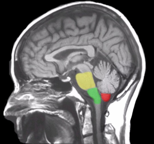
Chiari (key-arr-ee) Malformation is a brain malformation in which the brain is too large, skull is too small, or some combination on the both, causing the cerebellar tonsils (and in some instances the brain stem) to slip through the skull and into the spinal chord.
Chiari is most typically a congenital effect. There are two main types (though they aren't the only ones). The most common of the two us Chiari 1, in which only the cerebellar tonsils are descended through the skull. The second most common, Chiari 2 (also known as Arnold-Chiari malformation) has more tissue herniation in the cerebellar tonsils and even the cerebellum, as well as brain stem herniation as well.
(See Below, the Cerebellar tonsils are marked in red while the brainstem is marked in green and yellow. This is considered a normal MRI)
Chiari malformation is likely to occur in 1 in 1,000 people, making it uncommon but not rare. The statistics are likely to be slightly higher than that for Chiari 1, as many people don't present symptomatically (and many incidents are only found in cases where the person was receiving radiological imaging for other instances such as head injury, so many people are unaware they had Chiari to begin with).
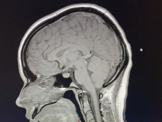
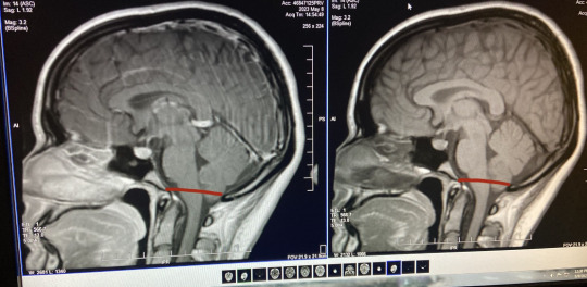
The only way to diagnose Chiari is through radiological imaging (many arguing upright MRI specifically is the only proper way to view the real level pf herniation). Herniation is measured down from the McRae line to the lowest point on the cerebellar tonsils. Depending on the accuracy of the machine (and which imaging tool is used) herniation can appear at different levels at different times. (See below, my first MRI looks markedly less in comparison to my second MRI, which features a roughly drawn on McRae line. In the second image I was noted to have a 7-8mm herniation.)
Symptoms
Chiari is marked by a number of symptoms and commorbidities, even moreso depending on the type you have. Symptoms can occur at any level of herniation. Some people with Chiari can have a 3mm descent and have debilitating symptoms, while some may have a 15 mm descent and be completely asymptomatic. The most common of these is occipital headaches & migraines, ranging from mild to severe, but many more are possible. These range from...
Balance Issues
Dizziness & Vertigo
Neck & Shoulder Pain
Difficulty Swallowing
Sore Throat
Sleep Apnea
Nausea & Vomitting
Tinnitus & Hearing Loss
Blurred Vision, Visual Snow, & Vision Loss
Muscle Weakness
Numbness or Pins & Needles (Caused by Nerve Damage)
Poor Motor Skills
Fatigue
Cognitive Difficulties (including but not limited to Brain Fog, Memory Problems, Confusion, & Difficulty Speaking)
Insomnia
Photophobia/Light Sensitivity
Syncope, Fainting, & Drop Attacks
Seizures
Dysautonomia
Since the cerebellar tonsils block the opening to the base of the skull, Chiari can halt the proper flow of CSF (Cerebral Spinal Fluid) between the brain and the spinal chord. Because of this, Syringomylia (cysts filled with CSF formed on the spine called Syrinxs) is considered common with Chiari. Other common disorders with Chiari are Scoliosis, EDS & Cervicocranial Instability, POTS, Tethered Spinal Chord Syndrome, Spina bifida, & Hydrocephalus.
So What's the Solution?
Well, the only known solution for Chiari as of right now is surgery. This surgery is called posterior fossa decompression-- in which a small portion of the base of the skull is removed from the Chiari patient to relieve pressure and give more room for the brain. The surgeon can then do for sone patients a duraplasty, in which the dura (or opening of the brain) is cut open and a patch of tissue is sewed into the incision to make the dura bigger and give even more room for the brain. Surgery can also be done as a preventative measure against syrinxes for those without them. In the case the patient also has a syrinx, more surgical procedure can be done to drain the cyst. In patients with EDS, special procedures must be made to avoid surgical complications and making things worse.
Surgery is not guaranteed to completely alleviate symptoms, but typically helps with some. However, due to large misunderstanding and disagreement on proper diagnostic traits of Chiari from doctors (most typically neurologists and neurosurgeons) many may be denied surgery for a number of years, and Chiari Diagnosis can take on an average of 4 years to officially receive.
Some go years experiencing symptoms and having "low lying cerebellar tonsils" (or similar language, such as incidental tonsillar ectopia) noted on their radiology reports without doctors officially recognizing it as Chiari. In this time many are misdiagnosed with other disorders such as chronic headaches, multiple sclerosis, fibromyalgia, and more before finally finding a doctor who will listen. Many will brush off the radiological findings as just a difference in your brain being formed at birth before admitting the symptoms can be due to Chiari. It can take years of your own patient advocacy before someone finally listens.
This is why awareness to it is so important, in hopes of reaching other people and doctors and forming a stronger understanding of the condition from information found by experts on it and those with Chiari themselves. With more awareness comes more accessibility to treatment and surgery so those who are symptomatic can hopefully find some relief. So this month send a little love & luck to those with Chiari!
#chiari#chiari malformation#chiari awareness#neurological disability#disabled#disability#disability awareness
34 notes
·
View notes
Text

Restless Foot Syndrome
Central Processing Unit Syndrome
Asperger Syndrome
Chronic Intestinal Pseudo Obstruction
Mowat Wilson Syndrome
Pitt Hopkins Syndrome
Charge Syndrome
Oppositional Defiant Disorder
Progressive Disease
Trisomy
Shortened Achilles Tendon
Arterial Venous Malformation
Schizophrenia
Anxiety
Stormme Syndrome
Panhypopituitarism
Adrenal Insufficiency
Bicuspid Aortic Valve
Chiari Malformation
Sensorineural Hearing Loss
Unknown Genetic Syndrome
Congenital Cytomegalovirus Infection
Catastrophic Epilepsy
Chromosomal Condition
Arthogryposis
Charcot Marie Tooth Disease
Disease Tuberose Sclerosis
End Stage Heart and Lung Disease
Spina Bifida
Hydrocephalus
Shaken Baby Syndrome
Cornelia De Lange Syndrome
Parry Romberg Syndrome
3 notes
·
View notes
Text
Brain Aneurysm: Causes, Symptoms, Risk Factors, and Treatment Options

A brain aneurysm, also known as an intracranial or cerebral aneurysm, occurs when a weak spot in the wall of a blood vessel in the brain bulges and fills with blood. It resembles a small, berry-shaped sac hanging from the artery. When a brain aneurysm ruptures or leaks, it can cause a life-threatening condition known as a hemorrhagic stroke, which requires immediate medical attention.
Causes of Brain Aneurysm
Brain aneurysms often develop in the arteries located at the base of the brain, especially at branching points where arterial walls are weakest. These weak areas are more susceptible to rupture.
Signs and Symptoms of a Brain Aneurysm
Common signs and symptoms include:
Severe headache
Nausea and vomiting
Stiff neck
Double or blurred vision
Sensitivity to light
Seizures
Drooping eyelids
Loss of consciousness
Confusion
A leaking aneurysm may cause a sudden, severe headache, while an unruptured aneurysm can cause localized pain, double vision, and facial numbness.
Risk Factors for Brain Aneurysm
Certain factors can increase the likelihood of developing a brain aneurysm, including:
Age (more common in adults)
Gender (women are more susceptible)
Smoking
High blood pressure (hypertension)
Drug use (e.g., cocaine)
Excessive alcohol consumption
Genetic conditions like Ehlers-Danlos syndrome
Polycystic kidney disease
Congenital conditions, such as brain AVM (arteriovenous malformation)
Family history of aneurysms
Complications from a Brain Aneurysm
When a brain aneurysm ruptures, it causes bleeding that can damage or destroy surrounding brain cells, increasing pressure inside the skull. This disruption of blood and oxygen flow can result in unconsciousness or even death. Other complications may include:
Rebleeding
Vasospasm (narrowing of blood vessels)
Hydrocephalus (fluid buildup in the brain)
Hyponatremia (low sodium levels)
Diagnosis of Brain Aneurysm
At Yashoda Hospital and Research Centre, Ghaziabad, various diagnostic tools are used to detect brain aneurysms, especially in patients with a family history of the condition. These may include:
CT Scan: Produces 3D images of the brain.
MRI: Uses radio waves and magnetic fields to capture brain images.
Cerebrospinal Fluid Test: Checks for blood in the fluid surrounding the brain and spine.
Cerebral Angiogram: Detects aneurysms by imaging the brain's blood vessels.
Treatment for Brain Aneurysm
Treatment options depend on factors like the size, location, and whether the aneurysm has ruptured. Urgent treatment is required for ruptured aneurysms, with several surgical options available:
Surgical Clipping: A metal clip is applied at the base of the aneurysm to halt blood flow.
Endovascular Coiling: A catheter is inserted into the artery, and tiny platinum coils are placed inside the aneurysm to block blood flow.
Flow Diverter Surgery: A stent is placed in the artery to redirect blood flow away from the aneurysm.
Symptom Management and Preventive Care
To manage symptoms and prevent complications, doctors may recommend:
Pain relief medications (e.g., acetaminophen) for headaches
Calcium channel blockers to prevent blood vessel narrowing
Anti-seizure medications for seizures
Drainage procedures (ventricular or lumbar catheters) to reduce pressure on the brain
Shunt surgery for fluid drainage
Rehabilitative therapy to help regain lost skills due to brain damage
By recognizing the signs and symptoms early, and understanding the risk factors, timely intervention can prevent life-threatening complications from a brain aneurysm.
Conclusion:
Early diagnosis is key to effectively treating a brain aneurysm. Being vigilant and recognizing the warning signs can make a significant difference. Immediate medical attention and timely intervention for a ruptured aneurysm greatly improve survival rates and recovery outcomes. While recovery from an unruptured aneurysm is typically fast, a ruptured aneurysm may require multiple surgeries, and the recovery period can vary based on the extent of damage. Prompt action is crucial for better long-term prognosis.
Dr. (Brig.) Yadvendra Singh Sirohi is a highly distinguished neurologist with over 24 years of experience, having trained and excelled at leading national institutions. Known for his clinical expertise, compassion, and commitment, he serves as a Senior Consultant in Neurology at Yashoda Hospital & Research Centre, Ghaziabad. Dr. Sirohi is adept at managing a wide range of neurological conditions with exceptional skill and care.
Dr. Shishir Kumar is a highly skilled neurosurgeon, neuro interventionist, and endoscopic spine surgeon with over 9 years of experience in the Delhi/NCR region. He has completed fellowships in neuro intervention, endovascular surgery, and minimally invasive spine surgery. With his extensive training, Dr. Kumar provides comprehensive neurosurgical care, focusing on patient education and a collaborative treatment approach. He currently practices at Yashoda Hospital & Research Centre, Nehru Nagar, Ghaziabad.
Dr. Atul Gupta is a highly experienced neurosurgeon with 29 years of expertise. He specializes in complex procedures, including brain tumor surgeries, cerebrovascular surgeries, epilepsy treatments, and spine surgeries.
Dr. Puneet Malik is a skilled neurosurgeon specializing in complex brain and spinal cord surgeries, particularly for patients experiencing severe numbness. He is adept at procedures such as clot removal, tumor excision, and halting brain bleeding, all performed with exceptional precision and care.
0 notes
Text
Neurosurgeon in Gaya and Bihar — Arsh Hospital Expertise in Neurosurgery

Arsh Hospital Neurosurgery Department
Located in Gaya, Arsh Hospital is renowned for providing specialized neurosurgical care that is both accessible and effective. The hospital’s Neurosurgery Department is equipped with state-of-the-art diagnostic and surgical equipment, allowing doctors to perform intricate procedures with precision. From routine brain surgeries to emergency trauma care, Arsh Hospital covers all aspects of neurosurgical treatment under one roof, setting it apart as a leading center for neurosurgery in Bihar.
Top Neurosurgeons at Arsh Hospital, Gaya
Arsh Hospital is home to some of the best neurosurgeons in Bihar, each with years of experience and specialized training.
Dr. Ranjan Kumar Jena is one of the most respected neurosurgeons in the region, with expertise in treating complex brain and spinal conditions. His meticulous surgical skills and deep knowledge have earned him a stellar reputation.
Dr. Ashish Kumar Jha, another leading neurosurgeon at Arsh Hospital, specializes in minimally invasive neurosurgical procedures and has successfully treated numerous patients suffering from spinal and brain disorders.
Both of these specialists, along with other qualified neurosurgeons, work together to provide a comprehensive range of services aimed at improving patient outcomes.
Common Neurosurgical Conditions Treated
At Arsh Hospital, the neurosurgery team handles a variety of neurological conditions, including:
Brain Tumors and Cranial Injuries: From benign tumors to malignant brain cancers, Arsh Hospital’s neurosurgeons are skilled in performing delicate brain surgeries.
Spinal Cord Disorders and Injuries: Patients suffering from herniated discs, spinal fractures, or degenerative spinal diseases are treated using the latest surgical techniques.
Cerebrovascular Diseases and Stroke Management: The hospital offers advanced treatment for stroke patients, including emergency intervention and post-stroke care.
Pediatric Neurosurgery: Children with congenital neurological disorders, such as hydrocephalus, receive specialized care from the pediatric neurosurgery team.
Advanced Neurosurgical Procedures Offered
Arsh Hospital is known for its ability to perform advanced neurosurgical procedures, some of which include:
Minimally Invasive Brain Surgery: Using cutting-edge techniques, surgeons at Arsh Hospital can operate on brain tumors and other conditions with minimal incisions, leading to quicker recovery times.
Spinal Fusion and Decompression Surgery: These surgeries are commonly performed on patients with chronic spinal issues, offering long-term pain relief and improved mobility.
Aneurysm Clipping and Endovascular Procedures: For patients with aneurysms or vascular malformations, the hospital provides both surgical and non-surgical interventions.
Neurotrauma Care at Arsh Hospital
Emergencies such as traumatic brain injuries require immediate attention, and Arsh Hospital’s 24/7 emergency neurotrauma services are designed to handle these critical situations. The hospital is equipped with a state-of-the-art Intensive Care Unit (ICU) that offers round-the-clock monitoring and care for patients recovering from neurotrauma.
Post-Surgical Care and Rehabilitation
Recovery after neurosurgery is a critical phase, and Arsh Hospital takes a holistic approach to post-surgical care. Patients are provided with personalized rehabilitation programs, including physical therapy and occupational therapy, to help them regain strength and function. The goal is to offer a smooth and steady recovery that allows patients to return to their normal lives.
Technology and Infrastructure Supporting Neurosurgery
At Arsh Hospital, neurosurgeons are supported by advanced imaging and diagnostic tools such as MRI, CT scans, and intraoperative monitoring systems. These technologies enable precise diagnosis and guide surgeons during complex procedures. In addition, the hospital’s surgical suites are equipped with the latest medical devices, ensuring high success rates and minimal complications during surgery.
Why Choose Arsh Hospital for Neurosurgery in Bihar?
There are several reasons why patients choose Arsh Hospital for Neurosurgeon in Bihar:
Multidisciplinary Approach: The hospital takes a team-based approach to patient care, with neurosurgeons working closely with neurologists, radiologists, and other specialists.
Expertise and Experience: With renowned neurosurgeons like Dr. Ranjan Kumar Jena and Dr. Ashish Kumar Jha leading the department, patients are in the hands of experts.
Compassionate Care: Beyond technical expertise, the hospital is known for its compassionate approach to patient care, ensuring that each patient feels supported throughout their treatment.
Success Stories and Patient Testimonials
Patients treated at Arsh Hospital have shared heartwarming stories of recovery and success. Many have praised the hospital’s expertise, state-of-the-art facilities, and the dedication of its medical staff. These testimonials reflect the hospital’s commitment to providing high-quality neurosurgical care.
Future of Neurosurgery at Arsh Hospital
As the demand for neurosurgical services continues to grow in Bihar, Arsh Hospital plans to expand its neurosurgery department further. By investing in research, training, and the latest technology, the hospital aims to remain at the forefront of neurosurgical innovation in the region.
How to Book a Consultation with a Neurosurgeon
Booking a consultation at Arsh Hospital is straightforward. You can contact the hospital directly through their helpline or schedule an appointment online via their website. The hospital’s team will assist you in getting a consultation with the best available neurosurgeon based on your needs.
Conclusion
For anyone in Gaya or Bihar seeking specialized neurosurgical care, Arsh Hospital offers an unmatched level of expertise, cutting-edge treatments, and compassionate care. Whether you need emergency neurotrauma care or complex brain surgery, the hospital’s team of experienced neurosurgeons is equipped to handle it all.
0 notes
Text
Pediatric Neurosurgeon in Pimpri Chinchwad: Expert Care at Neuronest Clinic
When it comes to the health and well-being of children, specialized medical care is crucial. Pediatric neurosurgery, which focuses on diagnosing and treating neurological disorders in children, requires a high level of expertise and compassion. Neuronest Clinic in Pimpri Chinchwad is a leading healthcare provider in this field, offering world-class pediatric neurosurgical care. This blog explores the importance of pediatric neurosurgery and why Neuronest Clinic is the top choice for parents seeking the best care for their children.
Understanding Pediatric Neurosurgery
Pediatric neurosurgery involves the surgical treatment of neurological disorders in children, from newborns to adolescents. These conditions can range from congenital anomalies and brain tumors to epilepsy and spinal disorders. The complexity of these cases requires a specialized approach, tailored to the unique needs of young patients whose bodies and brains are still developing.
Why Choose Neuronest Clinic for Pediatric Neurosurgery?
Neuronest Clinic is dedicated to providing exceptional care for children with neurological conditions. Here are the key reasons why it stands out as the premier choice for pediatric neurosurgery in Pimpri Chinchwad:
1. Expert Pediatric Neurosurgeons
Neuronest Clinic boasts a team of highly skilled and experienced pediatric neurosurgeons. These specialists have undergone extensive training in both neurosurgery and pediatrics, equipping them with the knowledge and skills needed to address the unique challenges of treating children. Their expertise ensures that young patients receive the best possible surgical care.
2. State-of-the-Art Facilities
The clinic is equipped with cutting-edge technology and advanced surgical tools, essential for performing precise and effective neurosurgical procedures. The state-of-the-art facilities at Neuronest Clinic enable the medical team to diagnose and treat complex neurological conditions with the highest level of accuracy and safety.
3. Comprehensive Multidisciplinary Care
Pediatric neurosurgery often requires a multidisciplinary approach. At Neuronest Clinic, a team of specialists, including neurologists, pediatricians, anesthesiologists, and rehabilitation therapists, work together to provide comprehensive care. This collaborative approach ensures that every aspect of the child’s health is addressed, from diagnosis to recovery.
4. Child-Friendly Environment
Understanding the emotional and psychological needs of young patients is crucial in pediatric care. Neuronest Clinic is designed to be a child-friendly environment, aiming to reduce anxiety and make children feel comfortable. The compassionate staff at the clinic are trained to handle pediatric patients with care and empathy, making the experience as stress-free as possible for both the children and their parents.
5. Focus on Family Support
Neuronest Clinic recognizes the importance of involving families in the treatment process. Parents are provided with detailed information about their child’s condition, the proposed surgical procedure, and the expected outcomes. The clinic also offers support services to help families cope with the challenges of dealing with a child’s neurological disorder.

Conditions Treated by Pediatric Neurosurgeons at Neuronest Clinic
Neuronest Clinic’s pediatric neurosurgeons are equipped to treat a wide range of neurological conditions in children, including:
Congenital Anomalies: Conditions such as spina bifida, encephalocele, and craniosynostosis, which are present at birth and require surgical intervention.
Brain and Spinal Tumors: Removal of benign and malignant tumors affecting the brain and spinal cord.
Hydrocephalus: Treatment of excessive accumulation of cerebrospinal fluid in the brain.
Epilepsy: Surgical options for children with drug-resistant epilepsy.
Traumatic Brain Injuries: Management of head injuries resulting from accidents or falls.
Chiari Malformations: Treatment of structural defects in the brain and spinal cord.
The Surgical Process: What to Expect
The surgical process at Neuronest Clinic begins with a thorough evaluation and diagnosis, using advanced imaging techniques to pinpoint the issue. The pediatric neurosurgeon will then discuss the recommended surgical plan with the family, explaining the procedure, potential risks, and benefits. On the day of surgery, the child will be cared for by a team of specialists, ensuring their safety and comfort throughout the process. Post-operative care includes monitoring, pain management, and rehabilitation to support the child’s recovery and long-term well-being.
Conclusion
For families in Pimpri Chinchwad seeking expert care for their children’s neurological conditions, Neuronest Clinic offers unparalleled pediatric neurosurgical services. With a team of dedicated specialists, state-of-the-art facilities, and a compassionate approach, the clinic ensures that young patients receive the best possible care. Trust Neuronest Clinic to provide the expertise and support needed to navigate the challenges of pediatric neurosurgery and help your child achieve a healthier future.
0 notes
Text
Advancing Brain Health: Neurosurgery Excellence
1. The Brain: Our Command Center
Understanding Neurosurgery
The brain—the epicenter of our thoughts, emotions, and bodily functions—requires specialized care. At Orthomed Multispeciality Hospital, their neurosurgery team combines expertise, precision, and compassion to address a wide range of brain-related conditions.
2. Comprehensive Brain Services
From Diagnostics to Treatment
Orthomed’s neurosurgeons offer a holistic approach:
Brain Tumor Surgery: Precise removal of brain tumors, whether benign or malignant.
Cerebrovascular Surgery: Treating aneurysms, arteriovenous malformations (AVMs), and strokes.
Functional Neurosurgery: Addressing movement disorders like Parkinson’s disease.
3. Minimally Invasive Techniques
Navigating Brain Pathways
Orthomed’s neurosurgery team employs minimally invasive approaches:
Endoscopic Brain Surgery: Using tiny incisions and specialized tools for tumor removal.
Stereotactic Radiosurgery: Precise radiation targeting for brain tumors.
4. Spinal Cord Surgery
Brain-Body Connection
Orthomed’s neurosurgeons specialize in spinal cord procedures:
Spinal Tumor Resection: Safely removing tumors affecting the spinal cord.
Spinal Fusion: Stabilizing the spine to alleviate pain and improve function.
5. Traumatic Brain Injury Care
Urgent Interventions
Orthomed’s neurosurgery team responds swiftly to head injuries:
Craniotomy: Surgical repair of skull fractures.
Intracranial Pressure Monitoring: Vital for severe head trauma cases.
6. Pediatric Neurosurgery
Nurturing Young Minds
Orthomed’s pediatric neurosurgeons address congenital brain conditions:
Hydrocephalus: Managing excess cerebrospinal fluid.
Spina Bifida Repair: Correcting neural tube defects.
7. Rehabilitation and Support
Beyond Surgery
Orthomed’s neurosurgery care extends to rehabilitation:
Neurorehabilitation: Tailored programs for brain injury recovery.
Pain Management Clinics: Addressing chronic pain.
8. Patient-Centric Approach
Compassion and Expertise
Orthomed Multispeciality Hospital’s neurosurgery team understands the impact of brain health on overall well-being. They collaborate with patients, families, and other specialists to provide personalized care.
Conclusion: Navigating Brain Health with Orthomed
Orthomed Multispeciality Hospital ensures that Chennai residents receive advanced neurosurgical care, empowering them to lead healthier lives—one neuron at a time.
Remember, Orthomed Multispeciality Hospital is your partner in health. Explore their services on their website here. Feel free to delve deeper into any of these topics or ask for more information! 🌟
0 notes
Text
What is Hydrocephalus? What is the Main Cause of Hydrocephalus
Medical science is complex, it includes studying topics like diseases, causes, and treatments to help people live healthy lives. One such topic that is crucial to understand in detail is Hydrocephalus.
So, let’s understand the basics about the subject and then we will discuss how you can grab in-depth information and study resources about the subject.
What is Hydrocephalus?
Hydrocephalus is a chronic neurological disorder where the fluid (Cerebrospinal fluid) builds inside the brain.
Though the Cerebrospinal fluid is already present inside the brain and acts as a caution, the excess can permanently damage the brain. It puts pressure on the skull and squashes surrounding brain tissue, which causes problems with physical and mental development. If untreated, it is usually fatal.
The condition is primarily common in infants or people older than 60. The common symptoms of hydrocephalus in infants include:
Increase in the size of an infant's head
A bulge or tense soft spot on the top of the head
Nausea and vomiting
Sleepiness or sluggishness
Poor eating
Seizures
Eyes fixed downward
Problems with muscle tone and strength.
The following are the more common symptoms of hydrocephalus among adults 60 and older:
Loss of bladder control
Memory loss
Progressive loss of reasoning skills.
Trouble walking
Poor coordination or balance.
Remember: The systems among toddlers, children, and middle-aged adults can differ.
Types of Hydrocephalus
The following are the different types of Hydrocephalus:
Congenital hydrocephalus: It is caused by a brain malformation or birth defect that causes excessive cerebrospinal fluid (CSF) to accumulate in brain cavities, called subarachnoid space.
Acquired hydrocephalus: Usually resulting from a stroke, brain tumour, meningitis, or a severe head injury.
Communicating hydrocephalus: A condition when the flow of CSF is blocked after leaving the ventricles. Because CSF can still flow between the brain’s ventricles, it is also called “communicating.”
Non-communicating hydrocephalus: Also known as obstructive hydrocephalus, this type occurs when the narrow pathways connecting the ventricles become blocked.
Normal pressure hydrocephalus: NPH affects primarily those aged 60 and up. It can arise following a stroke, injury, infection, surgery, or hemorrhage.
Hydrocephalus ex-vacuo: Occurs after a stroke, traumatic brain injury, or degenerative disease. In this type, as brain tissue shrinks, the brain's ventricles become larger.
Now that we know the symptoms and types of hydrocephalus, let's dive in to learn about the causes so we can understand the problem better.
What is the Main Cause of Hydrocephalus
There are two distinct cause categories into which hydrocephalus can be classified:
Congenital
Acquired
Congenital hydrocephalus is the result of both genetic and environmental factors working together during foetal development. The following are the main reasons for congenital hydrocephalus:
Spina bifida and other brain and spinal cord (neural tube) defects.
A narrowing of the small passage between the third and fourth ventricles of the brain (aqueductal stenosis).
Complications of premature birth, such as bleeding within ventricles.
Mom has an infection during pregnancy.
On the other hand, Acquired hydrocephalus develops after birth and can affect people of all ages. Below, we have listed the most common causes of acquired hydrocephalus:
Brain or spinal cord tumours
Head trauma
Stroke
Meningitis or other infections of your brain or spinal cord.
After gaining a basic understanding of the subject, let us give you a solution that will help you get more details of the topic: Pediatrics MD Course!

Enroll in an online pediatrics course.
Without the assistance of a professional, it can be challenging to find the study material and understand the basics of the subject.
That is why Dr. Piyush Gupta designed this course for students preparing for an MD in pediatrics.
The course includes pediatrics video lectures along with other crucial resources.
So start your journey toward a good hold on the subject, and enroll in the course now!
#pediatrics md#md in pediatrics#Dr. Piyush Gupta#online pediatrics course#pediatrics video lectures#neurological disorder#cerebrospinal fluid#obstructive#communicating#hypersecretory#NPH
0 notes
Text
Ultrasound Scans in Pediatrics: Ensuring Healthy Growth and Development
At Ovum Hospital, we prioritize the health and well-being of children, understanding that early detection and monitoring of medical conditions are crucial for ensuring healthy growth and development. One of the most valuable diagnostic tools in pediatric medicine is the ultrasound scan. This non-invasive imaging technique is essential for diagnosing a range of conditions and monitoring various aspects of a child's health. Here’s an in-depth look at the importance of ultrasound scans in pediatrics.
What is an Ultrasound Scan?
An ultrasound scan, also known as sonography, uses high-frequency sound waves to create images of structures within the body. Unlike X-rays or CT scans, ultrasounds do not use ionizing radiation, making them a safer option for imaging, especially in children. Ultrasound scans are performed using a device called a transducer, which emits sound waves and captures the echoes as they bounce back from internal tissues and organs. These echoes are then converted into real-time images.
Common Pediatric Conditions Diagnosed with Ultrasound
Ultrasound scans are incredibly versatile and can be used to diagnose a wide range of conditions in children, including:
Abdominal Issues: Ultrasounds can detect problems in the abdominal organs such as the liver, kidneys, spleen, and intestines. Conditions like appendicitis, gallstones, kidney stones, and liver disease can be accurately diagnosed with an ultrasound scan.
Congenital Anomalies: Prenatal ultrasounds are crucial for detecting congenital anomalies before birth. Postnatal ultrasounds continue to monitor these conditions, such as congenital heart defects or kidney malformations, ensuring early intervention.
Head and Brain Issues: In infants, ultrasounds can evaluate the brain's structure and check for conditions like hydrocephalus, where there is an accumulation of cerebrospinal fluid, or any other brain abnormalities.
Musculoskeletal Disorders: Ultrasounds can assess soft tissue injuries, joint problems, and developmental dysplasia of the hip in infants, helping to ensure proper growth and mobility.
Thyroid Problems: Ultrasound scans are used to examine the thyroid gland, checking for abnormalities such as goiters, nodules, or other thyroid-related issues.
The Benefits of Ultrasound Scans in Pediatrics
Safety: One of the most significant advantages of ultrasound scans is their safety profile. Since they do not use ionizing radiation, they are safe for repeated use, making them ideal for monitoring chronic conditions or performing follow-up exams.
Non-Invasive: Ultrasounds are non-invasive, which means they do not require any incisions or injections. This aspect is particularly beneficial for children, who may be more sensitive to invasive procedures.
Real-Time Imaging: Ultrasound scans provide real-time images, allowing for immediate assessment and diagnosis. This real-time capability is especially useful in emergency situations where quick decision-making is required.
Versatility: Ultrasounds can be used to examine almost any part of the body. From the abdomen to the brain, and from muscles to joints, the versatility of ultrasound scans makes them indispensable in pediatric care.
Cost-Effective: Compared to other imaging modalities like MRI or CT scans, ultrasound scans are generally more affordable. This cost-effectiveness makes them accessible and a preferred first-line diagnostic tool in many cases.
How Ultrasound Scans Contribute to Healthy Growth and Development
Early Detection and Intervention: Early detection of abnormalities or conditions through ultrasound scans can lead to timely intervention and treatment. For example, identifying a congenital heart defect early on can allow for surgical correction before it causes significant health issues.
Monitoring Growth and Development: Regular ultrasound scans can monitor a child’s growth and development, ensuring that organs are developing correctly and identifying any potential issues early.
Guiding Treatment Plans: Ultrasound scans provide detailed information that can guide treatment plans. Whether it's determining the exact location of a kidney stone or assessing the extent of an abdominal infection, the information gathered from an ultrasound scan is invaluable for creating effective treatment strategies.
Reassurance for Parents: For parents, having access to a safe and reliable diagnostic tool like ultrasound provides reassurance. Knowing that any potential health issue can be detected and monitored without exposing their child to harmful radiation offers peace of mind.
Choosing the Right Facility for Pediatric Ultrasound Scans
When it comes to ensuring the best care for your child, choosing the right medical facility is crucial. At Ovum Hospital, we pride ourselves on being a leading provider of pediatric healthcare services, including ultrasound scans. Our state-of-the-art equipment and experienced team of pediatric specialists ensure that every ultrasound scan is performed with the highest level of precision and care.
If you are looking for a reliable place for an ultrasound scan Bangalore, Ovum Hospital is your best choice. Our commitment to providing comprehensive and compassionate care makes us a trusted name in pediatric healthcare.
Conclusion
Ultrasound scans are a cornerstone of pediatric diagnostics, offering a safe, non-invasive, and effective way to monitor and ensure healthy growth and development in children. At Ovum Hospital, we utilize advanced ultrasound technology to provide accurate diagnoses and guide treatment plans, reinforcing our reputation as a top gynecologist hospital Bangalore residents trust. If you need an ultrasound scan Bangalore for your child, trust Ovum Hospital to deliver the highest standard of care.
0 notes
Text
Pediatric Neurosurgery in Borivali West: Expert Care for Your Child's Neurological Needs

When it comes to your child’s health, especially concerning neurological issues, you want the best care available. Pediatric neurosurgery in Borivali West is a highly specialized field focusing on diagnosing and treating neurological conditions in children. In Borivali West, Dr. Bhavesh Doshi offers unparalleled expertise and compassionate care for young patients facing these complex challenges.
Understanding Pediatric Neurosurgery
Pediatric neurosurgery involves surgical procedures to address neurological disorders in children, ranging from newborns to teenagers. These conditions may include congenital anomalies, brain tumors, spinal cord problems, and traumatic injuries. The primary goal is to restore normal function or improve the quality of life for the affected child.
Why Choose Dr. Bhavesh Doshi in Borivali West?
1. Expertise and Experience: Dr. Bhavesh Doshi is a leading pediatric neurosurgeon in Borivali West, known for his extensive experience and successful outcomes. With a deep understanding of the delicate and intricate nature of pediatric neurosurgery, he ensures that each child receives the best possible care tailored to their unique needs.
2. Comprehensive Care: At Dr. Doshi’s clinic, the focus is on comprehensive care that addresses the physical, emotional, and developmental needs of young patients. From the initial diagnosis to post-surgical recovery, every step is carefully planned and executed to ensure the best results.
3. State-of-the-Art Facilities: The clinic is equipped with advanced technology and state-of-the-art facilities, ensuring that all procedures are performed with the highest standards of safety and precision. This commitment to excellence ensures that children receive the best possible care in a comfortable and supportive environment.
Common Pediatric Neurosurgical Procedures
1. Brain Tumor Surgery: Brain tumors in children can be benign or malignant. Early diagnosis and surgical intervention are crucial for successful treatment. Dr. Doshi uses advanced imaging techniques and minimally invasive surgical methods to remove tumors with precision.
2. Hydrocephalus Treatment: Hydrocephalus, characterized by an accumulation of cerebrospinal fluid in the brain, requires timely intervention to prevent brain damage. Shunt systems and endoscopic third ventriculostomy (ETV) are commonly used procedures to treat this condition.
3. Spinal Cord Surgery: Conditions such as tethered spinal cord, spina bifida, and scoliosis may require surgical correction. Dr. Doshi’s expertise ensures that these delicate procedures are performed with utmost care to minimize risks and promote recovery.
4. Trauma Surgery: Accidents and injuries can lead to severe neurological damage. Prompt surgical intervention can significantly improve outcomes for children suffering from traumatic brain or spinal cord injuries.
Holistic Approach to Pediatric Care
At Dr. Bhavesh Doshi’s clinic, the approach to pediatric neurosurgery is holistic. This means considering not just the immediate surgical needs but also the long-term developmental and psychological well-being of the child. The team works closely with pediatricians, neurologists, physiotherapists, and psychologists to provide integrated care that supports overall recovery and development.
Support for Families
Navigating a child’s neurological condition can be overwhelming for families. Dr. Doshi and his team offer extensive support and guidance, ensuring that parents are well-informed and involved in every decision-making process. Educational resources, counseling, and follow-up care are integral parts of the patient journey, providing families with the tools they need to support their child’s recovery and well-being.
Book an Appointment
If your child is experiencing neurological issues, don’t wait to seek expert care. Dr. Bhavesh Doshi is here to help. With a proven track record and a compassionate approach, he is dedicated to improving the lives of young patients through exceptional pediatric neurosurgery in Borivali West.
Ensure your child receives the best possible care for their neurological needs with Dr. Bhavesh Doshi. Contact us at 9820565205 today to learn more and take the first step towards a healthier future for your child or click here for Direction to reach Clinic.
#gynecologist in borivali#maternity hospital in borivali#pediatric surgeon in mumbai#gynecologist in mira road#best gynecologist in borivali#pediatric surgeon in mumbai.#gynecologist in mumbai#dr. bhavesh doshi#dhanvantari hospital#abortion clinic in mumbai
0 notes
Text
Find Me Friday: Erik & Liam!
Logo that says Reece’s Rainbow Special Needs Adoption Support in blue, below a blue & yellow paint stroke rainbow graphic with a yellow Ukrainian trident symbol on the right half.
In this series, each Friday, I want to share a different child or group of children with you who are available for adoption and listed through the adoption advocacy website Reece’s Rainbow. All the kids who are listed…

View On WordPress
#accessible post#adoption advocacy#arthrogryposis#available to large families#available to married couples#available to older parents#available to single dads#available to single moms#CHD#congenital heart defect#craniofacial disability#disability adoption#education#Erik#Find Me Friday#FSP#hydrocephalus#Liam3#medical history#Noonan syndrome#photo descriptions#physiotherapy#pulmonary stenosis#Reece&039;s Rainbow#short stature#waiting children
0 notes
Text
Encephalocele - What You Need to Know?

Encephalocele is a birth disorder that requires surgical intervention to treat the cause. As a developmental disorder, your newborn child's brain tissue grows along with an opening in the skull. Such conditions can be mild or life-threatening depending on the developmental stage.
Such conditions are often regarded as neural tube defects. That is because the neural tube is the early form of the fetal brain and spinal cord when the baby develops inside the uterus. After the first few weeks of conceiving such an opening began to close. But when the neural tube fails to close completely, you should see a reliable neuro pediatrician in Siliguri. Know the details below.
Encephalocele Symptoms
The probable signs of encephalocele vary depending on the location of the baby's skull as well as how much brain tissue is consumed. Some common signs are:
Visual problems
Severe headache
Abnormal heart size during birth
Nasal obstruction or breathing problem
Facial malformations
The body movements are uncoordinated
Leakage of spinal fluid from the ear or nose
Normally, a baby with encephalocele will look like a semi-closed skull. This causes a sac or bulge of brain tissues wrapped with skin coming out of the skull opening. The skin on the sac can also have hair grown on it depending on the location of the skull opening.
Causes of Encephalocele
There's no environmental cause for encephalocele, as the condition is congenital. Some causes can be:
Genetical change during conceiving
Roberts syndrome
Knobloch syndrome
A neurobrain condition such as type 3 Chiari malformation
When the expecting mom is suffering from infections like rubella, cytomegalovirus, toxoplasmosis, etc.
Treatment For Encephalocele
In the early stages, encephalocele can be diagnosed with ultrasounds in the mother's womb itself. They may also suggest prenatal MRI image testing of the baby. The confirmation of the diagnosis is only possible after the baby is born. The most recommended treatment for encephalocele is surgery to repair the skull.
A systematic procedure is conducted to repair the baby's skull and remove the brain tissue growing outside the skull. As the part of the brain growing outside the skull is nonfunctional therefore it can be removed. If the opening is small, then the outside brain tissues can gently be moved back into the skull, and then repair the opening. If possible then such surgeries can also restore the facial malformations or hydrocephalus.
The surgery is conducted on a newborn baby within the first few months. However, the child once they grow may need additional supportive care for related conditions like seizures, vision problems, etc.
Therefore, encephalocele is extremely rare, and only demands surgical treatment to repair the skull opening. The experienced neuro-pediatrician in Siliguri takes immense care while treating the concern and even provides supportive care after the surgery.
0 notes
Text
Managing Hydrocephalus: Expert Care with Brain and Spine Surgeon in Jaipur
Introduction to Hydrocephalus
Hydrocephalus is a neurological condition characterized by an abnormal accumulation of cerebrospinal fluid (CSF) within the brain's ventricles, leading to increased pressure inside the skull. This increased pressure can result in various symptoms, ranging from headaches and nausea to more severe cognitive and motor impairments if left untreated. Effective management of hydrocephalus requires a multidisciplinary approach, often involving neurosurgeons specializing in brain and spine surgeries.

Understanding the Causes of Hydrocephalus
Hydrocephalus can develop at any age and may be congenital or acquired. Congenital hydrocephalus occurs when there is an obstruction in the flow of CSF during fetal development or shortly after birth. This obstruction can result from genetic factors, neural tube defects, or infections during pregnancy. Acquired hydrocephalus, on the other hand, can develop later in life due to trauma, tumors, infections such as meningitis, or bleeding within the brain.
Diagnosis and Assessment
Prompt diagnosis and accurate assessment of hydrocephalus are crucial for effective management. Patients presenting with symptoms suggestive of increased intracranial pressure, such as headaches, vomiting, visual disturbances, and changes in mental status, require thorough neurological evaluation. Imaging studies, including CT scans and MRI, help visualize the brain structures and identify any abnormalities in CSF circulation or ventricular enlargement indicative of hydrocephalus.
Treatment Options
The primary goal of hydrocephalus treatment is to alleviate symptoms by reducing intracranial pressure and restoring normal CSF circulation. The choice of treatment depends on various factors, including the underlying cause, the severity of symptoms, and the patient's age and overall health.
Surgical Intervention:
In many cases, surgical intervention is necessary to manage hydrocephalus effectively. Ventriculoperitoneal (VP) shunting is a common surgical procedure performed to divert excess CSF from the brain's ventricles to the abdominal cavity, where it can be reabsorbed. Another surgical option is endoscopic third ventriculostomy (ETV), which involves creating a new pathway for CSF drainage by making a hole in the floor of the third ventricle. Both procedures aim to restore normal CSF dynamics and relieve intracranial pressure.
Non-Surgical Management:
In some instances, non-surgical approaches may be considered, particularly for patients with milder symptoms or those deemed poor candidates for surgery. These approaches may include close observation, medication to reduce CSF production, or periodic drainage of CSF through lumbar punctures.
Long-Term Care and Follow-Up
Hydrocephalus is often a chronic condition that requires long-term management and regular follow-up care. Patients who undergo surgical treatment, such as VP shunting or ETV, require close monitoring to assess shunt function, detect any complications, and adjust treatment as needed. Neurological assessments, imaging studies, and evaluations of cognitive and motor function are essential components of long-term care to optimize patient outcomes and quality of life.
Preventing Complications and Recurrence
Preventing complications and recurrence of hydrocephalus involves ongoing patient education, adherence to treatment regimens, and proactive management of associated conditions. Patients and caregivers should be vigilant about monitoring symptoms of shunt malfunction, such as headaches, nausea, vomiting, and changes in mental status, and seek prompt medical attention if any concerning signs occur. Moreover, maintaining a healthy lifestyle, including regular exercise, balanced nutrition, and proper hydration, can support overall brain health and reduce the risk of complications.
Expert Care with Brain and Spine Surgeon in Jaipur
For individuals living with hydrocephalus in Jaipur and surrounding areas, access to specialized care from a skilled brain and spine surgeon in jaipur is essential. Dr. Himanshu Gupta, a renowned neurosurgeon with expertise in treating hydrocephalus, offers comprehensive evaluation, personalized treatment plans, and compassionate care to patients of all ages. With a focus on delivering optimal outcomes and improving patients' quality of life, Dr. Gupta collaborates closely with multidisciplinary teams to provide integrated care tailored to each patient's unique needs.
Conclusion
Hydrocephalus is a complex neurological condition that requires expert management and ongoing support to optimize outcomes and enhance patients' quality of life. Through a combination of surgical and non-surgical interventions, along with proactive long-term care and preventive measures, individuals living with hydrocephalus can effectively manage their condition and lead fulfilling lives. With the expertise of a dedicated brain and spine surgeon like Dr. Himanshu Gupta in Jaipur, patients can receive the specialized care and support they need to navigate their journey with hydrocephalus successfully.
Understanding the Causes of Hydrocephalus
Hydrocephalus can arise from a variety of causes, including congenital factors, such as developmental abnormalities that obstruct CSF flow, or acquired conditions, such as infections, tumors, or bleeding within the brain. In infants, congenital hydrocephalus may be linked to conditions like spina bifida or aqueductal stenosis, where the channels for CSF drainage are narrowed or blocked. In adults, hydrocephalus can develop due to head injuries, brain tumors, or infections like meningitis.
Recognizing the Symptoms of Hydrocephalus
The symptoms of hydrocephalus can vary depending on the age of the individual and the severity of the condition. In infants, symptoms may include an enlarged head, bulging fontanelle (soft spot), irritability, poor feeding, and developmental delays. In adults and older children, symptoms may manifest as headaches, nausea, vomiting, blurred vision, difficulty walking, cognitive impairment, and urinary incontinence. Prompt recognition and diagnosis of these symptoms are crucial for timely intervention and management.
Diagnosis and Treatment Options
Diagnosing hydrocephalus typically involves a combination of medical history review, physical examination, and neuroimaging studies, such as MRI or CT scans, to visualize the brain's ventricular system and assess CSF flow dynamics. Once diagnosed, the goal of treatment is to alleviate symptoms, reduce intracranial pressure, and prevent further neurological damage.
One of the primary treatment approaches for hydrocephalus involves surgical intervention to establish alternative pathways for CSF drainage. Common surgical procedures include ventriculoperitoneal (VP) shunt placement, where a thin tube is inserted into the brain's ventricles to divert excess CSF into the abdominal cavity, or endoscopic third ventriculostomy (ETV), where a small opening is created in the floor of the third ventricle to allow CSF to flow directly out of the brain.
Expert Care with Brain and Spine Surgeon in Jaipur
Dr. Himanshu Gupta, a renowned neurosurgeon specializing in brain and spine surgeries in Jaipur, offers expert care for patients with hydrocephalus. With extensive experience and advanced surgical skills, Dr. Gupta provides personalized treatment plans tailored to each patient's unique needs and medical history. His multidisciplinary approach involves collaboration with neurologists, radiologists, and other healthcare professionals to ensure comprehensive and holistic care.
Dr. Gupta's practice is equipped with state-of-the-art facilities and advanced neuroimaging technology, enabling accurate diagnosis and precise surgical interventions. He adopts minimally invasive techniques whenever possible to minimize surgical risks, reduce recovery times, and optimize patient outcomes. Whether performing VP shunt placements, ETV procedures, or other neurosurgical interventions, Dr. Gupta prioritizes patient safety and comfort throughout the treatment journey.
Preventing Hydrocephalus and Promoting Brain Health
While some cases of hydrocephalus may be unavoidable due to congenital or acquired factors, certain preventive measures can help reduce the risk of developing the condition. Pregnant women can take steps to promote fetal brain development by maintaining a healthy lifestyle, attending prenatal care appointments, and following medical advice regarding vaccinations and infection prevention.
Additionally, practicing safety measures to prevent head injuries, such as wearing helmets during recreational activities and avoiding risky behaviors, can help minimize the likelihood of traumatic hydrocephalus. Early detection and treatment of infections, tumors, or other conditions that may lead to hydrocephalus are also essential for preventing complications and preserving brain function.
Conclusion
Hydrocephalus is a complex neurological condition that requires expert medical care and specialized treatment approaches. With the expertise of professionals like Dr. Himanshu Gupta, patients in Jaipur and beyond can receive comprehensive management for hydrocephalus, including advanced surgical interventions and personalized care plans. By raising awareness, promoting early diagnosis, and advocating for preventive measures, we can improve outcomes for individuals affected by hydrocephalus and enhance overall brain health in our communities.
#brain and spine surgeon in jaipur#spine surrgeon in jaipur#best neurosurgeon in jaipur#brain tumor surgeon in jaipur
0 notes
Text
Brain surgery training from an avatar
New Post has been published on https://sunalei.org/news/brain-surgery-training-from-an-avatar/
Brain surgery training from an avatar

Benjamin Warf, a renowned neurosurgeon at Boston Children’s Hospital, stands in the MIT.nano Immersion Lab. More than 3,000 miles away, his virtual avatar stands next to Matheus Vasconcelos in Brazil as the resident practices delicate surgery on a doll-like model of a baby’s brain.
With a pair of virtual-reality goggles, Vasconcelos is able to watch Warf’s avatar demonstrate a brain surgery procedure before replicating the technique himself and while asking questions of Warf’s digital twin.
“It’s an almost out-of-body experience,” Warf says of watching his avatar interact with the residents. “Maybe it’s how it feels to have an identical twin?”
And that’s the goal: Warf’s digital twin bridged the distance, allowing him to be functionally in two places at once. “It was my first training using this model, and it had excellent performance,” says Vasconcelos, a neurosurgery resident at Santa Casa de São Paulo School of Medical Sciences in São Paulo, Brazil. “As a resident, I now feel more confident and comfortable applying the technique in a real patient under the guidance of a professor.”
Warf’s avatar arrived via a new project launched by medical simulator and augmented reality (AR) company EDUCSIM. The company is part of the 2023 cohort of START.nano, MIT.nano’s deep-tech accelerator that offers early-stage startups discounted access to MIT.nano’s laboratories.
In March 2023, Giselle Coelho, EDUCSIM’s scientific director and a pediatric neurosurgeon at Santa Casa de São Paulo and Sabará Children’s Hospital, began working with technical staff in the MIT.nano Immersion Lab to create Warf’s avatar. By November, the avatar was training future surgeons like Vasconcelos.
“I had this idea to create the avatar of Dr. Warf as a proof of concept, and asked, ‘What would be the place in the world where they are working on technologies like that?’” Coelho says. “Then I found MIT.nano.”
Capturing a Surgeon
As a neurosurgery resident, Coelho was so frustrated by the lack of practical training options for complex surgeries that she built her own model of a baby brain. The physical model contains all the structures of the brain and can even bleed, “simulating all the steps of a surgery, from incision to skin closure,” she says.
She soon found that simulators and virtual reality (VR) demonstrations reduced the learning curve for her own residents. Coelho launched EDUCSIM in 2017 to expand the variety and reach of the training for residents and experts looking to learn new techniques.
Those techniques include a procedure to treat infant hydrocephalus that was pioneered by Warf, the director of neonatal and congenital neurosurgery at Boston Children’s Hospital. Coelho had learned the technique directly from Warf and thought his avatar might be the way for surgeons who couldn’t travel to Boston to benefit from his expertise.
To create the avatar, Coelho worked with Talis Reks, the AR/VR/gaming/big data IT technologist in the Immersion Lab.
“A lot of technology and hardware can be very expensive for startups to access as they start their company journey,” Reks explains. “START.nano is one way of enabling them to utilize and afford the tools and technologies we have at MIT.nano’s Immersion Lab.”
Coelho and her colleagues needed high-fidelity and high-resolution motion-capture technology, volumetric video capture, and a range of other VR/AR technologies to capture Warf’s dexterous finger motions and facial expressions. Warf visited MIT.nano on several occasions to be digitally “captured,” including performing an operation on the physical baby model while wearing special gloves and clothing embedded with sensors.
“These technologies have mostly been used for entertainment or VFX [visual effects] or CGI [computer-generated imagery],” says Reks, “But this is a unique project, because we’re applying it now for real medical practice and real learning.”
One of the biggest challenges, Reks says, was helping to develop what Coelho calls “holoportation”— transmitting the 3D, volumetric video capture of Warf in real-time over the internet so that his avatar can appear in transcontinental medical training.
The Warf avatar has synchronous and asynchronous modes. The training that Vasconcelos received was in the asynchronous mode, where residents can observe the avatar’s demonstrations and ask it questions. The answers, delivered in a variety of languages, come from AI algorithms that draw from previous research and an extensive bank of questions and answers provided by Warf.
In the synchronous mode, Warf operates his avatar from a distance in real time, Coelho says. “He could walk around the room, he could talk to me, he could orient me. It’s amazing.”
Coelho, Warf, Reks, and other team members demonstrated a combination of the modes in a second session in late December. This demo consisted of volumetric live video capture between the Immersion Lab and Brazil, spatialized and visible in real-time through AR headsets. It significantly expanded upon the previous demo, which had only streamed volumetric data in one direction through a two-dimensional display.
Powerful impacts
Warf has a long history of training desperately needed pediatric neurosurgeons around the world, most recently through his nonprofit Neurokids. Remote and simulated training has been an increasingly large part of training since the pandemic, he says, although he doesn’t feel it will ever completely replace personal hands-on instruction and collaboration.
“But if in fact one day we could have avatars, like this one from Giselle, in remote places showing people how to do things and answering questions for them, without the cost of travel, without the time cost and so forth, I think it could be really powerful,” Warf says.
The avatar project is especially important for surgeons serving remote and underserved areas like the Amazon region of Brazil, Coelho says. “This is a way to give them the same level of education that they would get in other places, and the same opportunity to be in touch with Dr. Warf.”
One baby treated for hydrocephalus at a recent Amazon clinic had traveled by boat 30 hours for the surgery, according to Coelho.
Training surgeons with the avatar, she says, “can change reality for this baby and can change the future.”
0 notes
Last Seen Blogs

facetimeart
FacetimeART

the-strength-todo-this-all-again
You make me sick

thefisica
FISICA SECUNDARIA GENERAL NO.18
dantefrits
Portgas

averyangryhuman-blog
Fuck if I know