#diverticula
Note
quick say something funny
Sea spider
Order of marine arthropods
Sea spiders are marine arthropods of the order Pantopoda (lit. ‘all feet’), belonging to the class Pycnogonida, hence they are also called pycnogonids (/pɪkˈnɒɡənədz/; named after Pycnogonum, the type genus; with the suffix -id). They are cosmopolitan, found in oceans around the world. The over 1,300 known species have legs ranging from 1 mm (0.04 in) to over 70 cm (2.3 ft). Most are toward the smaller end of this range in relatively shallow depths; however, they can grow to be quite large in Antarctic and deep waters.
Although "sea spiders" are not true spiders, or even arachnids, their traditional classification as chelicerates places them closer to true spiders than to other well-known arthropod groups, such as insects or crustaceans. This is in dispute, however, as genetic evidence suggests they may be the sister group to all other living arthropods.
Description

Anatomy of a pycnogonid: a: head; b: thorax; c: abdomen 1: proboscis; 2: chelifores; 3: palps; 4: ovigers; 5: egg sacs; 6a–6d: four pairs of legs
Sea spiders have long legs in contrast to a small body size. The number of walking legs is usually eight (four pairs), but the family Pycnogonidae have species with five pairs, and the families Colossendeidae and Nymphonidae have species with both five and six pairs. Seven species distributed among four genera (Decolopoda, Pentacolossendeis, Pentapycnon, and Pentanymphon) have five pairs, and two species in two genera (Dodecolopoda and Sexanymphon) have six pairs. Pycnogonids do not require a traditional respiratory system. Instead, gasses are absorbed by the legs and transferred through the body by diffusion. A proboscis allows them to suck nutrients from soft-bodied invertebrates, and their digestive tract has diverticula extending into the legs.

A pycnogonid grazing on a hydroid
Certain pycnogonids are so small that each of their very tiny muscles consists of a single cell, surrounded by connective tissue. The anterior region (cephalon) consists of the proboscis, which has fairly limited dorsoventral and lateral movement, the ocular tubercle with eyes, and up to four pairs of appendages. The first of these are the chelifores, followed by the palps , the ovigers, which are used for cleaning themselves and caring for eggs and young as well as courtship, and the first pair of walking legs. Nymphonidae is the only family where both the chelifores and the palps are functional. In the others the chelifores or palps, or both, are reduced or absent. In some families, also the ovigers can be reduced or missing in females, but are always present in males. In those species that lack chelifores and palps, the proboscis is well developed and flexible, often equipped with numerous sensory bristles and strong rasping ridges around the mouth. The last segment includes the anus and tubercle, which projects dorsally.
In total, pycnogonids have four to six pairs of legs for walking. A cephalothorax and a much smaller unsegmented abdomen make up the extremely reduced body of the pycnogonid, which has up to two pairs of dorsally located simple eyes on its non-calcareous exoskeleton, though sometimes the eyes can be missing, especially among species living in the deep oceans. The abdomen does not have any appendages, and in most species it is reduced and almost vestigial. The organs of this chelicerate extend throughout many appendages because its body is too small to accommodate all of them alone.
The morphology of the sea spider creates an efficient surface area-to-volume ratio for respiration to occur through direct diffusion. Oxygen is absorbed by the legs and is transported via the hemolymph to the rest of the body. The most recent research seems to indicate that waste leaves the body through the digestive tract or is lost during a moult. The small, long, thin pycnogonid heart beats vigorously at 90 to 180 beats per minute, creating substantial blood pressure. The beating of the sea spider heart drives circulation in the trunk and in the part of the legs closest to the trunk, but is not important for the circulation in the rest of the legs. Hemolymph circulation in the legs is mostly driven by the peristaltic movement in the part of the gut that extends into every leg, a process called gut peristalsis. These creatures possess an open circulatory system as well as a nervous system consisting of a brain which is connected to two ventral nerve cords, which in turn connect to specific nerves.
Reproduction and development
All pycnogonid species have separate sexes, except for one species that is hermaphroditic. Females possess a pair of ovaries, while males possess a pair of testes located dorsally in relation to the digestive tract. Reproduction involves external fertilisation after "a brief courtship" [citation needed]. Only males care for laid eggs and young.
The larva has a blind gut and the body consists of a head and its three pairs of cephalic appendages only: the chelifores, palps and ovigers. The abdomen and the thorax with its thoracic appendages develop later. One theory is that this reflects how a common ancestor of all arthropods evolved; starting its life as a small animal with a pair of appendages used for feeding and two pairs used for locomotion, while new segments and segmental appendages were gradually added as it was growing.
At least four types of larvae have been described: the typical protonymphon larva, the encysted larva, the atypical protonymphon larva, and the attaching larva. The typical protonymphon larva is most common, is free living and gradually turns into an adult. The encysted larva is a parasite that hatches from the egg and finds a host in the shape of a polyp colony where it burrows into and turns into a cyst, and will not leave the host before it has turned into a young juvenile.
Little is known about the development of the atypical protonymphon larva. The adults are free living, while the larvae and the juveniles are living on or inside temporary hosts such as polychaetes and clams. When the attaching larva hatches it still looks like an embryo, and immediately attaches itself to the ovigerous legs of the father, where it will stay until it has turned into a small and young juvenile with two or three pairs of walking legs ready for a free-living existence.
Distribution and ecology

A pycnogonid in its natural habitat
These animals live in many different parts of the world, from Australia, New Zealand, and the Pacific coast of the United States, to the Mediterranean Sea and the Caribbean Sea, to the north and south poles. They are most common in shallow waters, but can be found as deep as 7,000 metres (23,000 ft), and live in both marine and estuarine habitats. Pycnogonids are well camouflaged beneath the rocks and among the algae that are found along shorelines.
Sea spiders either walk along the bottom with their stilt-like legs or swim just above it using an umbrella pulsing motion. Sea spiders are mostly carnivorous predators or scavengers that feed on cnidarians, sponges, polychaetes, and bryozoans. Although they can feed by inserting their proboscis into sea anemones, which are much larger, most sea anemones survive this ordeal, making the sea spider a parasite rather than a predator of anemones.
Classification
The class Pycnogonida comprises over 1,300 species, which are normally split into eighty-six genera. The correct taxonomy within the group is uncertain, and it appears that no agreed list of orders exists. All families are considered part of the single order Pantopoda.
Sea spiders have long been considered to belong to the Chelicerata, together with horseshoe crabs, and the Arachnida, which includes spiders, mites, ticks, scorpions, and harvestmen, among other, lesser-known orders.
A competing theory proposes that pycnogonida belong to their own lineage, distinct from chelicerates, crustaceans, myriapods, or insects. This theory contends that the sea spider's chelifores, which are unique among extant arthropods, are not in any way homologous to the chelicerae in real chelicerates, as was previously supposed. Instead of developing from the deutocerebrum, they can be traced to the protocerebrum, the anterior part of the arthropod brain and found in the first head segment that in all other arthropods give rise to the eyes only. This is not found anywhere else among arthropods, except in some fossil forms like Anomalocaris, indicating that the Pycnogonida may be a sister group to all other living arthropods, the latter having evolved from some ancestor that had lost the protocerebral appendages. If this is confirmed, it would mean the sea spiders are the last surviving (and highly modified) members of an ancient stem group of arthropods that lived in Cambrian oceans. However, a subsequent study using Hox gene expression patterns consistent with a developmental homology between chelicerates and chelifores, with chelifores innervated from a deuterocerebrum that has been rotated forwards; thus, the protocerebral Great Appendage clade does not include the Pycnogonida.[self-published source?]
Recent work places the Pycnogonida outside the Arachnomorpha as basal Euarthropoda, or inside Chelicerata (based on the chelifore-chelicera putative homology).
Group taxonomy
According to the World Register of Marine Species, the order Pantopoda is subdivided as follows:
suborder Eupantopodida
including the following superfamilies:
Ascorhynchoidea Pocock, 1904
Colossendeidoidea Hoek, 1881
Nymphonoidea Pocock, 1904
Phoxichilidoidea Sars, 1891
Pycnogonoidea Pocock, 1904
Rhynchothoracoidea Fry, 1978
suborder Stiripasterida Fry, 1978 including the following family:
Austrodecidae Stock, 1954
suborder incertae sedis, including the following extinct genera:
Flagellopantopus Poschmann & Dunlop, 2005 †
Palaeothea Bergstrom, Sturmer & Winter, 1980 †
family Pantopoda incertae sedis, including the following genera:
Alcynous Costa, 1861 (nomen dubium)
Foxichilus Costa, 1836 (nomen dubium)
Oiceobathys Hesse, 1867 (nomen dubium)
Oomerus Hesse, 1874 (nomen dubium)
Paritoca Philippi, 1842 (nomen dubium)
Pephredro Goodsir, 1842 (nomen dubium)
Phanodemus Costa, 1836 (nomen dubium)
Platychelus Costa, 1861 (nomen dubium)
This taxonomic classification replaces the older version in which Pantopoda is subdivided into families as follows:
Ammotheidae
Austrodecidae
Callipallenidae
Colossendeidae
Endeididae
Nymphonidae
Pallenopsidae
Phoxichilidiidae
Pycnogonidae
Rhynchothoracidae
Fossil record
Although the fossil record of pycnogonids is scant, it is clear that they once possessed a coelom, but it was later lost, and that the group is very old.[citation needed]
The earliest fossils are known from the Cambrian 'Orsten' of Sweden, the Silurian Coalbrookdale Formation of England and the Devonian Hunsrück Slate of Germany. Some of these specimens are significant in that they possess a longer 'trunk' behind the abdomen and in two fossils the body ends in a tail; something never seen in living sea spiders.
In 2013, the first fossil pycnogonid found within an Ordovician deposit was reported from William Lake in Manitoba.
In 2007, remarkably well preserved fossils were exposed in fossil beds at La Voulte-sur-Rhône, south of Lyon in south-eastern France. Researchers from the University of Lyon discovered about 70 fossils from three distinct species in the 160-million-year-old Jurassic La Voulte Lagerstätte. The find will help fill in an enormous gap in the history of these creatures.
77 notes
·
View notes
Text
PAP is screening only, not indicated in work up of post-menopausal bleeding
General EMStripe <4 is <1% for endometrial cancer
>4mm then EMBiopsy is indicated
Polps with cancer can be missed on EMB
Therefore remove since you can't guarantee it's not cancer
Other special cases
Tamoxifen (1% increased cancer risk). Uterus looks odd on US, need to do a sonohystogram and Hysteroscopy
Recurrent episodes -> Go to biopsy
Risk factors (Consider biopsy)
Risk Factors for early menopause
Smoking #1 risk factor for early menopause
Null parity
Family history
Chemotherapy
Causes of PMB - > generally do the full work up, assess for cancer, don't stop at vaginal atrophy
Uterine Atrophy
Very thin lining, can be unstable (lack of estrogen)
Exacerbated by blood thinners
Most common etiology with "negative" work up
Uterine Atrophy
Loss of rugation, frail skin, dysparenuia, speckled (bursting of blood vessels)
Consider
Uterine track atrophy
Urethral Caruncles, diverticulae
Tx vaginal estrogen (esterase cream preferred), capsules, rings (lasts 90 days, not great if sexually active, but good if already using a Pessary and/or have recurrent UTIs)
4mg imvexxy (Lowest estrogen dose)
Nightly for 2 weeks, then night twice a week
Assess at 2-3 months
Endometrial polyps
Proliferative
Carcinoma/Hyperplasia
Risk- Increasing age, nulliparity, obesity, increased estrogen exposure, tamoxifen
EIN -> Send to GYN/Onc for biopsy on follow up
Simple Hyperplasia
Stop estrogen, weight loss (obesity),
Pre-cancerous
Treat with local progesterone therapy (Merina IUD) /or monitor /or biopsy
"you don't have to do anything, but it sucks to watch someone get cancer"
Fibroid
3-5% chance of malignancy in the post-menopausal women
If US negative, get saline infusion sonohysterogram (In office), saves the women from Hysteroscopy (In OR)(Go right to this if concern for cancer)
Other
HRT
Pessary
Injury
Prolapse/ulceration
Silver nitrate, estrogen, pessary (or surgery)
Non-GYN Causes of PMB
Hematuria
Hemorrhoids
Hematochezia
Excoriations
2 notes
·
View notes
Text
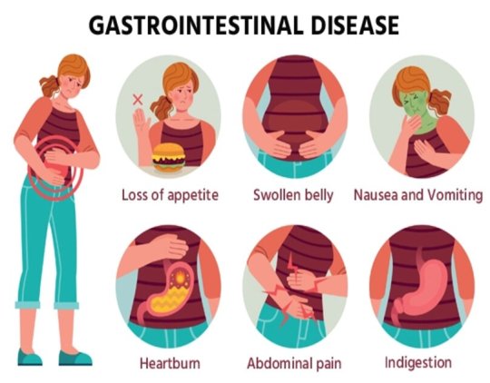
Common Conditions Treated by Gastrointestinal Surgeons: Exploring Digestive Disorders
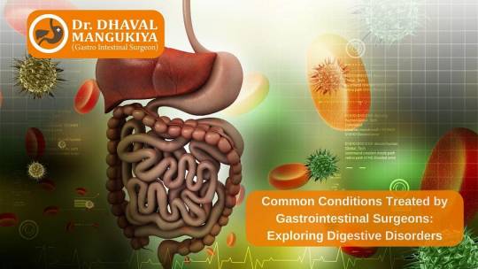
Digestive disorders encompass a wide range of conditions that affect the gastrointestinal (GI) tract, including the oesophagus, stomach, small intestine, colon, rectum, and anus. While many digestive issues can be managed through nonsurgical interventions, some may require the expertise of gastrointestinal surgeons to provide surgical treatment and relief. In this blog, Dr Dhaval Mangukiya, one of the best laparoscopic surgeons in Surat, shares information about the most common conditions treated by gastrointestinal surgeons, shedding light on these complex disorders and the surgical interventions used to address them.
Gallstones and Gallbladder Disease
Gallstones are hardened deposits that form in the gallbladder, a small organ located beneath the liver. When gallstones become symptomatic or lead to complications such as gallbladder inflammation (cholecystitis) or blockage of the bile ducts, surgical intervention may be necessary. Some of the best gastrointestinal surgeons in Surat, like Dr Dhaval Mangukiya, perform procedures such as laparoscopic cholecystectomy to remove the gallbladder and alleviate symptoms associated with gallstones and gallbladder disease.
Appendicitis
Appendicitis is inflammation of the appendix, a small pouch attached to the large intestine. If left untreated, appendicitis can lead to a ruptured appendix, a life-threatening condition requiring emergency surgery. Gastrointestinal surgeons perform an appendectomy to remove the inflamed appendix and prevent complications. Laparoscopic appendectomy, a minimally invasive procedure, is commonly used by the best gastro surgeons in Surat for uncomplicated cases of appendicitis, resulting in shorter recovery times and less postoperative pain.
Hernias
Hernias occur when organs or tissues protrude through weakened areas of the abdominal wall. Common types of hernias include inguinal hernias (occurring in the groin area), umbilical hernias (around the belly button), and hiatal hernias (involving the diaphragm and upper stomach). Gastrointestinal surgeons perform hernia repair surgeries to strengthen the abdominal wall and reposition herniated organs or tissues. Depending on the type and severity of the hernia, surgical techniques may include open repair or laparoscopic techniques with mesh reinforcement.
Gastroesophageal Reflux Disease (GERD)
GERD is a chronic digestive disorder characterized by the reflux of stomach acid into the oesophagus, leading to symptoms such as heartburn, regurgitation, and chest pain. While GERD can often be managed with lifestyle modifications and medications, some individuals may require surgical intervention to alleviate symptoms and prevent complications such as Barrett’s oesophagus or oesophageal cancer. Gastrointestinal surgeons may perform procedures such as fundoplication to reinforce the lower oesophageal sphincter and reduce reflux.
Diverticulitis
Diverticulitis is inflammation or infection of small pouches (diverticula) that form in the walls of the colon. Severe cases of diverticulitis, characterized by symptoms such as abdominal pain, fever, and bowel changes, may require surgical treatment. Gastrointestinal surgeons perform procedures such as bowel resection to remove the affected portion of the colon and repair any perforations or fistulas. In some cases, a temporary colostomy or ileostomy may be necessary to divert stool away from the affected area during the healing process.
Colorectal Cancer
Colorectal cancer is cancer that develops in the colon or rectum, often starting as precancerous polyps that can be detected and removed during screening colonoscopies. When colorectal cancer is diagnosed, surgical removal of the tumour and surrounding tissue is typically the primary treatment. Gastrointestinal surgeons perform procedures such as colectomy or rectal resection to remove the cancerous tissue and may also perform lymph node dissection to assess the spread of cancer.
Inflammatory Bowel Disease (IBD)
Inflammatory bowel disease (IBD), including Crohn’s disease and ulcerative colitis, is characterized by chronic inflammation of the digestive tract. While medical management is often the first-line treatment for IBD, Dr Dhaval Mangukiya, one of the best stomach surgeons in Surat, says surgery may be necessary in cases of severe disease, complications, or failure of medical therapy. Gastrointestinal surgeons perform procedures such as bowel resection, strictureplasty, or ileal pouch-anal anastomosis (IPAA) to remove diseased segments of the intestine, alleviate symptoms, and improve quality of life for individuals with IBD.
Conclusion
Gastrointestinal surgeons play a critical role in the management of common digestive disorders. These surgeons may sometimes have to resort to surgical interventions to alleviate symptoms, prevent complications, and improve quality of life for individuals affected by these conditions. From gallbladder disease to colorectal cancer, surgical treatment options offer hope and healing for patients facing a wide range of gastrointestinal challenges.
0 notes
Photo
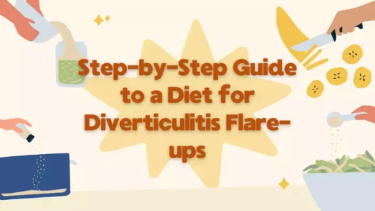
Diverticulitis can be a painful condition, and managing it through a proper diet is crucial during flare-ups. This step-by-step guide will help you navigate the complexities of a diet tailored for diverticulitis, providing practical tips and insights. Understanding Diverticulitis and its Triggers Diverticulitis is the inflammation or infection of small pouches (diverticula) that can form in the walls of your intestines, particularly the colon. A Diet for Diverticulitis plays a pivotal role in managing symptoms and preventing flare-ups. Here's a breakdown of the factors that trigger diverticulitis: Low-Fiber Diet Conundrum: A diet lacking in fiber is a common precursor to diverticulitis. Fiber-rich foods help maintain bowel regularity, preventing the formation of diverticula. When fiber intake is insufficient, constipation may occur, putting strain on the intestines and leading to diverticulitis. Inflammatory Foods Impact: Certain foods can exacerbate inflammation and trigger diverticulitis flare-ups. These include processed foods, red meat, and dairy. Understanding the inflammatory impact of these items is crucial for devising an effective diet plan. Crafting a Diverticulitis-Friendly Diet Plan Now that we've outlined the triggers, let's delve into a step-by-step guide to creating a diet that supports diverticulitis management. 1. Embrace High-Fiber Foods A diet rich in fiber is your first line of defense against diverticulitis flare-ups. Incorporate these high-fiber options into your daily meals: Whole Grains: Opt for whole grains like brown rice, quinoa, and whole wheat bread. These provide essential nutrients and promote healthy bowel movements. Fruits and Vegetables: Load up on colorful fruits and vegetables such as berries, apples, spinach, and broccoli. These not only offer fiber but also contribute vital vitamins and antioxidants. Legumes: Beans, lentils, and chickpeas are excellent sources of fiber. They add variety to your diet and help maintain digestive health. 2. Prioritize Lean Proteins While red meat can exacerbate inflammation, opt for lean proteins that support your digestive system: Fish: Fatty fish like salmon, mackerel, and trout are rich in omega-3 fatty acids, which possess anti-inflammatory properties. Skinless Poultry: Choose skinless chicken or turkey for a lean protein source. These options are gentler on your digestive system. Plant-Based Proteins: Explore plant-based proteins such as tofu, tempeh, and legumes. They provide protein without the inflammatory impact of certain animal products. 3. Mindful Hydration Adequate hydration is crucial for managing diverticulitis. Incorporate these hydration practices into your daily routine: Water Intake: Drink at least 8 glasses of water per day to prevent constipation and maintain overall digestive health. Herbal Teas: Consider incorporating soothing herbal teas like peppermint or chamomile. These can aid digestion and alleviate discomfort. 4. Limit Trigger Foods To prevent diverticulitis flare-ups, it's essential to identify and limit trigger foods: Processed Foods: Cut down on processed foods high in added sugars and unhealthy fats. These can contribute to inflammation and digestive distress. Red Meat and Dairy: Limit the intake of red meat and dairy products, as they can be inflammatory for some individuals. 5. Small, Frequent Meals Instead of three large meals, opt for smaller, more frequent meals throughout the day: Portion Control: Smaller portions are easier for your digestive system to handle, reducing the risk of irritation and inflammation. Snack Wisely: Choose healthy snacks like nuts, seeds, or yogurt to maintain steady energy levels without burdening your digestive tract. 6. Supplement Wisely Certain supplements can complement your diverticulitis-friendly diet: Probiotics: Consider incorporating probiotics to promote a healthy balance of gut bacteria, aiding digestion and reducing inflammation. Fiber Supplements: If getting enough fiber from food is challenging, talk to your healthcare provider about fiber supplements to support your digestive health. Conclusion Crafting a diet for diverticulitis is a proactive step in managing symptoms and preventing flare-ups. By prioritizing high-fiber foods, lean proteins, mindful hydration, and limiting trigger foods, you can support your digestive health and promote overall well-being. Remember to consult with your healthcare provider to tailor these dietary recommendations to your specific needs. With a thoughtful approach to your diet, you can navigate diverticulitis flare-ups more effectively and enhance your quality of life. FAQs 1. What is Diverticulitis, and why is diet crucial during flare-ups? Diverticulitis is the inflammation or infection of small pouches in the walls of the colon, called diverticula. During flare-ups, a specific diet helps manage symptoms, promote healing, and prevent complications. 2. What are the key dietary guidelines for managing Diverticulitis flare-ups? a. High-Fiber Foods: Why are they important? A diet rich in fiber aids in regular bowel movements, preventing constipation and reducing pressure on the diverticula. It also promotes a healthier gut microbiome, crucial for diverticulitis management. b. Hydration: How does it play a role? Hydration is essential for maintaining soft stools and making bowel movements easier. It also helps flush out toxins, reducing the risk of infection in the inflamed diverticula. c. Low-FODMAP Diet: When and why is it recommended? In some cases, a low-FODMAP diet can be beneficial during flare-ups. It limits certain fermentable carbohydrates, reducing gas production and bloating, and providing relief from symptoms. 3. Can I eat seeds and nuts during a diverticulitis flare-up? While traditionally advised to avoid seeds and nuts, recent studies suggest that they might not pose a significant risk during flare-ups. Moderation is key; consider incorporating ground seeds and nuts for added nutritional benefits. 4. How does stress impact diverticulitis, and can diet help manage stress? a. Understanding the Stress-Diverticulitis Connection Stress doesn't directly cause diverticulitis, but it can exacerbate symptoms. Stress management techniques, coupled with a balanced diet, contribute to overall well-being, potentially reducing the severity of flare-ups. b. Incorporating Stress-Reducing Foods Certain foods, like those rich in omega-3 fatty acids (found in fatty fish like salmon), can have a calming effect on the nervous system, assisting in stress management. 5. Are there specific foods to avoid during a diverticulitis flare-up? a. Red Meat and Processed Foods: Why should they be limited? High intake of red meat and processed foods may contribute to inflammation. Choosing lean proteins and whole foods reduces the risk of aggravating diverticulitis symptoms. b. Dairy and Diverticulitis: What's the connection? While dairy is generally well-tolerated, some individuals may be sensitive. Monitor your body's response and opt for lactose-free or low-lactose alternatives if needed. 6. Is there a recommended meal plan for diverticulitis flare-ups? a. Sample Day: Balancing Nutrients Breakfast: Oatmeal with berries and almond milk. Lunch: Grilled chicken salad with mixed greens and olive oil dressing. Snack: Greek yogurt with sliced banana. Dinner: Baked salmon with quinoa and steamed vegetables. b. Fluid Intake throughout the Day Include at least eight cups of water, herbal teas, and clear broths to stay adequately hydrated. 7. Can I continue my regular exercise routine during a diverticulitis flare-up? a. Moderate Exercise Benefits Engaging in moderate exercise can enhance digestion and reduce stress. Consider activities like walking or gentle yoga, but listen to your body and avoid strenuous exercises during flare-ups. b. Adjusting Exercise Intensity If your symptoms are severe, it's advisable to scale back on the intensity of your workouts temporarily. Gradually reintroduce more vigorous exercises as your symptoms subside. 8. How long should I follow a specific diet during diverticulitis flare-ups? The duration of a specialized diverticulitis diet varies among individuals. Consult with a healthcare professional for personalized advice. Once symptoms subside, gradually reintroduce a broader range of foods while monitoring your body's response. 9. Are supplements recommended for managing diverticulitis symptoms? a. Probiotics: Supporting Gut Health Probiotic supplements can aid in maintaining a healthy gut microbiome. Discuss with your healthcare provider to determine the most suitable probiotic strains for your specific needs. b. Fiber Supplements: When to Consider If it's challenging to meet daily fiber requirements through diet alone, fiber supplements may be beneficial. However, consult with your healthcare provider before incorporating them into your routine. 10. Can diverticulitis be prevented through diet and lifestyle changes? a. Long-Term Dietary Habits Adopting a diet rich in fiber, staying hydrated, and managing stress can contribute to preventing diverticulitis. Maintaining a healthy lifestyle, including regular exercise and avoiding excessive red meat consumption, plays a crucial role in prevention. b. Regular Check-ups and Monitoring Regular medical check-ups allow for early detection and management of any digestive issues. If you have a history of diverticulitis, staying vigilant and proactive in your healthcare is key to preventing flare-ups. In conclusion, a well-balanced diet, tailored to manage diverticulitis flare-ups, plays a pivotal role in alleviating symptoms and promoting overall gut health. Remember to consult with healthcare professionals for personalized advice, and listen to your body's signals throughout the process.
0 notes
Text
Exploring the Depths of Gastrointestinal Radiology: Techniques, Innovations, and Insights
Gastrointestinal radiology, a specialized branch of medical imaging, plays a crucial role in diagnosing and treating diseases of the digestive tract. This field leverages various imaging techniques to visualize the structure and function of the gastrointestinal (GI) system, providing invaluable insights that guide clinical decision-making. In this blog, we'll delve into the core techniques, recent innovations, and key insights within gastrointestinal radiology.
Core Techniques in Gastrointestinal Radiology
1. X-Ray Imaging
X-rays are the cornerstone of gastrointestinal radiology. Techniques such as fluoroscopy, barium studies, and plain radiographs allow for the visualization of the esophagus, stomach, and intestines. Barium swallow and barium enema are commonly used to detect structural abnormalities, such as strictures, diverticula, and tumors.
2. Computed Tomography (CT)
CT scans provide detailed cross-sectional images of the abdomen and pelvis, offering a comprehensive view of the GI tract and surrounding organs. CT enterography and CT colonography are specialized techniques that enhance visualization of the small intestine and colon, respectively. These methods are essential for detecting inflammatory diseases, such as Crohn's disease, and identifying neoplasms.
3. Magnetic Resonance Imaging (MRI)
MRI, particularly magnetic resonance enterography (MRE), is increasingly used in gastrointestinal radiology. MRE offers high-contrast images without radiation exposure, making it ideal for patients requiring frequent imaging, such as those with inflammatory bowel disease (IBD). MRI is also valuable in liver imaging, providing detailed information about hepatic lesions and vascular structures.
4. Ultrasound
Ultrasound is a non-invasive, real-time imaging technique commonly used to evaluate the liver, gallbladder, and biliary system. Endoscopic ultrasound (EUS) combines endoscopy and ultrasound to obtain high-resolution images of the GI tract and surrounding tissues, facilitating the diagnosis of pancreatic and esophageal disorders.
5. Nuclear Medicine
Nuclear medicine techniques, such as positron emission tomography (PET) and single-photon emission computed tomography (SPECT), are used to assess metabolic activity and detect malignancies. These modalities are particularly useful in staging cancers and evaluating treatment response.
Innovations in Gastrointestinal Radiology
The field of gastrointestinal radiology is continually evolving, with technological advancements enhancing diagnostic accuracy and patient care. Some notable innovations include:
1. Artificial Intelligence (AI) and Machine Learning
AI and machine learning are revolutionizing radiology by improving image analysis and interpretation. Algorithms can assist radiologists in detecting subtle abnormalities, reducing diagnostic errors, and increasing efficiency. AI-driven tools are particularly promising in screening for colorectal cancer and assessing liver fibrosis.
2. Advanced Imaging Techniques
Innovations such as dual-energy CT, diffusion-weighted MRI, and contrast-enhanced ultrasound offer enhanced tissue characterization and better differentiation between benign and malignant lesions. These advanced techniques provide more precise diagnostic information, aiding in early disease detection and personalized treatment planning.
3. Minimally Invasive Procedures
Interventional radiology (IR) techniques, such as percutaneous biopsies, ablations, and drainage procedures, are becoming more refined. These minimally invasive procedures, often guided by imaging, reduce the need for surgical interventions, decrease patient recovery time, and minimize complications.
Insights and Clinical Applications
Gastrointestinal radiology provides critical insights that impact patient management across various conditions:
1. Inflammatory Bowel Disease (IBD)
Imaging plays a vital role in diagnosing and monitoring IBD, including Crohn's disease and ulcerative colitis. MRI and CT enterography are essential for evaluating disease extent, detecting complications, and guiding therapeutic decisions.
2. Gastrointestinal Cancers
Early detection and accurate staging of GI cancers are paramount for successful treatment. CT, MRI, and PET scans are integral in identifying tumors, assessing their spread, and planning surgical or therapeutic interventions. Regular imaging follow-ups help monitor treatment efficacy and detect recurrences.
3. Liver Disease
Imaging modalities are indispensable in evaluating liver diseases such as cirrhosis, hepatitis, and hepatocellular carcinoma. MRI and ultrasound are particularly valuable for assessing liver morphology, vascular structures, and detecting focal lesions.
4. Functional Disorders
Radiological techniques, including motility studies and defecography, aid in diagnosing functional GI disorders such as achalasia, gastroparesis, and chronic constipation. These studies provide insights into the dynamic processes of the GI tract, guiding appropriate therapeutic interventions.
Conclusion
Gastrointestinal radiology is a dynamic and integral part of modern medicine, offering essential diagnostic and therapeutic capabilities. The continuous advancements in imaging technology and the integration of AI are enhancing the precision and efficacy of GI radiology, ultimately improving patient outcomes. As we continue to explore the depths of gastrointestinal radiology, the future holds promise for even more innovative solutions to complex clinical challenges.
Important Information:
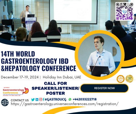
Conference Name: 14th World Gastroenterology, IBD & Hepatology Conference
Short Name: 14GHUCG2024
Dates: December 17-19, 2024
Venue: Dubai, UAE
Email: [email protected]
Visit: https://gastroenterology.universeconferences.com/
Call for Papers: https://gastroenterology.universeconferences.com/submit-abstract/
Register here: https://gastroenterology.universeconferences.com/registration/
Exhibitor/Sponsor: https://gastroenterology.universeconferences.com/exhibit-sponsor-opportunities/
Call Us: +12073070027
WhatsApp Us: +442033222718
0 notes
Text
Introduction
Diverticular disease refers to the presence of small pouches, called diverticula, that form in the lining of the digestive system, most commonly in the colon. These pouches can become inflamed or infected, leading to a condition known a...
#Mirari #MirariDoctor #MirariColdPlasma #ColdPlasma
0 notes
Text
AEW Dynamite 5/1/24
The Bucks have ascended to new heights of causing trouble on purpose
Oooh and now Swerve vs Bucks? FINED
CHRISTIAN IS BACK
Okay so do we know the status of Swerve's dad
Oooh Swerve attacking Nick Wayne is paying off, his story continues to be the sins of his past coming back to haunt him
NOT SWERVE'S HAIR
Okay so Swerve now knows what it's like to have an opponent threaten his child and what if this leads to an eventual team up with him and Hangman
LOL saw someone say that this is Buddy enacting Judgement Day vengeance on behalf of Rhea
Okay if Buddy spitting up blood is a work then this adds to my theory of Cope becoming a vampire again
LIVEN THE FUCK UP, CROWD
Malakai and Buddy goading Cope to hit Buddy YES
RIP Zay, I was enjoying having Private Party back
Oooh Skye vs Willow for Rampage!
Orange is sadly walking to the ring and I am preparing myself to be emotionally destroyed
Orange you got bedhead
THIS CAN ABSOLUTELY NOT BE TRUE
IT NEEDS TO BE FAKE AND CHICK WILL HEAL WITH THE POWER OF FRIENDSHIP AND ALL WILL BE RIGHT IN THE WORLD
STAY AWAY FROM MY PRECIOUS BOY YOU LARGE EVIL THUMB
Oh no Jericho is in trunks why are we being punished like this
Jericho's boobs are purple
Dammit I'm really enjoying this 😠
Stokely is an absolute treasure.
Large men time
Brian Cage is not wearing a cape like he was in the match image, I feel cheated
Yay Mariah but also I'm distracted by pho
Do you think Excalibur has a tumblr?
Aww Toni is protective of Mariah 😭
KENNY TIME
So fun fact, risk of forming diverticula is higher in people with EDS. Technically I have them but thankfully have only ever had very very mild diverticulitis attacks and not for a few years.
OKADA AND KENNY FACE TO FACE
THIS EPISODE HAS ME SO EMOTIONAL
Jack Perry how dare you do a sweet baby kitten smile while hurting Kenneth
I'VE MISSED KENNY'S TITS SO MUCH
NOOOOOO
oh goddammit
1 note
·
View note
Text
7 Symptoms of Diverticulitis ( Causes Treatment and Remedies)
7 Symptoms of Diverticulitis (+ Causes, Treatment, and Remedies)
https://theheartysoul.com/diverticulitis-symptoms-remedies/
Diverticulitis is a condition characterized by the inflammation or infection of diverticula. These are small, bulging pouches that can develop in the lining of the digestive system. These pouches are most commonly found in the large intestine, specifically the sigmoid colon. While many individuals with diverticula experience no symptoms, when these pouches become inflamed or […]
The post 7 Symptoms of Diverticulitis (+ Causes, Treatment, and Remedies) appeared first on The Hearty Soul.
via The Hearty Soul https://theheartysoul.com/
April 24, 2024 at 07:30AM
0 notes
Text
Barriga inchada - possíveis causas e tratamento

Um estômago inchado é também chamado de barriga inchada e é frequentemente acompanhado por dor abdominal ou cãibras no estômago.
Esta condição muito comum é geralmente de curta duração e desaparece por si só, por vezes após a passagem do vento (flatulência).
Embora todos tenham gás no intestino, algumas pessoas são mais sensíveis ao gás que lhes passa pelo intestino. Isto pode causar sintomas de dor, arrotos excessivos e flatulência. O seu estômago pode também sentir-se duro, inchado, esticado e desconfortável, bem como parecer inchado.
Um estômago inchado não é normalmente motivo de preocupação, mas pode ser um sinal de um problema de saúde subjacente.
Se tiver alguma preocupação sobre um estômago inchado, marque uma consulta com o seu médico de clínica geral.
https://www.youtube.com/watch?v=-r3CsTqhFPU
Causas da barriga inchada
O seu intestino contém sempre gás, a maior parte do qual vem do ar de engolir quando come e bebe.
No entanto, o gás também é produzido por bactérias que se alimentam dos alimentos no seu intestino - certos alimentos estimulam mais a produção de gás por bactérias do que outros.
Quando se senta, o gás no seu intestino passa normalmente através do seu esófago e da sua boca (arrotar).
Quando deitado, este gás geralmente passa para o seu estômago, o que pode causar um estômago inchado (inchaço abdominal) depois de comer e um abdómen duro e inchado.
O gás também pode sair do seu corpo ao passar o vento, vulgarmente conhecido como flato, onde o gás entra no seu intestino delgado e passa para fora da sua passagem de costas (ânus).
Várias condições de saúde possíveis podem causar um estômago inchado ou uma barriga inchada, incluindo:
Uma condição que afecta o sistema digestivo - isto inclui:
Doença celíaca - o seu sistema imunitário ataca erradamente o seu tecido intestinal quando come glúten
Constipação Diverticulite - infecção de pequenas bolsas (diverticula) na parede do seu intestino
Síndrome do intestino irritável (SII) - uma condição que causa dor e cólicas estomacais, inchaço, gás e um abdómen inchado
Síndrome do intestino curto - geralmente uma consequência de cirurgia para remover porções significativas do seu intestino, onde não é possível absorver nutrientes suficientes dos alimentos através do intestino delgado
Crescimento excessivo de bactérias intestinais - crescimento excessivo de bactérias que ocorrem naturalmente no intestino delgado
Uma intolerância alimentar ou uma intolerância a certas substâncias naturais, como a lactose, frutose ou glúten - se for intolerante à lactose, não consegue digerir lactose e, portanto, pode desenvolver inchaço, gás e um estômago inchado dentro de duas horas após comer ou beber lactose, que é um açúcar encontrado em produtos lácteos
Ascite - acumulação de líquido no abdómen, normalmente causado por problemas hepáticos, por exemplo cirrose hepática onde o fígado desenvolve muito tecido cicatrizado; ascite começa sem sintomas visíveis, mas com o tempo o seu abdómen vai ficar cada vez mais inchado, o que causa desconforto
Cálculos biliares - caroços duros e gordurosos na vesícula biliar
Pancreatite - inflamação do pâncreas
Escleroderma - uma condição auto-imune que causa endurecimento e aperto da sua pele e tecido conjuntivo.

Se certos alimentos parecerem desencadear o seu inchaço (por exemplo, feijão, brócolos e couve), evite ou reduza a quantidade destes que come.
Se pensa que tem uma intolerância alimentar, manter um diário alimentar pode ajudá-lo a identificar alimentos desencadeantes, tais como produtos lácteos, se for intolerante à lactose, ou trigo e glúten, se tiver uma sensibilidade ao glúten.
No entanto, não corte completamente um grupo alimentar inteiro da sua dieta, uma vez que isto pode afectar negativamente a sua saúde.
Em vez disso, fale com o seu médico de clínica geral ou um nutricionista sobre o ajuste seguro da sua dieta.
Dependendo dos seus outros sintomas, também pode tentar:
- Evitar bebidas gaseificadas e beber através de uma palhinha para evitar engolir demasiado ar ao beber.
- Mudar a forma como come - isto inclui mastigar correctamente os alimentos e com a boca fechada para evitar engolir demasiado ar
- Comer lentamente - comer refeições mais pequenas, mas mais frequentes Sentar-se quando come sem bater ou palpitar
- Reduzir o risco de obstipação através de uma dieta rica em fibras e exercício físico regular, por exemplo, andar a pé durante 20-30 minutos quatro vezes por semana
- Parar de mastigar pastilha elástica para reduzir a quantidade de ar que engole

Probióticos
Os probióticos são úteis porque reduzem a produção de gás intestinal, restabelecem o equilíbrio da flora bacteriana, promovem a divisão celular das paredes do cólon, reduzem a fermentação bacteriana dos alimentos e reduzem a hipermotilidade gastrointestinal.
Tudo isto, como resultado, contribui para aliviar os sintomas irritantes de uma barriga inchada, síndrome do cólon irritável e outras doenças inflamatórias.
Em geral, os probióticos são encontrados nos alimentos. Na realidade, são bactérias "boas" que restabelecem um equilíbrio adequado da flora intestinal, combatendo a acção de germes e bactérias que atacam o revestimento.
Encontram-se disponíveis em iogurte, leite ou queijo. Em alguns casos, porém, como a disbiose intestinal (síndrome do intestino irritável, gastrite, etc.), os probióticos nos alimentos não são suficientes.
O que podemos concluir?
A razão da sensação de “barriga inchada” é muitas vezes complexa e causada por vários fatores.
De forma a compreender a sua verdadeira origem, é indispensável conseguir informação clínica detalhada sobre o indivíduo como: qual o timing do inchaço abdominal, qual a história clínica e cirúrgica do doente, se faz algum tipo de suplementação ou medicação e os seus hábitos alimentares.
Há diversas causas para ficar de barriga inchada. Algumas são relativamente benignas e transitórias e podem estar relacionadas com a dieta de uma pessoa.
Outras podem indicar um estado de saúde subjacente que requer tratamento.
Deve consultar o seu médico se sofrer de inchaço grave, persistente, ou recorrente do estômago. Um médico trabalhará para diagnosticar a causa e fornecer tratamentos apropriados.
Read the full article
0 notes
Text
Complications Of Constipation If Left Untreated
Most of us have been constipated at some point during our lives. It’s inconvenient, but at least it doesn’t happen often. For some people, though, constipation is a regular part of their lives. These women and men have, on average, three or fewer bowel movements per week, leaving them feeling bloated, crampy, and uncomfortable. Sometimes constipation remedies itself with a few changes to diet or water intake, but if constipation is prolonged, your risk of developing complications, like hemorrhoids, increases. Chronic constipation is defined as having fewer than three bowel movements per week, passing hard stools, or difficulty and pain when passing stools. In this Blog, a leading Surgical Gastroenterologist in Hyderabad Dr. N.S. Babu Will explain how untreated constipation problems cause complications.
Let’s see one by one
However chronic constipation can cause complications.
Piles: Piles, also known as hemorrhoids, are a common complication of constipation. When you are constipated, the excreta becomes hard and delicate to pass. This can result in straining during bowel motions, which puts pressure on the veins in the rectal area. Over time, this expanded pressure can cause the veins to swell and bulge, leading to hemorrhoids. Piles can cause pain, itching, and bleeding, especially during bowel movements. They can be internal, located inside the rectum, or external, located under the skin around the anus. In severe cases, hemorrhoids may need to be treated with medicine or surgery.
Anal Fissures: Anal fissures are slight slits in the skin around the anus that can come due to various factors, involving constipation. When you are constipated, the coprolite becomes hard and difficult to pass, leading to straining during bowel motions. This straining can affect slits in the delicate skin of the anus, resulting in anal fissures. Anal fissures can be quite painful and may affect bleeding during bowel motions. They can also conduct to discomfort and itching in the anal area. It’s essential to address constipation to prevent the evolution or worsening of anal fissures. Increasing fiber input, staying hydrated, and exercising regularly can help soften the coprolite and make bowel motions easier, reducing the threat of fissures.
Rectal Prolapse: Rectal prolapse occurs when the rectum protrudes from the anus, either partly or completely. habitual constipation, involving constant straining during bowel movements, can weaken the muscles supporting the rectum, boosting the trouble. Symptoms of rectal prolapse carry a bulge or swell from the anus, difficulty controlling bowel movements, and a feeling of incomplete emptying.
Fecal Impaction: Fecal impaction is a tough complication of constipation where a large, hard mass of dropping becomes jammed in the rectum and can’t be expelled. This condition usually occurs when constipation is left untreated, and the dropping becomes too large and hard to pass easily. Fecal impaction can cause abdominal pain, bloating, and a feeling of fullness or pressure in the rectum. In severe cases, it can result in complications similar to bowel inhibition or the expansion of ulcers in the rectum.
Diverticulitis: Diverticulitis is a condition where small, pouching pouches( diverticula) in the digestive region become inflamed or infected. Constipation can be a contributing factor to the expansion of diverticulitis. When constipated, the increased pressure in the colon can result in the formation of diverticula. Hard droppings can also get trapped in these pouches, leading to inflammation or infection.
How to get rid of Constipation?
To alleviate constipation, try adding your fiber input through fruits, vegetables, and whole grains, which can support soft dropping and promote bowel motions. Stay doused by drinking plenty of water throughout the day. Regular physical exercise can also stimulate bowel motions. Establishing a regular restroom routine and not neglecting the appetite to have a bowel motion can support this.
In some cases, over-the-counter laxatives or dropping softeners may be helpful. However, if you witness severe pain or bleeding, seek medical guidance from the best colorectal surgeon in Hyderabad, Dr. N. S. Babu for effective treatment and proper care
1 note
·
View note
Text
Wondering about the relationship between back pain and diverticulitis? Explore the fascinating insights into "Does Diverticulitis Lead to Back Pain?" Gain a deeper understanding of how your digestive health might be influencing your spinal well-being. Dive into the exploration. Read now to know more.
0 notes
Text
Gastrointestinal Expert in Mumbai: Dr. Chintamani Godbole
Common Gastrointestinal Diseases: Overview and Symptoms
Healthcare providers encounter various gastrointestinal (GI) diseases affecting the digestive system. Here are some prevalent conditions:
1. GERD (Gastroesophageal Reflux Disease): Chronic condition with stomach acid flowing back into the esophagus, causing heartburn and regurgitation.
2. Peptic Ulcer Disease: Sores on the stomach, upper small intestine, or esophagus due to stomach acid erosion.
3. IBD (Inflammatory Bowel Disease): Chronic inflammation in the intestines, such as Crohn's disease and ulcerative colitis, leading to abdominal pain, diarrhea, and weight loss.
4. IBS (Irritable Bowel Syndrome): Functional disorder causing abdominal pain, bloating, and bowel habit changes without structural abnormalities.
5. Celiac Disease: Autoimmune disorder damaging the small intestine due to gluten ingestion, leading to nutrient malabsorption.
6. Gallstones: Hardened deposits in the gallbladder causing pain, nausea, and digestive issues.
7. Hepatitis: Liver inflammation often due to viral infections (Hepatitis A, B, C) or factors like alcohol consumption.
8. Pancreatitis: Inflammation of the pancreas, associated with abdominal pain and digestive enzyme imbalances, can be acute or chronic.
9. Diverticulitis: Inflammation or infection of diverticula in the colon walls.
10. Colorectal Cancer: Cancer developing in the colon or rectum, often starting as polyps that can be detected and removed early.
Healthcare providers tailor diagnoses and treatments based on the specific disease and its severity. If experiencing digestive issues, consult a professional for proper evaluation and management.
For more information, consult Dr. Chintami Godbole one of the best Gastrointestinal Doctor in Mumbai or you can Contact us on 84518 65944.
#colorectal surgeon in mumbai.#colorectalcancer#gastrointestinal surgeon in mumbai#colorectal surgeon in mumbai#colon cancer treatment in mumbai#gastrointestinal doctor in mumbai#dr. chintamani godbole#drchintamanigodbole#colorectalsurgery#colorectalsurgeon
0 notes
Text
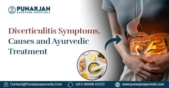
Diverticulitis Symptoms, Causes And Ayurvedic Treatment
Everything You Need To Know About Diverticulitis
Meet Diverticulitis: an unpleasant visitor, notorious for causing discomfort and frustration. It’s like that unwelcome house guest who overstays their welcome, only, it’s hanging out in your digestive tract. Less than ideal, isn’t it? But don’t panic! Understanding Diverticulitis can help you recognize its signals and nip it in the bud. So, let’s delve into Diverticulitis, from what it is to how you can prevent it.
#DiverticulitisAwareness#DigestiveHealth#AyurvedicTreatment#HolisticWellness#NaturalRemedies#GutHealth#HealthyLiving#AyurvedaCare#WellnessJourney#HealthTips
0 notes
Text
Diverticulitis Symptoms, Causes And Ayurvedic Treatment
Meet Diverticulitis: an unpleasant visitor, notorious for causing discomfort and frustration. It’s like that unwelcome house guest who overstays their welcome, only, it’s hanging out in your digestive tract. Less than ideal, isn’t it? But don’t panic! Understanding Diverticulitis can help you recognize its signals and nip it in the bud. So, let’s delve into Diverticulitis, from what it is to how you can prevent it.
Let’s imagine your colon is a calm, serene stream. Now, picture a tiny pouch or bulge growing on the side of the stream—kind of like an outgrowth. That’s a diverticulum. When these diverticula become inflamed or infected, we get what’s known as Diverticulitis, a digestive disease that often lays claim to the large intestine.
For more information visit: https://www.punarjanayurveda.com/diverticulitis-symptoms-causes-and-ayurvedic-treatment/
0 notes
Text
Journey Through the Gut: Understanding Small Bowel and Colonic Diseases
Introduction:
The gastrointestinal tract, often referred to as the gut, plays a crucial role in digestion and nutrient absorption. Within this intricate system, the small bowel and colon are vital components, responsible for processing food, extracting nutrients, and eliminating waste. However, these organs are susceptible to a myriad of diseases and conditions that can impact their function and overall health. In this blog, we'll explore some common disorders affecting the small bowel and colon, their symptoms, diagnosis, and treatment options.
Inflammatory Bowel Disease (IBD):
Inflammatory Bowel Disease encompasses a group of chronic inflammatory conditions affecting the gastrointestinal tract, including Crohn's disease and ulcerative colitis. These diseases cause inflammation and damage to the lining of the intestines, leading to symptoms such as abdominal pain, diarrhea, rectal bleeding, and weight loss. Diagnosis typically involves a combination of clinical evaluation, imaging studies such as endoscopy and colonoscopy, and laboratory tests. Treatment aims to reduce inflammation, control symptoms, and prevent complications through medications, lifestyle modifications, and in some cases, surgery.
Irritable Bowel Syndrome (IBS):
Irritable Bowel Syndrome is a common functional gastrointestinal disorder characterized by abdominal pain or discomfort, bloating, and changes in bowel habits without evidence of underlying structural abnormalities. While the exact cause of IBS remains unclear, factors such as diet, stress, and altered gut motility may contribute to its development. Diagnosis is based on symptom criteria, and management focuses on symptom relief through dietary modifications, stress management techniques, medications, and lifestyle changes.
Gastrointestinal Infections:
Gastrointestinal infections, commonly referred to as gastroenteritis, can affect both the small bowel and colon, causing symptoms such as diarrhea, nausea, vomiting, abdominal cramps, and fever. These infections are often caused by bacteria, viruses, or parasites transmitted through contaminated food or water. Diagnosis may involve stool tests to identify the causative organism. Treatment aims to alleviate symptoms and prevent dehydration through fluid replacement and, in some cases, antimicrobial therapy.

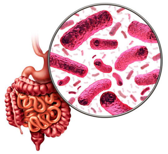
Diverticular Disease:
Diverticular disease refers to the presence of diverticula, small pouches that protrude through the weak areas of the colon wall. While many individuals with diverticula remain asymptomatic, some may experience symptoms such as abdominal pain, bloating, changes in bowel habits, and rectal bleeding, particularly if complications such as diverticulitis or diverticular bleeding occur. Diagnosis is typically made through imaging studies such as CT scans or colonoscopy. Treatment focuses on symptom management, dietary modifications, and, in severe cases, surgical intervention to address complications.
Understanding small bowel and colonic diseases involves exploring a range of conditions that affect the gastrointestinal (GI) tract, including inflammatory, infectious, functional, and neoplastic disorders. Here’s a detailed look into some of the most common and significant diseases affecting the small bowel and colon:
Small Bowel Diseases
Celiac Disease:
Definition: An autoimmune disorder where ingestion of gluten leads to damage in the small intestine.
Symptoms: Diarrhea, weight loss, and malabsorption.
Diagnosis: Serologic tests for antibodies (tTG-IgA) and a confirmatory biopsy of the small intestine.
Treatment: Strict gluten-free diet (MDLinx) (MGMA Homepage).
Crohn’s Disease:
Definition: A type of inflammatory bowel disease (IBD) that can affect any part of the GI tract but commonly affects the terminal ileum.
Symptoms: Abdominal pain, diarrhea, weight loss, and fatigue.
Diagnosis: Endoscopy, imaging studies, and biopsy.
Treatment: Anti-inflammatory medications, immunosuppressants, and biologic agents (MDLinx).
Small Bowel Obstruction:
Definition: A blockage that prevents the contents of the intestine from passing through.
Symptoms: Severe abdominal pain, vomiting, and inability to pass gas or stool.
Diagnosis: Clinical evaluation, abdominal X-rays, and CT scans.
Treatment: Nasogastric suction, IV fluids, and sometimes surgical intervention (MDLinx).
Irritable Bowel Syndrome (IBS):
Definition: A functional GI disorder characterized by a group of symptoms that occur together, including repeated pain in the abdomen and changes in bowel movements.
Symptoms: Abdominal pain, bloating, diarrhea, and constipation.
Diagnosis: Based on symptoms and the exclusion of other diseases.
Treatment: Dietary modifications, stress management, and medications to manage symptoms (MDLinx).
Colonic Diseases
Ulcerative Colitis:
Definition: A chronic inflammatory condition limited to the colon and rectum.
Symptoms: Bloody diarrhea, abdominal pain, and urgency.
Diagnosis: Colonoscopy with biopsy.
Treatment: Anti-inflammatory drugs, immunosuppressants, and biologic therapies (MDLinx) (MGMA Homepage).
Colorectal Cancer:
Definition: Cancer that starts in the colon or rectum.
Symptoms: Changes in bowel habits, blood in stool, weight loss, and fatigue.
Diagnosis: Colonoscopy, imaging tests, and biopsy.
Treatment: Surgery, chemotherapy, radiation therapy, and targeted therapy (MDLinx) (MGMA Homepage).
Diverticulosis and Diverticulitis:
Definition: Diverticulosis is the presence of diverticula (small bulging pouches) in the colon, which can become inflamed or infected (diverticulitis).
Symptoms: Diverticulosis often has no symptoms. Diverticulitis can cause severe abdominal pain, fever, and changes in bowel habits.
Diagnosis: CT scan and clinical evaluation.
Treatment: High-fiber diet for diverticulosis; antibiotics, and sometimes surgery for diverticulitis (MDLinx) (MGMA Homepage).
Irritable Bowel Syndrome (IBS):
Definition: Similar to its manifestation in the small bowel, it also affects the colon.
Symptoms: Abdominal pain, bloating, diarrhea, and constipation.
Diagnosis: Based on symptoms and exclusion of other diseases.
Treatment: Dietary changes, medications, and mental health therapy (MDLinx).
Diagnostic Techniques
Endoscopy and Colonoscopy: These are crucial for visualizing the GI tract, taking biopsies, and diagnosing conditions like IBD, celiac disease, and colorectal cancer.
Imaging Studies: CT scans, MRI, and X-rays help in diagnosing obstructions, diverticulitis, and other structural abnormalities.
Laboratory Tests: Blood tests, stool tests, and serologic tests are used to identify infections, inflammation, and specific antibodies for celiac disease.
Conclusion:
The small bowel and colon are integral components of the gastrointestinal tract, susceptible to a wide range of diseases and conditions that can significantly impact health and quality of life. Understanding the symptoms, diagnosis, and treatment options for these disorders is essential for effective management and optimal outcomes. Through advancements in medical research, diagnostic techniques, and therapeutic interventions, healthcare providers continue to strive towards improving outcomes for individuals affected by small bowel and colonic diseases, guiding them on their journey towards digestive health and well-being. Understanding small bowel and colonic diseases requires a comprehensive approach involving accurate diagnosis and tailored treatment strategies. Gastroenterologists utilize a range of diagnostic tools to identify these conditions and manage them effectively with medical, dietary, and sometimes surgical interventions.
For more detailed information, consider exploring these sources:
MDLinx on Gastroenterology
Gastroenterology & Hepatology Journal
Medical Group Management Association (MGMA)
Important Information:
Conference Name: 14th World Gastroenterology, IBD & Hepatology Conference
Short Name: 14GHUCG2024
Dates: December 17-19, 2024
Venue: Dubai, UAE
Email:�� [email protected]
Visit: https://gastroenterology.universeconferences.com/
Call for Papers: https://gastroenterology.universeconferences.com/submit-abstract/
Register here: https://gastroenterology.universeconferences.com/registration/
Exhibitor/Sponsor: https://gastroenterology.universeconferences.com/exhibit-sponsor-opportunities/
Call Us: +12073070027
WhatsApp Us: +442033222718
0 notes
Text
Meet Diverticulitis: an unpleasant visitor, notorious for causing discomfort and frustration. It’s like that unwelcome house guest who overstays their welcome, only, it’s hanging out in your digestive tract. Less than ideal, isn’t it? But don’t panic! Understanding Diverticulitis can help you recognize its signals and nip it in the bud. So, let’s delve into Diverticulitis, from what it is to how you can prevent it.
0 notes
Last Seen Blogs

eternally-flustered-lee
Way Too Ticklish

gokuista
❀ 淡水水母 ❀

chibibungoukappa
Chibi Bungou Kappa

callanthas
Beautiful guys

tellmehowlostareyou
"Estamos enamoradas"