#malleolable
Text
Lesbian Pussy tribing & humping //full vid on OF
Nice chick dominates boy with her feet whilst jerking him off
SOY MUJER MADURA, Y FOLLANDO CON MI
rican mami anii gets pussy slow stroked by bbc
Brutal amateur bondage and humiliation of slave Louise Red in homemade bdsm and crying masochist punishment of kinky brunette tied and punished
Lesbian Linda Sweet and Rebecca Black toyed their assholes
Hot sexy Anita bhabi fucking in Bathroom
Sultry Tranny Nicolly Pantoja Catches a Peeping Tom and Makes Him Blow Her
Wife flashed boobs to stranger, he fucked all here holes, husband films
Bajo falda pendeja descuidada
#malleolable#ablauts#popishly#granomerite#torgoch#windburned#merwoman#thirty-third#quadrupled#associators#thutter#windings#myotome#arterial#tear-owned#violetta#polyptoton#arquerite#anadromous#upclimb
0 notes
Text
Spor Anatomisi 2022-2023 Vize Soruları
Spor Anatomisi 2022-2023 Vize Soruları
1. Medial malleol hangi kemiğe aittir?
Cevap : tibia
Medial malleol, tibia (kaval kemiği) adı verilen kemiğe aittir. Ayak bileğinde, tibianın alt ucunda yer alır ve ayak bileğinin iç tarafında, ayak bileği eklemine katılan bir çıkıntı oluşturur. Bu çıkıntı, ayak bileği eklemi stabilitesinde önemli bir rol oynar ve ayak bileği hareketlerine katkıda…
View On WordPress
0 notes
Photo
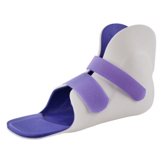
Mega Protez Ortez Yapım ve Uygulama Merkezi - SMO AFO - (Supra Malleoler) Valgus Ateli #smo #afo #supramalleoler #valgusateli #atel #protez #ortez #megaprotez
8 notes
·
View notes
Text
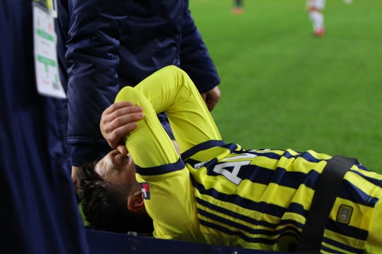
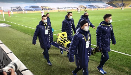
Mesut Özil'in sakatlık sonucu açıklandı
Mesut Özil’in sakatlık sonucu açıklandı
Antalyaspor maçında sakatlık yaşayan ve sedye ile sahadan ayrılan Fenerbahçe’nin yıldız futbolcusu Mesut Özil’in en az 1 ay sahalardan uzak kalma olasılığı olduğu bildirildi.
Fenerbahçe Kulübünden yapılan açıklamada, Mesut Özil’in Acıbadem Altunizade Hastanesi’nde çekilen MR’ında; ayak bileği iç ve dış yan bağlarında kısmi yırtıklar ile ayak bileği iç-dış malleol kemiklerinde yoğun ödem…
View On WordPress
0 notes
Photo
Süper Lig'de oynanan Fenerbahçe-Antalyaspor mücadelesinin 65. dakikasında girdiği ikili mücadele sonucunda yerde kalan Mesut Özil, sahayı sedye ile terk etmişti. Hastaneye kaldırılan oyuncunun, çekilen MR'ında; ayak bileği iç ve dış yan bağlarında kısmi yırtıklar ile ayak bileği iç-dış malleol kemiklerinde yoğun ödem gözlendiği aktarıldı.
0 notes
Text
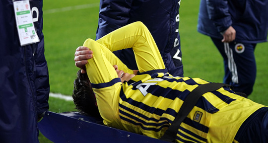
Mesut Özil'in ayak bileğinde kısmi yırtık ve ödem tespit edildi
Mesut Özil’in ayak bileğinde kısmi yırtık ve ödem tespit edildi
Kulübün internet sitesinde yapılan açıklamada, karşılaşmada aldığı darbe sonrası ayak bileği burkulan Mesut Özil’in çekilen MR’ında; ayak bileği iç ve dış yan bağlarında kısmi yırtıklar ile ayak bileği iç-dış malleol kemiklerinde yoğun ödem gözlendiği bildirildi.
Oyuncunun tedavisine başlandığı kaydedildi.
Haberin Kaynağı
View On WordPress
0 notes
Photo

I'd like to share with you all my latest experience with X39... I fell down the stairs, causing a fracture to the distal epiphysis of the perone with a fracture to the third malleol... I came to the emergency room with a pair of icewaves, put immediately after the fall (I always carry them in my purse). During grafting I used X39 and glutathione... and after only 5 sessions of physical therapy I'm walking again! Attaching some pictures... Thanks Dr. David, for the passion and love of His life mission.. a life for all of us always on the Wave. @lifewave_by_defran https://www.instagram.com/p/CKkeBxHjGf8/?igshid=e5eo3z8rha76
0 notes
Text
What is the Pain of the Calcaneal Spine?
Pain and discomfort with the walking of calcane spines is a common reason for medical consultation. Located on the bone that makes up the heel of the foot, this pain is related to the tension of the arch and prevents it from sagging.
What are the signs of this pathology, Doctor?
People with this condition sometimes describe this pain as a strong feeling of warmth in the heel. The pain usually appears as soon as you get up in the morning, when you stand up. The pain tends to subside or even disappear after a certain amount of walking and activity. But it often reappears vividly and acutely, as soon as walking or activity is resumed after a certain period of rest.
This pain can be along the inner edge or up the inner side of the calcaneus by 1 cm or even 2 cm. The pain can be felt in the inner plantar and/or in the middle of the heel. Podiatrists say there is a “trigger point” of pain. The point is located in the extension of the internal malleole beyond the inner edge of the heel about 1 cm in plantar. This pressure is exerted just at the aponeurotic attachment. You have to put your foot and toes in dorsiflexion; strong pressure recreates the pain.
The diagnosis is essentially clinical, hence the need to consult your treating physician, in the face of disabling pain in the heel, which does not pass. On examination, a strong pressure of the thumb on the center of the heel will awaken the pain. The pain obtained by applying strong pressure with the finger along the entire inner edge of the fascia with the dorsal bending foot confirms the presence of associated inflammation (“fasciitis”).
Although the evidence of a calcaneal spine visible on X-rays confirms the diagnosis, a normal X-ray will not eliminate a calcaneal spine at its onset. Calcaneal spurs rarely are fuzzy and show a simple fluffy-looking bone deformation that can direct other inflammatory problems such as ankylosing Spondylitis or Rheumatoid Arthritis or even a Drop.
However, these inflammatory joint diagnoses can usually be distinguished from local causes of heel pain, by the presence of local heat and swelling more or less marked.
How to treat this pathology?
First of all, some preventive measures are needed: The following tips will prevent the onset of plantar fasciitis and its recurrence, as well as the Lenoir spine that may be associated with it.
Do regular exercises to relax the Achilles tendon as well as the calf and foot muscles and be careful when playing sports. In addition to proper footwear, it is important to consider the following recommendations:
Doing stretching and warm-up exercises before any physical activity is a little demanding and prolonged.
Gradually increase distances when jogging.
Avoid running for long on sloping terrain, on hard (asphalt) or uneven surfaces. Prefer, if possible, dirt roads.
Respect his need for rest.
Wear shoes that support the arch and absorb shocks depending on the type of work or physical activity.
Replace your shoes at the first sign of wear. As for running shoes, they need to be renewed, as the pads wear out.
Avoid standing for too long, especially if you are wearing hard-soled shoes.
Medical treatments
Treatments almost always work well, but it can take several months to achieve a full recovery
Application of ice
Apply a bag of ice to relieve inflammation for 5 to 15 minutes. Avoid applying the bag directly to the skin. Settle down so that the feet are higher than the body. The best time to apply ice is at the end of the day or after physical activity.
Exercises
The general practitioner, podiatrist (podiatrist) or physiotherapist may recommend stretching exercises of the Achilles tendon and plantar fascia, which promotes both healing and prevention of recurrence.
Sitting
Place a tissue on the floor, then grasp it with your toes. Do this several times.
Place a bottle or tennis ball under the arch. Once the pain has subsided, it is a matter of rolling a golf ball directly under the heel.
Put a towel under your foot as if you were holding it in a scarf, then lengthen the leg while holding the towel securely. Pull on the towel to bring the foot back to you, then release.
Standing
Stand in front of a wall at a distance of about 60 cm. Then lay the palms of your hands against the wall. Then do the next two exercises one after the other and several times
While flexing the left leg forward, slide the right foot backwards, keeping it completely on the foot pillow heel protector.
Fold the knee so that it is aligned with the toes to stretch the Achilles tendon. Stay in this position for 30 to 60 seconds.
0 notes
Text
Fluoroscopic Guide With Amniofix Injection For Level 3 OA ...
Fluoroscopic Guide With Amniofix Injection For Level 3 OA …
Fluoroscopic Guided Eye Infusion with Amniofix for Grade 3 OA following Neglect Bi-Malleol fracture years ago … patient is too young for TAR and original arthrodesis is not an option at this point 💉💉 #lafootanklesurgeons #lafootanklespecialists #ankle #injury #trauma #fracture #arthritis #ocd #pain #footpain #toes #stemcell #regenerativemedicine #amniofix
Source
View On WordPress
#aerobics#amniofix#ankle#anti aging food#anti aging medicine#arthritis#back pain#Fluoroscopic#footpain#fracture#Guide#Injection#injury#lafootanklespecialists#lafootanklesurgeons#Level#OA#ocd#pain#RegenerativeMedicine#stemcell#toes#trauma
0 notes
Text
Traore ameliyat edildi
Kulüp Doktoru Yrd.Doç.Dr. Galip Bilen Kürklü, yeşil beyazlı oyuncu Abdou Razack Traore’nin Trabzon’da ve Konya’da kontrollerinin yapıldığını belirterek, “Traore’nin sol ayak bileğinde darbeye bağlı iç malleol kemikte parçalı ve ayrık kırıklar tespit edildi. Bağların durumu da çekilecek MR sonucunda belli olacak. Ama tedavi sürecini de göz önüne aldığımızda oyuncumuz Abdou Razack Traore’nin…
View On WordPress
0 notes
Photo

I'd like to share with you all my latest experience with X39... I fell down the stairs, causing a fracture to the distal epiphysis of the perone with a fracture to the third malleol... I came to the emergency room with a pair of icewaves, put immediately after the fall (I always carry them in my purse). During grafting I used X39 and glutathione... and after only 5 sessions of physical therapy I'm walking again! Attaching some pictures... Thanks Dr. David, for the passion and love of His life mission.. a life for all of us always on the Wave. @lifewave_by_defran https://www.instagram.com/p/CKcimpojbHy/?igshid=p6toiq1u14e1
0 notes
Photo

Ayakbileği ateşli silah yaralanması sonrasında kemik içi mermi çekirdeği ile çoklu kırık(medial-posterior malleol kırığı) ve eklem(syndesmotik) ayrışması ile başvuran hastamızda uyguladığımız 2 seanslı mermi çıkarımı-gergi bantlama-posterior malleol plaklama ve syndesmoz vidası ile eklem stabilizasyonu ameliyatlarımız (İrmet Hospital)
0 notes
Last Seen Blogs
liliumontherun
Lilium
astralsi
scorched

christinaayvazian
Untitled

soy-sauce-and-mothra
What is happening

kiss-the-apex
you have all the weapons you need