#myotome
Text
Magical king Arthur he got other magic in place of chaos

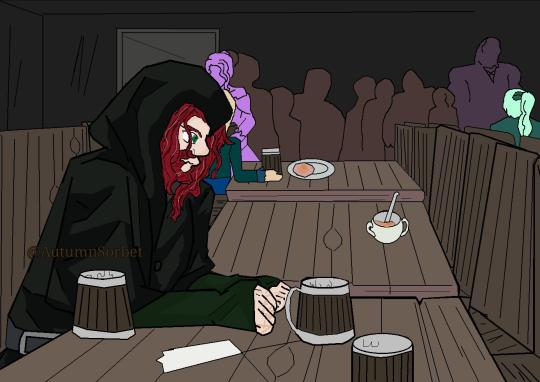
Art dump sorry
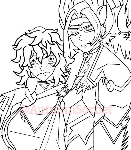
This one from a fanfiction I really like it's about King Arthur on a03
My otome / rwby and nanatsu no taizai au art
Lancelot (my oc version not the one from 4kota bluelance is 20-26)
, Arthur (au version he's 21-26 )and (my version of Guinevere she's 20-28)

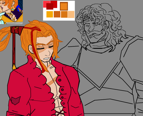
King Arthur and Oscar from RWBY
Otome /hime RWBY au oscar


, some unfinished art of Arthur and one of the nights of the round table he's an older night in his late 60s he's also a night who served under Arthur's father King Uther
Originally Arthur shirt is going to be purple with silver buttons and then I realized it looked too much like Merlin's outfit XD
#rwby#art is mine#autumn sorbet art#rwby art#rwby designs#rwby fan art#rwby9#vacuo#rwby10#rwbyv10#arthurian literature#Arthur pendragon#nanatsu no taizai au#nanatsu no taizai#myotome#my-otome#my hime#ztypehime#honkai star rail#rwby oscar#oscarpine#Arthurpendragon#king arthur#Arthur pendragon blog#Arthur blog#blog#oscar blog#au#au blog#rwby fanart
4 notes
·
View notes
Text
Lesbian Pussy tribing & humping //full vid on OF
Nice chick dominates boy with her feet whilst jerking him off
SOY MUJER MADURA, Y FOLLANDO CON MI
rican mami anii gets pussy slow stroked by bbc
Brutal amateur bondage and humiliation of slave Louise Red in homemade bdsm and crying masochist punishment of kinky brunette tied and punished
Lesbian Linda Sweet and Rebecca Black toyed their assholes
Hot sexy Anita bhabi fucking in Bathroom
Sultry Tranny Nicolly Pantoja Catches a Peeping Tom and Makes Him Blow Her
Wife flashed boobs to stranger, he fucked all here holes, husband films
Bajo falda pendeja descuidada
#malleolable#ablauts#popishly#granomerite#torgoch#windburned#merwoman#thirty-third#quadrupled#associators#thutter#windings#myotome#arterial#tear-owned#violetta#polyptoton#arquerite#anadromous#upclimb
0 notes
Text
boards on wednesday and then I’m gonna be SO annoying yall don’t even know what’s coming… I have so many wips sitting inside my head rn that I need to get onto the page the second I have time again
#brain is mush rn. only things I know are posterior tibial tendonitis and myotomes and parkinson’s disease and every single muscle in the bod#anyways. catch me wednesday afternoon when I finally can make space in my brain for bug and cat show#I ALSO WILL CATCH UP ON REQUESTS!!! I think there’s currently 3 in my inbox I’m planning to get to… eventually…#ramblings
2 notes
·
View notes
Text
i hate memorising the myotomes what’s the point unless you’re a neurologist 🫠🫠 it’s literally just numbers
#like even if you know the nerve and the nerve roots you don’t know WHICH of possible nerve roots it is#ffs#dermatomes are okay because it’s just remembering where to poke sequentially#but myotomes… fuck that#walks into examining room and knocks myself out with the hammer
2 notes
·
View notes
Text
youtube
Does the pain originate from the extremity or the spine? with Dr., Nick Rainey
Determining whether pain originates from the extremity (such as an arm or leg) or the spine can be challenging and often requires a thorough assessment by a healthcare professional. Pain can be referred, meaning that it is felt in a different location than its actual source.
#upper spine pain#advanced spine and pain#severe leg pain#severe spinal stenosis#failed back syndrome#decompress spine#nevro spinal cord stimulator#neck decompression#spine problems#spine and pain center#lumbar facet syndrome#spine and pain management#l4 l5 stenosis treatment#spinal stenosis therapies#spinal muscles#myotome testing#myotome weakness#the spine?#Does the pain originate from the extremity or the spine? with Dr.#Nick Rainey#Youtube
0 notes
Text
Lerning is sometimes tough for Scarlett Fever
Nude bugil girl show cam
Ace Rockwood the guy from American Pornstar fucks gay twink in Detroit
Blowjob, fuck and cumshot on a German teen babe
Store officer fucks latina teens pussy
webcam live sex with me
I will feed you cum until you are addicted to it CEI
Amazing post op babe fingering herself
Lynx fucking fornite
Rough POV blowjob deepthroat mouthfuck with slim busty blonde MILF Masha Markova
#noticing#Transitional#roadman#gymnospore#Aarhus#metroscope#ocelli-#Wiltshire#myotomic#Antheus#carragheen#pseudocoel#textposts#Torricelli#featherbone#activewear#undefensible#amylogenesis#modalism#moiler
0 notes
Text

الرد على شبهة وجود خطأ علمي في الآية الكريمة (فكسونا العظام لحماً)
بقلم : عبدالله محمد (طبيب أسنان)
نص الشبهة؛
يقول الملحد لقد أخطأ قرآنكم في ذكر تخلق العضلات بعد العظام لان كليهما ينشآن من طبقة الميزوديرم (mesoderm) وبالتالي يتكونا في نفس الوقت ، فلا تسبق العظام تكون العضلات (اللحم)
الرد:
ملحوظة قبل البداية : اللحم هو العضلات وليس الجلد فحين تشتري لحما من الجزار يعطيك من العضلات وليس من الجلد
أولا سنتكلم عن تكوين الجنين :
تشارك ثلاث طبقات في تكوينه و هي : الاكتوديرم (ectoderm)و الميزوديرم (mesoderm) و الاندوديرم (endoderm)، و ما يهمنا في هذا الموضوع هي طبقة الميزوديرم التي تتكون منها العظام و العضلات.
ادعى الملحدون أن العظام و العضلات يتكونان في نفس الوقت بحجة أنهما يتكونان من نفس الطبقة، و لكنه لم يدرك أننا لو تتبعنا نمو أنسجة الجسم سنجد : القلب و الغدد التناسلية و الغضاريف و العظام و العضلات وغيرها وغيرها من الاعضاء (1)، و كل ذلك لا يتكون في نفس الوقت.
و نسأله : ما رأيك الآن؟! هل القلب يتكون مع الاعضاء التناسلية؟ هل الغدد تتكون مع العظام؟! هل الغضاريف تتكون مع العظام؟!
كل هذه الأنسجة تتكون من نفس طبقة الميزوديرم (mesoderm)، ولا تتكون في نفس الوقت فهذه حجة سخيفة تدل سطحية تفكيرك و معلوماتك ، فأنت تضع استنتاجا زائفا و أنت لست أهلا للاستنتاج اصلا.
على سبيل المثال، الغضاريف تتكون في منتصف الأسبوع الخامس (2) بينما العظام يبدأ تكوينها عند السادس، و العضلات ما بين منتصف السادس حتى الثامن. و هذا يعني أن الغضاريف تتكون من نفس الطبقة و لكن لا تتكون في نفس الوقت.
و إذا ما طلبت منهم مرجعا علميا لإثبات كلامهم هذا لا تجد إلا قولهم "يخرجان من نفس الطبقة، إذاً يخرجان في نفس الوقت!!!" وقد اثبتنا بطلان هذه الحجة
ثم لو سلمنا لكلامهم فهذا لا صلة له بمعنى الآية، فالآية تقول : فكسونا العظام لحما، "فكسونا" و بالتالي فالآية تتكلم عن مرحلة الكسو، اي مرحلة ارتباط العضلات بالعظام، و ليس مرحلة تكون العضلات نفسها.
لأن العضلات تتكون منفصلة وكذلك العظام منفصلة ثم بعد تكونهمها تقوم الأربطة بربط العضلات بالعظام فالآية تتحدث عن عميلة الربط هذه وليس عن تكون العضلات نفسها
ولاحظ أن الله عز و جل ((لم)) يقل : ثم كسونا العظام لحماً !!، بل قال : فكسونا، و الفاء في اللغة تدل على السرعة في الفعل الذي يليها.
و لكن سنتنزل و نفترض أن الآية تتكلم عن تكوين العضلات.
هذا المرجع الواسع المتخصص في علم الأجنة (3) يذكر ان بداية تكون العظام يحدث عند بداية الاسبوع السادس فيقول حرفيا "at sixth week : mesenchymal condensation for appendicular skeletal bone" فنلاحظ هنا انه يتحدث عن عظم حقيقي "bone" بينما في الاسبوع السادس ايضا يحدث فقط بداية تكون الخلايا التي تشكل العضلات وليس العضلات نفسها فيقول حرفيا "segmental elaboration of somite myotome occurs at sixth week" بينما تشكل العضلات يظهر عند الاسبوع الثامن فيقول حرفيا "at eighth week : individual muscles develop" فهنا يتحدث عن عضلات حقيقية "muscles". في الأسبوع الثامن . فاذهبوا وحاكموا العالم صاحب هذا المرجع الطبي !! او لا تصدعوا رؤوسنا ، و على الرغم أن هذا ليس معنى الآية، و لكن لو اعتبرنا أنه معناها فنحن أيضا نمتلك الأدلة على أن خلايا العظام أسبق من حيث التمييز.
و إذا ما عدنا للمعنى الصحيح للآية نجدها تتكلم عن مرحلة الكسو أو مرحلة ارتباط العضلات بالعظام.
في موقع EHD , أحد المواقع المتخصصة في علم الأجنة، سنجده يقول أن تكون العظام يحدث ما بين الأسبوع السادس و الأسبوع السابع فيقول حرفيا "Bone formation begins between 6 and 7 weeks, starting with the clavicle..."(4)، يعني قبل السادس لا يحدث تكامل للعظام. و لنرَ هذا الحديث الموافق لهذه المعلومة الطبية، يقول رسول الله ﷺ : "إذا مر بالنطفة اثنتان و أربعون ليلة بعث الله إليها ملكا، فصورها و خلق سمعها بصرها و جلدها و لحمها و عظمها" (5)، و هذه الأعضاء كلها لا يكتمل نموها إلا بعد الأسبوع السادس، أي بعد الاثنتين و أربعين ليلة، و قبل ذلك لا يكتمل نموها. لدرجة أن الجلد يستمر في النمو إلى ما بعد الأسبوع الحادي عشر و لا يكتمل تكوينه قبل الأسبوع السادس أبداً ، ملحوظة: حرف الواو في الحديث السابق لا يفيد و لا يستلزم الترتيب، و إنما تدل على العطف فقط ، وهذا الحديث دليل على إختلاف الجلد عن اللحم وانهما ليسا شيئاً واحداً
ملحوظة : قد تجد اختلاف في موعد تكون الأنسجة من مرجع لمرجع آخر وذلك لإختلاف العينات المفحوصة لدى صاحب كل مرجع لذلك فقد تختلف في بعض الايام التقريبية .
و بخصوص ارتباط العضلات بالعظام، نفس موقع يقول أنه بحلول منتصف الأسبوع السابع تقوم الأربطة بربط العضلات بالعظام فيقول حرفيا "From 7 to 7½ weeks, tendons attach leg muscles to bones...."(6).
و بهذا يتبين لنا أن اللفظ القرآني دقيق جدا حين قال فكسونا، و لم يقل ثم كسونا.
"فكسونا" تدل على السرعة، و بالفعل كل الفرق بينهما أقل من نصف أسبوع. و هذا كله مثبت بأدلة علمية من مواقع طبية متخصصة في علم الأجنة ((كما وضحنا)).
من ناحية أخرى، نجد أن العضلة لها مبدأ و منتهى، مبدأ العضلة يُسمى المبدأ العظمي (origin)، أي الطرف المتصل بالعظام. فكيف يكون للعضلة مبدأ و منتهى (أطراف) (insertion) قبل تكون العظمة التي من المفترض أنها ركيزة صلبة تتصل بها العضلة لكي تؤدي وظيفتها(7).
و هذا إثبات بديهي سهل من ناحية أخرى، فلا يمكن أن تتصل العضلة بالعظم إن كانت العظام لم تتكون بعد.
وبذلك يتبين جهل وتدليس من يزعم بوجود خطأ علمي في هذه الآية ويتبين الإعجاز والسبق العلمي في القرآن الكريم وهو ما يؤكد وحيه من خالق هذا الانسان
والسلام عليكم ورحمة الله وبركاته
المراجع :
(1) https://www.researchgate.net/.../Schematic-overview-of...
(2) https://www.ehd.org/dev_article_unit6.php
(3) Netter's Atlas of human Embryology ch:8 (musculoskeletal system)
(4) https://www.ehd.org/dev_article_unit7.php
صحيح مسلم 2645 (5)
(6) https://www.ehd.org/dev_article_unit8.php
(7) https://www.visiblebody.com/learn/muscular/muscle-movements
2 notes
·
View notes
Text
All the General Character lists
zoloje yay
Urochordata
Exclusively marine and cosmopolitan, found in all seas at all depths.
Mostly sedentary, some free-swimming.
Simple (solitary), aggregated in groups or composite (colonial)
Size (0.25-250mm), shape, and color variable.
Adult body degenerate, sac-like, unsegmented, without paired appendages and usually without tail.
Notochord present only in larval tail, hence "urochordata".
Respiration through test and gill-slits
Mostly hermaphrodite. Fertilization cross and external.
Development indirect including a free-swimming tailed larva with basic chordate characters. Metamorphosis retrogressive.
Asexual reproduction by budding common.
Cephalochordata
Marine, widely distributed in shallow waters
Mostly sedentary and buried with only anterior body end, projecting above bottom sand.
Body small, 5 to 8 cm long, slender, fish-like, metameric and transparent.
Head lacking. Body has trunk and tail.
Paired appendages lacking. Median fins present.
Exoskeleton absent. Epidermis single-layered.
Notochord rod-like, persistent, extending from rostrum to tail, hence the name Cephalochordata.
Sexes separate. Gonads numerous and metamerically repeated. Gonoducts lacking. No asexual reproduction.
Fertilization external in sea water.
Development indirect, including a free-swimming larva.
Cyclostomata
Body elongated, eel-like.
Median fins with cartilaginous fin rays, but no paired appendages. Tail diphycercal.
Skin soft, smooth containing unicellular mucous glands but no scales.
Trunk and tail muscles segmented into myotomes separated by myocommata.
Endoskeleton fibrous and cartilaginous. Notochord persists throughout life. Imperfect neural arches over notochord represent rudimentary vertebrae.
Jaws absent in group Agnatha
Mouth ventral, suctorial and circular, hence "cyclostomata".
Dorsal nerve cord with differentiated brain. 8-10 pairs of cranial nerves.
Sexes separate or united. Gonad single, large, without gonoduct.
Fertilization external. Development direct or with a prolonged larval stage.
Chordata
Aquatic, aerial or terrestrial. All free-living with no fully parasitic forms.
Body small to large, bilaterally symmetrical and metamerically segmented.
A postanal tail usually projects beyond the anus at some stage and may or may not persist in the adult.
Exoskeleton often present; well developed in most vertebrates.
Bodywall triploblastic with 3 germinal layers: ectoderm, mesoderm, and endoderm.
A skeleton rod, the notochord, present at some stage in life.
A cartilaginous or bony, living and jointed endoskeleton present in the majority of members.
Pharyngeal gill slits present at some stage; may or may not be functional.
Digestive system complete with digestive glands.
Sexes separate with rare exceptions.
Pisces
Aquatic, marine, or freshwater, herbivorous or carnivorous, cold-blooded, oviparous or ovoviviparous vertebrates.
Body usually spindle-shaped, streamlines and differentiated into head, trunk and tail. A neck is absent.
Locomotion by paired pectoral and pelvic fins along with median dorsal and caudal, supported by true dermal fin rays. Muscular tail used in propulsion.
Exoskeleton of dermal scales, denticles, or bony plates covering body surface.
Endoskeleton cartilaginous or bony. Jaws are hinged. Notochord more or less replaced by true vertebrae.
Respiration by gills. Gill-slits 5-7 pairs, naked or covered by operculum.
Heart 2 chambered, 1 auricle and 1 ventricle. Venous or single circuit. Sinus venosus and renal and portal systems present.
Kidneys mesonephric. Excretion ureotelic.
Brain with usual 5 parts. 10 pairs of cranial nerves.
Sexes separate. Gonads typically paired. Gonoducts open into cloaca or independently.
Amphibia
Aquatic or semiaquatic (freshwater), air and water breathing, carnivorous, cold-blooded, oviparous, tetrapod vertebrates.
Head distinct, trunk elongated. Neck and tail maybe present or absent.
Limbs usually 2 pairs (tetrapod), some limbless. Toes 4-5 (pentadactyl) or less. Paired fins absent. Median fins, if present, without fin rays.
Skin soft, moist, glandular. Pigment cells (chromatophores) present.
Exoskeleton absent. Digits clawless. Some with concealed dermal scales.
Endoskeleton mostly bony. Notochord does not persist. Skull with 2 occipital condyles.
Respiration by lungs, skin and mouth lining. Larvae with external gills which may persist in some aquatic adults.
Heart 3-chambered, 2 auricles and 1 ventricle. Sinus venosus present. Aortic arches 1-3 pairs. Renal and hepatic portal systems well developed.
Brain poorly developed. Cranial nerves 10 pairs.
Sexes separate. Male without copulatory organ. Gonoducts open into cloaca. Fertilization mostly external. Females mostly oviparous.
Development indirect.
Reptilia
Predominantly terrestrial, creeping or burrowing, mostly carnivorous, air-breathing, cold-blooded, oviparous, and tetrapodal vertebrates.
Body bilaterally symmetrical and divisible into 4 regions: head, neck, trunk, and tail.
Limbs 2 pairs, pentadactyl. Digits provided with horny claws. However, limbs absent in a few lizards and all snakes.
Exoskeleton of horny epidermal scales, shields, plates, and scutes.
Skin dry, cornified and devoid of glands.
Endoskeleton bony. Skull with one occipital condyle (monocondylar). A characteristic t-shaped interclavicle present.
Heart usually 3 chambered/partially 4 chambered, 4 chambered in crocodiles. Sinus venosus reduced. 2 systematic arches present. RBC oval and nucleated. Cold-blooded.
Respiration by lungs throughout life.
Brain with better development of cerebrum than in Amphibia. Cranial nerves 12 pairs.
Sexes separate. male usually with muscular copulatory organ.
Parental care usually absent.
Aves
Feather-clad, air-breathing, warm-blooded, oviparous, bipedal, flying vertebrates.
Limbs are two paired. Forelimbs are modified as wings for flying. Hindlimbs are large and variously adapted for walking, running, scratching, perching, food capture, swimming, or wading.
Pectoral muscles of flight are well developed.
Endoskeleton fully ossified, light but strong and without epiphyes. Long bones pneumatic or hollow and have no marrow. Usually, there is a fusion of bones.
Heart completely 4 chambered. There is neither sinus venosus nor truncus arteriosus. Only right aortic arch persists in adult. Renal portal system vestigial. RBCs nucleated.
Birds are the first vertebrates to have warm blood. Body temp. is regulated.
Respiration by compact, spongy, non-distensible lungs continuous with thin-walled air-sacs.
Brain large but smooth. Cerebrum, cerebellum, and optic loves greatly developed. Cranial nerves 12 pairs.
Sexes separate. Sexual dimorphism often well marked.
Parental care well marked.
Mammalia
Hair-clad, terrestrial, air-breathing, mostly warm-blooded, viviparous, tetrapod vertebrates.
Body distinctly divisible into head, neck, trunk, and tail.
Respiration always by lungs (pulmonary). Glottis protected by a fleshy and cartilaginous epiglottis. Larynx contains vocal cords.
Heart 4 chambered with double circulation. Only the left aortic arch present. Renal portal system absent. RBCs small, circular, and non-nucleated. Body temperature regulated.
Brain highly evolved. Both cerebrum and cerebellum large and convoluted. Optic lobes small and 4 in number. Corpus callosum present connecting both cerebral hemispheres. Cranial nerves 12 pairs.
Sexes separate. Sexual dimorphism generally well marked. Male has an erectile copulatory organ or penis. Testes commonly found in a bag or scrotum outside the abdomen. Eggs are small, with little yolk and no shell.
Fertilization internal, preceded by copulation.
After birth, young nourished by milk secreted from mammary glands of mother.
Parental care well-developed reaching its climax in humans.
Mammals show greatest intelligence among all animals.
1 note
·
View note
Text
IJMS, Vol. 25, Pages 3440: Systematic Identification of Long Noncoding #RNAs during Three Key Organogenesis Stages in Zebrafish
Thousands of l#ncRNAs have been found in zebrafish embryogenesis and adult tissues, but their identification and organogenesis-related functions have not yet been elucidated. In this study, high-throughput sequencing was performed at three different organogenesis stages of zebrafish embryos that are important for zebrafish muscle development. The three stages were 10 hpf (hours post fertilization) (T1), 24 hpf (T2), and 36 hpf (T3). L#ncRNA gas5, associated with muscle development, was screened out as the next research target by high-throughput sequencing and qPCR validation. The spatiotemporal expression of l#ncRNA gas5 in zebrafish embryonic muscle development was studied through qPCR and in situ hybridization, and functional analysis was conducted using CRISPR/Cas9 (clustered regularly interspaced short palindromic repeats/Cas9, CRISPR/Cas9). The results were as follows: (1) A total of 1486 differentially expressed l#ncRNAs were identified between T2 and T1, among which 843 l#ncRNAs were upregulated and 643 were downregulated. The comparison with T3 and T2 resulted in 844 differentially expressed l#ncRNAs, among which 482 l#ncRNAs were upregulated and 362 l#ncRNAs were downregulated. A total of 2137 differentially expressed l#ncRNAs were found between T3 and T1, among which 1148 l#ncRNAs were upregulated and 989 l#ncRNAs were downregulated, including l#ncRNA gas5, which was selected as the target gene. (2) The results of spatiotemporal expression analysis showed that l#ncRNA gas5 was expressed in almost all detected embryos of different developmental stages (0, 2, 6, 10, 16, 24, 36, 48, 72, 96 hpf) and detected tissues of adult zebrafish. (3) After l#ncRNA gas5 knockout using CRISPR/Cas9 technology, the expression levels of detected genes related to muscle development and adjacent to l#ncRNA gas5 were more highly affected in the knockout group compared with the control group, suggesting that l#ncRNA gas5 may play a role in embryonic muscle development in zebrafish. (4) The results of the expression of the skeletal myogenesis marker myod showed that the expression of myod in myotomes was abnormal, suggesting that skeletal myogenesis was affected after l#ncRNA gas5 knockout. The results of this study provide an experimental basis for further studies on the role of l#ncRNA gas5 in the embryonic skeletal muscle development of zebrafish. https://www.mdpi.com/1422-0067/25/6/3440?utm_source=dlvr.it&utm_medium=tumblr
0 notes
Text
Unintelligent design
It reminds me of the saga of the Hubble Space Telescope. You'll remember that, when it was launched in 1990, the Hubble was discovered to possess a major flaw. Owing to an undetected fault in the calibration apparatus when it was being ground and polished, the main mirror was slightly, but seriously, out of shape. The telescope was launched into orbit, and then discovered to be defective. In a daring and resourceful move, astronauts were dispatched to the telescope, and they succeeded in fitting it with what amounted to spectacles. The telescope thereafter worked very well, and further improvements were effected by three more servicing missions. The point I am making is that a major design flaw - catastrophic blunder, even - can be corrected by subsequent tinkering, whose ingenuity and intricacy can, under the right circumstances, perfectly compensate for the initial error. In evolution generally, major mutations, even if they cause improvements in generally the right direction, almost always require a lot of subsequent tinkering - a sweeping-up operation by lots of small mutations that come along later and are favoured by selection because they smooth out the rough edges left by the initial large mutation. [...]
A favourite example, ever since it was pointed out to me by Professor J. D. Currey when he tutored me as an undergraduate, is the recurrent laryngeal nerve. It is a branch of one of the cranial nerves, those nerves that lead directly from the brain rather than from the spinal cord. One of the cranial nerves, the vagus (the name means 'wandering' and it is apt), has various branches, two of which go to the heart, and two on each side to the larynx (voice box in mammals). On each side of the neck, one of the branches of the laryngeal nerve goes straight to the larynx, following a direct route such as a designer might have chosen. The other one goes to the larynx via an astonishing detour. It dives right down into the chest, loops around one of the main arteries leaving the heart (a different artery on the left and right sides, but the principle is the same), and then heads back up the neck to its destination.
If you think of it as the product of design, the recurrent laryngeal nerve is a disgrace. But, like the eye, it makes perfect sense the moment you forget design and think history instead. To understand it, we need to go back in time to when our ancestors were fish. Fish have a two-chambered heart, unlike our four-chambered one. It pumps blood forward through a big central artery called the ventral aorta. The ventral aorta usually gives off six pairs of branches, leading off to the six gills on either side. The blood then passes up through the gills where it becomes richly laced with oxygen. Above the gills, it is collected by six more pairs of blood vessels into another big vessel running down the middle, called the dorsal aorta, which feeds the rest of the body. The six pairs of gill arteries are evidence of the segmented body plan of the vertebrates, which is clearer and more obvious in fish than it is in us. Fascinatingly, it is very obvious in human embryos, whose 'pharyngeal arches' are clearly derived from ancestral gills, as one can tell by looking at their detailed anatomy. [...]
All vertebrates have a segmented body plan, but in adult mammals as opposed to embryos this is readily apparent only in the spinal region, where the vertebrae and the ribs, the blood vessels, muscle blocks (myotomes) and nerves all follow a pattern of modular repetition from front to back. Every segment of the vertebral column has two big nerves sprouting from the spinal cord on either side, called the dorsal root and the ventral root. These nerves mostly do their business, whatever it is, in the vicinity of the vertebrae from which they spring, but some shoot off down the legs and some down the arms.
The head and neck too, follow the same segmented plan, but it is harder to discern, even in fish, because the segments, instead of being neatly laid out in a fore-and-aft array as they are in the spinal column, have become all jumbled up over evolutionary time. It was one of the triumphs of nineteenth- and early twentieth-century comparative anatomy and embryology to discern the ghostly footprints of segments in the head. For example, the first gill arch in jawless fishes like lampreys (and in embryos of jawed vertebrates) corresponds to the jaws in those vertebrates that have them (that is, all modern vertebrates except lampreys and hagfishes). Insects, too, and other arthropods such as crustaceans, as we saw in Chapter 10, have a segmented body plan. And it was a similar triumph to show that the insect head contains - again, all jumbled up - the first six segments of what, in their remote ancestors, would have been a train of modules just like the rest of the body. It was a triumph of late twentieth-century embryology and genetics to show that insect segmentation and vertebrate segmentation, far from being independent of each other as I was taught, are actually mediated by parallel sets of genes, the so-called hox genes, which are recognizably similar in insects and vertebrates and many other animals, and that the genes are even laid out in the correct serial order in the chromosomes! [...]
I won't go into the messy details of which of our big chest arteries are the survivors of which of the six numbered gill arteries. All that we need to know, to understand the history of our recurrent laryngeal nerves, is that in fish the vagus nerve has branches that supply the last three of the six gills, and it is natural for them, therefore, to pass behind the appropriate gill arteries. There is nothing 'recurrent' about these branches: they seek out their end organs, the gills, by the most direct and logical route. During the evolution of the mammals, however, the neck stretched (fish don't have necks) and the gills disappeared, some of them turning into useful things such as the thyroid and parathyroid glands, and the various other bits and pieces that combine to form the larynx. Those other useful things, including the parts of the larynx, received their blood supply and their nerve connections from the evolutionary descendants of the blood vessels and nerves that, once upon a time, served the gills in orderly sequence. As the ancestors of mammals evolved further and further away from their fish ancestors, nerves and blood vessels found themselves pulled and stretched in puzzling directions, which distorted their spatial relations one to another. The vertebrate chest and neck became a mess, unlike the tidily symmetrical, serial repetitiveness of fish gills. And the recurrent laryngeal nerves became more than ordinarily exaggerated casualties of this distortion.
The picture opposite, from a 1986 textbook by Berry and Hallam, shows how the laryngeal nerve lacks a detour in a shark. To illustrate the detour in a mammal, Berry and Hallam chose - what more striking example could there be? - a giraffe. In a person, the route taken by the recurrent laryngeal nerve represents a detour of perhaps several inches. But in a giraffe, it is beyond a joke - many feet beyond - taking a detour of perhaps 15 feet in a large adult! [...] On its downward journey, the nerve (at this point it is bundled in with the larger vagus nerve) passes within inches of the larynx, which is its final destination. Yet it proceeds down the whole length of the neck before turning round and going all the way back up again. [...] Quite apart from the waste of resources involved in making such a long nerve, I can't help wondering whether giraffe vocalizations are subject to a delay, like a foreign correspondent talking over a satellite link. [...] The important point is that this whole story of the detour is a splendid example of how very far living creatures are from having been well designed. And, for an evolutionist, the important question is why natural selection does not do as an engineer would: go back to the drawing board and rejig things in a sensible manner. It is the same question we are meeting over and over in this chapter, and I have attempted to answer it in various ways. The recurrent laryngeal lends itself to an answer in terms of what economists call 'marginal cost'. As the giraffe's neck slowly lengthened over evolutionary time, the cost of the detour - whether economic cost or cost in terms of 'stuttering' - gradually increased, with the emphasis on 'gradually'. The marginal cost of each millimetre of increase was slight. As the giraffe's neck began to approach its present impressive length, the total cost of the detour might have begun to approach the point where - hypothetically - a mutant individual would survive better if its descending laryngeal nerve fibres hived themselves off from the vagus bundle and hopped across the tiny gap to the larynx. But the mutation needed to achieve this 'hop across' would have to have constituted a major change - upheaval even - in embryonic development. Very probably, the necessary mutation would never happen to arise anyway. Even if it did, it might well have disadvantages - inevitable in any major upheaval during the course of a sensitive and delicate process. And even if these disadvantages might eventually have been outweighed by the advantages of bypassing the detour, the marginal cost of each millimetre of increased detour compared with the existing detour is slight. Even if a 'back to the drawing board' solution would be a better idea if it could be achieved, the competing alternative was just a tiny increase over the existing detour, and the marginal cost of this tiny increase would have been small. Smaller, I am conjecturing, than the cost of the 'major upheaval' required to bring about the more elegant solution. [...]
George C. Williams is one of the most respected of American evolutionary biologists (his quiet wisdom and craggy features recall one of the most respected of American presidents - who happens to have been born on the same day as Charles Darwin and was also renowned for quiet wisdom). Williams called attention to another detour, similar to that taken by the recurrent laryngeal nerve, but at the other end of the body. The vas deferens is the pipe that carries sperm from the testis to the penis. The most direct route is the fictitious one shown on the left-hand side of the diagram opposite. The actual route taken by the vas deferens is shown on the right of the diagram. It takes a ridiculous detour around the ureter, the pipe that carries urine from the kidney to the bladder. If this were designed, nobody could seriously deny that the designer had made a bad error.
But, just as with the recurrent laryngeal nerve, all becomes clear when we look at evolutionary history. The likely original position of the testes is shown in dotted lines. When, in the evolution of mammals, the testes descended to their present position in the scrotum (for reasons that are unclear, but are often thought to be associated with temperature), the vas deferens unfortunately got hooked the wrong way over the ureter. Rather than reroute the pipe, as any sensible engineer would have done, evolution simply kept on lengthening it - once again, the marginal cost of each slight increase in length of detour would have been small. Yet again, it is a beautiful example of an initial mistake compensated for in a post hoc fashion, rather than being properly corrected back on the drawing board. [...]
The human body abounds with what, in one sense, we could call imperfections but, in another sense, should be seen as inescapable compromises resulting from our long ancestral history of descent from other kinds of animal. Imperfections are inevitable when 'back to the drawing board' is not an option - when improvements can be achieved only by making ad hoc modifications to what is already there.
As a nice example, when our fish ancestors took to breathing air, they didn't modify their gills to make a lung (as do some modern air-breathing fish, such as the climbing perch Anabas). Instead, they modified a pouch of the gut. And later, by the way, the teleosts - which means just about any fish you are likely to meet, except sharks and their kind - modified the lung (which had previously evolved in ancestors that occasionally breathed air) to become yet another vital organ, which has nothing to do with breathing: the swim bladder.
The swim bladder is perhaps the major key to the teleosts' success, and it is well worth a digression to explain it. It is an internal bladder filled with gas, which can be sensitively adjusted to keep the fish in hydrostatic equilibrium at any desired depth. [...] When the fish wants to rise to a higher level in the water it releases molecules of gas from the blood into the bladder, thereby increasing the volume. When it wants to sink deeper, it absorbs molecules of gas from the bladder into the blood, thereby decreasing the volume [and increasing the pressure, following Boyle's Law] of the bladder. The swim bladder means that a fish doesn't have to do muscular work, as a shark does, in order to stay at a desired depth. It is at hydrostatic equilibrium at whatever depth it chooses. The swim bladder does that job, thereby freeing up the muscles for active propulsion. Sharks, by contrast, have to keep swimming all the time, otherwise they would sink to the bottom, admittedly slowly because they have special low-density substances in their tissues that keep them moderately buoyant. The swim bladder, then, is a coopted lung, which is itself a coopted gut pouch (not, as you might have expected, a coopted gill chamber). And in some fish, the swim bladder itself is yet further coopted into a hearing organ, a kind of eardrum. History is written all over the body, not just once but repeatedly, in exuberant palimpsest. [...]
Williams next mentions the pouch of that iconic Australian animal the koala, which - not a great idea in an animal that spends its time clinging to tree trunks - opens downwards, instead of upwards as in a kangaroo. Once again, the reason is a legacy of history. Koalas are descended from a wombat-like ancestor. Wombats are champion diggers, flinging great paws full of soil backwards like an excavator digging out a tunnel. Had this ancestor's pouch pointed forwards, its babies would have had eyes and teeth permanently filled with grit. So backwards it was and, when one day the creature moved up a tree, perhaps to exploit a fresh food source, the 'design' came with it, too complicated to change. As with the recurrent laryngeal nerve, it might theoretically be possible to change the embryology of the koala to turn its pouch the other way up. But - I'm guessing - the embryological upheaval attendant on such a major change would render the intermediates even worse off than koalas coping with the existing state of affairs. Another consequence of our own shift from quadruped to biped concerns the sinuses, which give such grief to many of us (including me at the moment of writing) because their drainage hole is in the very last place a sensible designer would have chosen. Williams quotes an Australian colleague, Professor Derek Denton: 'The big maxillary sinuses or cavities are behind the cheeks on either side of the face. They have their drainage hole in their top, which is not much of an idea in terms of using gravity to assist drainage of fluid.' In a quadruped, the 'top' is not the top at all but the front, and the position of the drainage hole makes much more sense: the legacy of history, yet again, is written all over us. [...]
To quote the American biologist Colin Pittendrigh, the whole thing is nothing but a 'patchwork of makeshifts pieced together, as it were, from what was available when opportunity knocked, and accepted in the hindsight, not the foresight, of natural selection'.
— Richard Dawkins, The Greatest Show on Earth
0 notes
Text

Oscar Pine from RWBY
Dressed in an outfit based on
Sakura from my otome sifr 0 uniform
Doing Christmas magic
This is in away connection to my main au were Ozpin and Oscar are separated from each other in V8 using the staff
Then Oscar learns he not from remnant he's from another magical world and it's like this big crossover a you thing I want to do with animes I like the main one being my otome ,and the cartoon / comic w.i.t.c.h. and maybe fairy tail
#rwby#art is mine#autumn sorbet art#rwby art#rwby designs#rwby fan art#rwby9#vacuo#rwby10#rwbyv10#oscar pine#mai otome#myotome#myhime#ztypehime#maiotome#my-otome#my otome#my otome sifr 0#only girls can be otome so in this version oscar trans#hope im not offending anyone or upseting anyone by drawing this#trans oscar pine#rwby trans#oscar rwby#female to male#transgender#oscar pine trans#lgbt
1 note
·
View note
Text
Beatport Best New Deep House: Top December

- Artists: Beatport
DATE CREATED: 2023-12-01
GENRES: Deep House
Tracklist :
1. Lauer - Enso(Original Mix)
2. Boo Williams - The Final Stop(Original Mix)
3. Politics Of Dancing - INTO THE CLOUDS(Original Mix)
4. Philippa - Hold(Original Mix)
5. Leon Phal - Vibing in Ay(Folamour Remix)
6. Aguila - Hazy Dew(Original Mix)
7. Thabo (DE) - Calamari(Kawaii San Remix)
8. Kuna Maze - Dawn(Byron the Aquarius Remix)
9. Module One, Soela - Let's Spend Some Time Outside(Original Mix)
10. Yambow, Vitaline - Put the Bassi Up(Yambow Remix)
11. Johnny Hulus - Backlog(Original Mix)
12. Ron Trent, Jerome Sydenham - Skylinez(Original Mix)
13. Bellaire, Contrecoeur - Take Me Higher(Original Mix)
14. Ka§par - Whatchadoo(Raw Mix)
15. Pornbugs - Cielos(Dachshund Remix)
16. The Policy - De Unie(Original Mix)
17. Discuji, Myotome - Timelines(Original Mix)
18. Offshore and Coen - Around Midnight(Original Mix)
19. Zopelar - Shibuya(Original Mix)
20. Demuja - Liquid Happiness(Original
Read the full article
0 notes
Text
Beatport Best New Deep House: Top December

- Artists: Beatport
DATE CREATED: 2023-12-01
GENRES: Deep House
Tracklist :
1. Lauer - Enso(Original Mix)
2. Boo Williams - The Final Stop(Original Mix)
3. Politics Of Dancing - INTO THE CLOUDS(Original Mix)
4. Philippa - Hold(Original Mix)
5. Leon Phal - Vibing in Ay(Folamour Remix)
6. Aguila - Hazy Dew(Original Mix)
7. Thabo (DE) - Calamari(Kawaii San Remix)
8. Kuna Maze - Dawn(Byron the Aquarius Remix)
9. Module One, Soela - Let's Spend Some Time Outside(Original Mix)
10. Yambow, Vitaline - Put the Bassi Up(Yambow Remix)
11. Johnny Hulus - Backlog(Original Mix)
12. Ron Trent, Jerome Sydenham - Skylinez(Original Mix)
13. Bellaire, Contrecoeur - Take Me Higher(Original Mix)
14. Ka§par - Whatchadoo(Raw Mix)
15. Pornbugs - Cielos(Dachshund Remix)
16. The Policy - De Unie(Original Mix)
17. Discuji, Myotome - Timelines(Original Mix)
18. Offshore and Coen - Around Midnight(Original Mix)
19. Zopelar - Shibuya(Original Mix)
20. Demuja - Liquid Happiness(Original
Read the full article
0 notes
Photo

Myotome of upper limb Comment with ⬇️⬇️any❓and give these a try!! 💠 .............................................................................. - Follow 👉 @runtowin.in - Follow 👉 @runtowin.in - Follow 👉 @runtowin.in - Follow 👉 @runtowin.in - Follow 👉 @runtowin.in 💠 ----Follow for more amazing updates and intresting posts 👍 🔹 ----If you I find intresting!!! Do not forget to like comment and shars 💕 🔹 --- TAG YOUR MATE WHO NEEDS TO SEE THIS POST 🔖 🔹 -- Like💚 -- Follows 💗 -- Comment 💬 ------------------------------------------------------------------------------- 🎇 Repost & credit: @runtowin.in ------------------------------------------------------------------------------- 💠 💠 💠 💠 💠 @runtowin.in, ❤️🙏 Turn on post notifications🔔 Save & share this post 👍 Please DM for credit & Removal☺ Follow @runtowin.in Give us a follow if we deserve 💌 Turn on your post Notification for learning something new every day ✅😍🥰 Follow for daily unique content 👇👇👇 @runtowin.in @runtowin.in @runtowin.in @runtowin.in @runtowin.in @pawangupta_97 #runtowin #run2win #NEET #NEETUG #NEETPG #medical #physio #Nursing #Pharma #MBBS #AIIMS #BAMS #BPT #healthcare #physiotherapy #physicaltherapy #physio #fisioterapia #rehab #fitness #rehabilitation #health #backpain #exercise #pain #chiropractic #healthylifestyle #chiropractor #sport #neckpain #painrelief (at Mumbai, Maharashtra) https://www.instagram.com/p/CpN_0clIcCl/?igshid=NGJjMDIxMWI=
#runtowin#run2win#neet#neetug#neetpg#medical#physio#nursing#pharma#mbbs#aiims#bams#bpt#healthcare#physiotherapy#physicaltherapy#fisioterapia#rehab#fitness#rehabilitation#health#backpain#exercise#pain#chiropractic#healthylifestyle#chiropractor#sport#neckpain#painrelief
0 notes
Photo
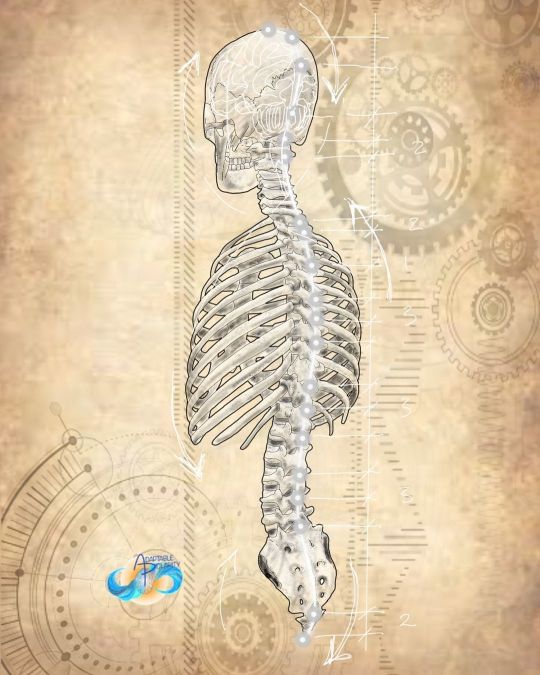
Axial Skeleton- Governor Vessel I have heard Ido Portal say, “you are your spine.” We have sayings, “spineless,” “having a spine. Countless yoga quotes about your spine and connecting to god or the infinite. I’ve seen Joseph Pilates quotes saying, “You’re only as old as your spine.” There are doctors fixated on unfixating your fixated spinal joints. And yet, how many take the time to listen to their spine? Axial Skeleton? How many only know, “ouch,” and not the finer freedom of spinal breathing. Growing hypothesis: within whatever dermatome or myotome one experiences discomfort, they will not breathe well at that level of the spine. We can learn to listen to our spines for ourselves and others. To feel the subtle things. #breath #breathing #breathoflifedaily #breathoflife #polarityoflife #spine #spinalbreathing #deeplistening #governingvessel #humanarchitecture #adaptablepolarity #cst #cranialsacraltherapy #craniosacraltherapy #craniosacral https://www.instagram.com/p/Cnr0aG3prGW/?igshid=NGJjMDIxMWI=
#breath#breathing#breathoflifedaily#breathoflife#polarityoflife#spine#spinalbreathing#deeplistening#governingvessel#humanarchitecture#adaptablepolarity#cst#cranialsacraltherapy#craniosacraltherapy#craniosacral
0 notes
Text
Findings in various levels of myelomeningocele
what is interesting is that myelomeningocele helped a lot in the study of the nerve root muscle supply by two means: first it gives a unique opportunity for stimulation of motor nerve roots and second to study of motor root lesions.
at least 50% of nerve supply to an individual muscle must be lost before clinical paralysis can be detected
infants born with myelomeningocele and treated conservatively are liable to remain paralyzed or become increasingly paralyzed. among the factors responsible for this deterioration may be traction exerted on the nerve roots passing from the plaque of spinal cord to the intervertebral foramina when the MM sac fools up with cerebrospinal fluid during the first week of life and the caudal roots tend to be more severely affected.
so lets go back to the title of this article, findings in various levels of myelomeningocele.
paralysis below T12: complete paralysis of lower limbs
paralysis below L1: weak to moderate hip flexion + weak sartorius = hip flexion and external rotation

paralysis below L2: strong hip flexion + moderate hip adduction = hip flexion and adduction

paralysis below L3: normal hip flexion + normal hip adduction + normal knee extension = hip flexion and adduction and knee extension
paralysis below L4: normal hip flexion + normal hip adduction + some hip abduction in flexion + normal knee extension + strong dorsiflexion and inversion = flexion and adduction and external rotation of hip + recurvatum of knee + calcaneo-varus foot
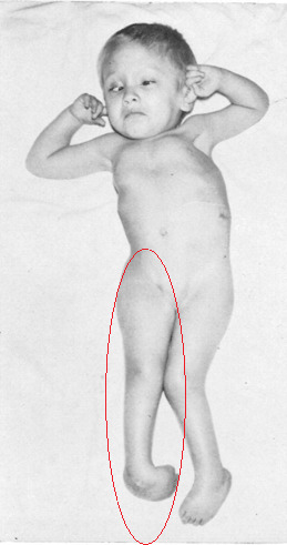

paralysis below L5: normal hip flexion + normal hip adduction + moderate abduction + no hip extension + normal knee extension + moderate knee flexion + normal inversion + moderate eversion + strong dorsiflexion + no planterflexion = hip flexion without adduction or abduction + semi-flexed knee + foot calcaneus

paralysis below S1: normal hip flexion + normal hip adduction + normal hip abduction + moderate hip extension + normal hip internal and external rotation + strong knee flexion + normal dorsiflexion + normal inversion + normal eversion+ moderate planterflexion + normal toes extension + strong great toe flexion + no foot intrinsic muscles (except abductor hallucis + flexor hallucis brevis) + weak flexor digitorum longus = clawing of toes and flattening of the skin of the sole of the foot

paralysis below S2: weak intrinsic muscles of foot = at clinical examination it is difficult to detect any abnormality but, with growth development of clawing of the toes.
so this study came with a conclusion regarding the innervation of muscles of the lower limbs

but newer studies using direct intraoperative nerve root stimulations during decompressive surgery concluded that a significant number of roots innervate a broader range of muscles than expected.
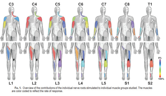
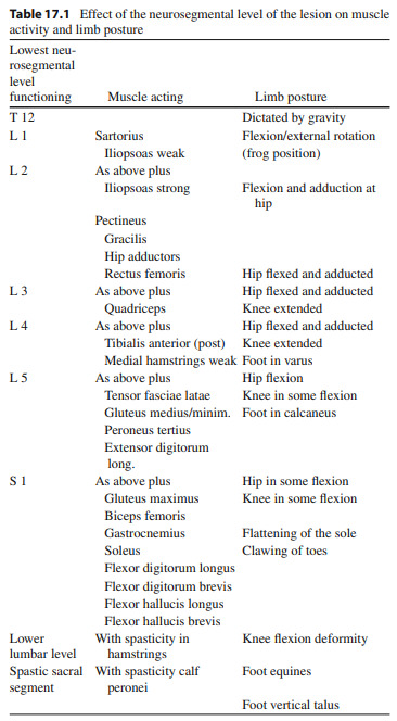
this table summarizes the neurological findings in MM
References:
(1) SHARRARD WJ. THE SEGMENTAL INNERVATION OF THE LOWER LIMB MUSCLES IN MAN. Ann R Coll Surg Engl. 1964 Aug;35(2):106-22. PMID: 14180405; PMCID: PMC2311748.
(2) Schirmer CM, Shils JL, Arle JE, Cosgrove GR, Dempsey PK, Tarlov E, Kim S, Martin CJ, Feltz C, Moul M, Magge S. Heuristic map of myotomal innervation in humans using direct intraoperative nerve root stimulation. J Neurosurg Spine. 2011 Jul;15(1):64-70. doi: 10.3171/2011.2.SPINE1068. Epub 2011 Apr 8. PMID: 21476796.
(3) Graham, H. K., & Parsch, K. (2009). Neural Tube Defects, Spina Bifida, and Spinal Dysraphism. Children’s Orthopaedics and Fractures, 265–286. doi:10.1007/978-1-84882-611-3_17
0 notes
Last Seen Blogs

seanconneryy
if you never shoot, never know
macklinsgf
Izzy.

idleminnies
[idle minnie]

littlehollyleaf
littlehollyleaf

auxanogram
18+ NSFW