#neurologyresident
Explore tagged Tumblr posts
Visit Tumblr Blog
Explore Tumblr blogs with no restrictions, modern design and the best experience.
Last Seen Tumblr Blogs


Fun Fact
There are dozens of funny blogs to kill time on Tumblr.
Link
Here’s one of our newest tutorial in Pathology and Pharmacology! Learn about Multiple Sclerosis and its treatments, check out our notes and flashcards, and see how our Drawing Pad works!
#ditki#neurology#multiplesclerosis#pathology#pharmacology#pathophysiology#acutemultplesclerosis#immunesystem#abpn#neurologycme#neurologist#neurologyresident#comlex#usmlereview#usmlestep1#usmlestep2ck#pathology tutorials#pharmacologytutorials
6 notes
·
View notes
Photo

Repost from @barrowneuro “In 2010, Barrow President and CEO Dr. Michael Lawton published a textbook on the tenets and techniques of surgical clipping for aneurysms, which are weakened areas of blood vessels that bulge outward and can bleed into the brain if left untreated. Named "Seven Aneurysms," the textbook features step-by-step strategies for handling the seven aneurysms most often seen by neurosurgeons. This illustration, from Ken Probst and courtesy of Thieme Medical Publishers, Inc. @thieme.ny shows the strategy for approaching a posterior communicating artery aneurysm.” #throwbackthursday #aneurysm #aneurysmclipping #clipping #cerebral #neuroscience #neurovascular #microsurgery #vasculardisease #neurosurgery #medicalillustration #medicalart #tbt #sciart #neurologyresident #neurosurgeon #residency #pathology #vascular #brainsurgery #photoshoppainting #digitalartwork #barrow #skuzz #textbook #medicalart #brains #neuro #scienceillustration #bloodvessels (at Barrow Neurological Institute) https://www.instagram.com/p/B2mewiznLbb/?igshid=1wvxqm9tr1zsj
#throwbackthursday#aneurysm#aneurysmclipping#clipping#cerebral#neuroscience#neurovascular#microsurgery#vasculardisease#neurosurgery#medicalillustration#medicalart#tbt#sciart#neurologyresident#neurosurgeon#residency#pathology#vascular#brainsurgery#photoshoppainting#digitalartwork#barrow#skuzz#textbook#brains#neuro#scienceillustration#bloodvessels
1 note
·
View note
Text
#علم#معلم#معلمات#علم_النفس#علمی#هل_تعلم#neurology#functionalneurology#neurologyresident#neurologylife#veterinaryneurology#pediatricneurology
0 notes
Photo
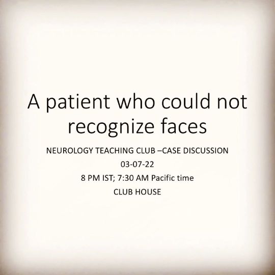
Neurology teaching club case presentation on clubhouse at 8 PM IST on 03/07/2022. A patient who could not recognise faces. #prosopagnosia #neurology #neuroscience #neurologyresident #neuroeducation #medicalstudent #medicaleducation #dementia #internalmedicineresidency #internalmedicine #bedsideteaching #clinicalcasediscussion #neurosurgery #medico #medicineresidency #medicineresidents https://www.instagram.com/p/CfijPGzLfPI/?igshid=NGJjMDIxMWI=
#prosopagnosia#neurology#neuroscience#neurologyresident#neuroeducation#medicalstudent#medicaleducation#dementia#internalmedicineresidency#internalmedicine#bedsideteaching#clinicalcasediscussion#neurosurgery#medico#medicineresidency#medicineresidents
0 notes
Text

Large hemispheric arachnoid cyst in an infant fenestrated and opened into basal cisterns and normal CSF absorption pathways for treatment. High-powered microscopic magnification showing
the delicate vasculature in a small baby.
#neurosurgery #neurosurgeon #neurosicence #brain #brains #brainsurgery #braintumor #brainpower #neuroanatomia #neurologia #neurología #neurologyresident #neurology #neurosurgeryblog #neurosurgerylife #neurocirurgia #neurociencias #arachnoidcyst #arachnoiditis #braincysts #pediatrician #pediatricneurosurgery #pediatricnurse #pediatrics #microsurgery
3 notes
·
View notes
Photo

Lobes of the brain Intro · The two hemispheres of the brain are divided into four lobes: o Frontal o Parietal o Temporal o Occipital · Each lobe carries a specific function. These will be explained in detail over the next few slides. Frontal Lobe · It’s located on the front section of the brain. · The frontal lobe is responsible for skills known as the Executive functions. These are: o Solving problems o Planning o Making decisions o Controlling our behaviour · It is considered as the emotional control center. Also, helps with attention skills and controlling movement. Parietal Lobe · It’s located behind the frontal lobes and above the temporal lobes. · The parietal lobe is responsible for processes information about the: o Temperature o Taste o Touch o Movement · Also, the parietal lobe allows us to perceive objects around our body. Therefore, we can move around without bumping into objects. This is known as Visuospatial Processing. Temporal Lobe · It’s located on the side section of the brain. · The temporal lobe is responsible for processes ..... References · https://courses.lumenlearning.com/waymaker-psychology/chapter/reading-parts-of-the-brain/ · https://headway.ie/about-the-brain/lobes-of-the-brain/ . . . #discoverphysiotherapy #physiotherapystudents #neuro #brain #lobes #lobe #frontal #parietal #occipital #temperature #temporary #temporal #touch #visual #sound #eye #eyes #visual #problemsolving #decisions #braincancerawareness #neurociencia #neurosurgeon #neuroblastoma #neuroformat #neuroanatomia #brainy #brain_waves #brainiac #brainyquote . @neurokinetictherapy @neuroorthobasedapproach @neurophysiowalesltd @neurofit360 @noigroup @neurorehab_geek @neurologicpt @neurosciencedubonheur @neuro.diversitea @neuropilates @integratedkineticneurology @neurologyresident @the_brain_doctor @neurology_review @neurologie.essen @loyolaneuro @neurobloggers @neurology_m @neuroacu @neurologysupportcentre @pennstateneuro https://www.instagram.com/p/CDQ3l_UHYwB/?igshid=1hza7bocyri3h
#discoverphysiotherapy#physiotherapystudents#neuro#brain#lobes#lobe#frontal#parietal#occipital#temperature#temporary#temporal#touch#visual#sound#eye#eyes#problemsolving#decisions#braincancerawareness#neurociencia#neurosurgeon#neuroblastoma#neuroformat#neuroanatomia#brainy#brain_waves#brainiac#brainyquote
0 notes
Photo

Cerebral Cortex What is the cerebral cortex? · Cortex – ‘the outer layer of the cerebrum (the cerebral cortex), composed of folded grey matter and playing an important role in consciousness.’ · The cerebral cortex is the outer layer of cells that surrounds the two cerebral hemispheres (or halves). It’s primarily grey matter. · The purpose of the cerebral cortex is to think and process information from our five senses. Closer look into the Cerebral cortex · The cerebral cortex’s surface is extensively folded, forming ridges called gyri (gyrus). · Valleys are formed due to the folding of the cerebral cortex. These valleys are called sulci (sulcus). · The folds increase the surface area of the cerebral cortex, this allows for more space for more neurons. · The two cerebral hemispheres are separated by a large sulcus called the medial longitudinal fissure. References · https://www.lexico.com/en/definition/cortex · https://brainmadesimple.com/cerebral-cortex-and-lobes-of-the-brain/ · https://www.neuroscientificallychallenged.com/blog/know-your-brain-cerebral-cortex . . . #discoverphysiotherapy #physioterapist #physio #physiotherapystudents #physioworld #physiostudent #physiotherapyday #physiotutors #brainpower #brain #brainfood #brainhealth #cerebral #gysi #cortex #cerebralcortex #hemisphere #neuro #neuroscience #neurology #thursday #thursdaymotivation . @neuroorthobasedapproach @neurokinetictherapy @neurophysiowalesltd @neurologicpt @neurorehab_geek @neuro @_neuroscience_ @physio_brain_hubz______ @integratedkineticneurology @neurology_review @neurobloggers @neuroacu @neurocliniclehi @neurologyresident https://www.instagram.com/p/CCs5nJZg1m4/?igshid=nrp4bxlxc7z2
#discoverphysiotherapy#physioterapist#physio#physiotherapystudents#physioworld#physiostudent#physiotherapyday#physiotutors#brainpower#brain#brainfood#brainhealth#cerebral#gysi#cortex#cerebralcortex#hemisphere#neuro#neuroscience#neurology#thursday#thursdaymotivation
0 notes