#retinotopy
Text
A connectomics-driven analysis reveals novel characterization of border regions in mouse visual cortex
Leveraging retinotopic maps to parcellate the visual cortex into its respective sub-regions has long been a canonical approach to characterizing the functional organization of visual areas in the mouse brain. However, with the advent of extensive connectomics datasets like MICrONS, we can now perform more granular analyses on biological neural networks, enabling us to better characterize the structural and functional profile of the visual cortex. In this work, we propose a statistical framework for analyzing the MICrONS dataset, focusing our efforts on the network encompassed by the retinotopically-induced V1, RL, and AL visual areas. In particular, we bridge the gap between connectomics and retinotopy by identifying several structural and functional differences between these regions. Most notably, by placing our attention on the borders between these regions, we demonstrate how connectomics, in some ways, supersedes retinotopy, providing evidence for two major findings. One, by comparing the V1-RL and RL-AL border regions, we show that not all borders in the visual cortex are the same with respect to structure and function. Two, we propose a novel interpretation for the V1-RL border region in particular, motivating it as a subnetwork that possesses heightened synaptic connectivity and more synchronous neural activity. Going one step further, we analyze structure and function in tandem by measuring information flow along synapses, demonstrating that the V1-RL border serves as a bridge for communication between the V1 and RL visual areas, offering justification as to why it presents itself uniquely with respect to both structure and function. http://dlvr.it/T7RcF3
0 notes
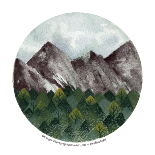
Photo
26
Through the spyglass on tumblr
@retinotopy on instagram
#through the spyglass#Daniel Smith#watercolor#background#background concept art#concept art#sennelier#holbein watercolor#mountains#scenery#sky#retinotopy#traditonal art#spyglass#spyglass series#landscape#watercolor art
11 notes
·
View notes
Text
Interesting Papers for Week 4, 2020
Structured inhibitory activity dynamics in new virtual environments. Arriaga, M., & Han, E. B. (2019). eLife, 8, e47611.
Lateral orbitofrontal cortex promotes trial-by-trial learning of risky, but not spatial, biases. Constantinople, C. M., Piet, A. T., Bibawi, P., Akrami, A., Kopec, C., & Brody, C. D. (2019). eLife, 8, e49744.
Dendritic NMDA receptors in parvalbumin neurons enable strong and stable neuronal assemblies. Cornford, J. H., Mercier, M. S., Leite, M., Magloire, V., Häusser, M., & Kullmann, D. M. (2019). eLife, 8, e49872.
The role of the nucleus accumbens in learned approach behavior diminishes with training. Dobrovitsky, V., West, M. O., & Horvitz, J. C. (2019). European Journal of Neuroscience, 50(9), 3403–3415.
Relation between gamma oscillations and neuronal plasticity in the visual cortex. Galuske, R. A. W., Munk, M. H. J., & Singer, W. (2019). Proceedings of the National Academy of Sciences, 116(46), 23317–23325.
Classical conditioning drives learned reward prediction signals in climbing fibers across the lateral cerebellum. Heffley, W., & Hull, C. (2019). eLife, 8, e46764.
An image-computable model for the stimulus selectivity of gamma oscillations. Hermes, D., Petridou, N., Kay, K. N., & Winawer, J. (2019). eLife, 8, e47035.
Every Local Minimum Value Is the Global Minimum Value of Induced Model in Nonconvex Machine Learning. Kawaguchi, K., Huang, J., & Kaelbling, L. P. (2019). Neural Computation, 31(12), 2293–2323.
Cerebellar climbing fibers encode expected reward size. Larry, N., Yarkoni, M., Lixenberg, A., & Joshua, M. (2019). eLife, 8, e46870.
Retinotopic specializations of cortical and thalamic inputs to area MT. Mundinano, I.-C., Kwan, W. C., & Bourne, J. A. (2019). Proceedings of the National Academy of Sciences of the United States of America, 116(46), 23326–23331.
Diversity of Ocular Dominance Patterns in Visual Cortex Originates from Variations in Local Cortical Retinotopy. Najafian, S., Jin, J., & Alonso, J.-M. (2019). Journal of Neuroscience, 39(46), 9145–9163.
Minimizing Precision-Weighted Sensory Prediction Errors via Memory Formation and Switching in Motor Adaptation. Oh, Y., & Schweighofer, N. (2019). Journal of Neuroscience, 39(46), 9237–9250.
Differential contributions of the two human cerebral hemispheres to action timing. Pflug, A., Gompf, F., Muthuraman, M., Groppa, S., & Kell, C. A. (2019). eLife, 8, e48404.
Dendritic Spikes Expand the Range of Well Tolerated Population Noise Structures. Poleg-Polsky, A. (2019). Journal of Neuroscience, 39(46), 9173–9184.
Functional Logic of Layer 2/3 Inhibitory Connectivity in the Ferret Visual Cortex. Scholl, B., Wilson, D. E., Jaepel, J., & Fitzpatrick, D. (2019). Neuron, 104(3), 451-457.e3.
Paradoxical Rules of Spike Train Decoding Revealed at the Sensitivity Limit of Vision. Smeds, L., Takeshita, D., Turunen, T., Tiihonen, J., Westö, J., Martyniuk, N., … Ala-Laurila, P. (2019). Neuron, 104(3), 576-587.e11.
Can Grid Cell Ensembles Represent Multiple Spaces? Spalla, D., Dubreuil, A., Rosay, S., Monasson, R., & Treves, A. (2019). Neural Computation, 31(12), 2324–2347.
Dopamine neuron ensembles signal the content of sensory prediction errors. Stalnaker, T. A., Howard, J. D., Takahashi, Y. K., Gershman, S. J., Kahnt, T., & Schoenbaum, G. (2019). eLife, 8, e49315.
Modular organization of cerebellar climbing fiber inputs during goal-directed behavior. Tsutsumi, S., Hidaka, N., Isomura, Y., Matsuzaki, M., Sakimura, K., Kano, M., & Kitamura, K. (2019). eLife, 8, e47021.
Reinforcement Learning in Spiking Neural Networks with Stochastic and Deterministic Synapses. Yuan, M., Wu, X., Yan, R., & Tang, H. (2019). Neural Computation, 31(12), 2368–2389.
#science#Neuroscience#computational neuroscience#Brain science#research#cognition#cognitive science#neurobiology#neurons#psychophysics#scientific publications
17 notes
·
View notes
Link
In physiology, tonotopy (from Greek tono = frequency and topos = place) is the spatial arrangement of where sounds of different frequency are processed in the brain. Tones close to each other in terms of frequency are represented in topologically neighbouring regions in the brain. Tonotopic maps are a particular case of topographic organization, similar to retinotopy in the visual system.
0 notes
Text
Diversity of Ocular Dominance Patterns in Visual Cortex Originates from Variations in Local Cortical Retinotopy
http://dlvr.it/RJClpr
0 notes
Photo
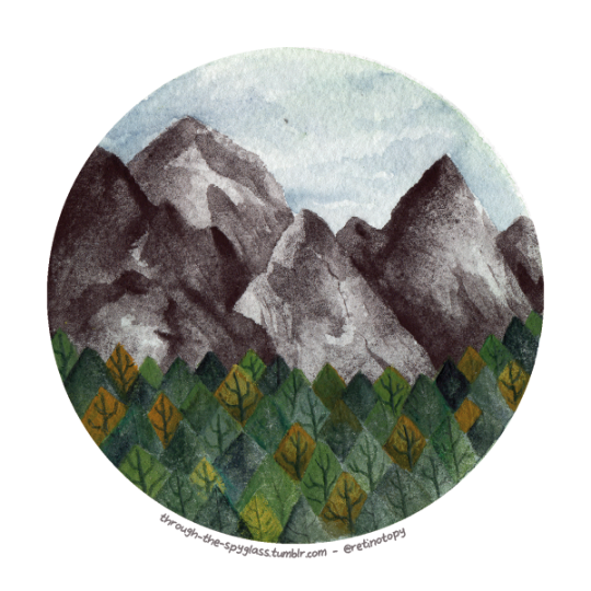
14
Through the spyglass on tumblr
@retinotopy on instagram
#through the spyglass#Daniel Smith#winsor and newton#watercolor#watercolour#watercolor pencils#fabercastell#Polychromos#albrechtdurer#traditonal art#background concept art#background#concept art#trees#mountains#sky#sennelier#watercolor painting#retinotopy#landscape#watercolor art
111 notes
·
View notes
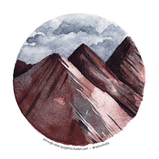
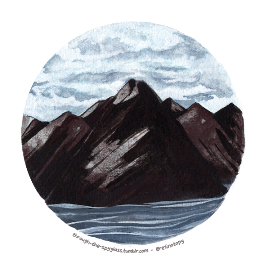
Photo
25
Through the spyglass on tumblr
@retinotopy on instagram
#through the spyglass#Daniel Smith#watercolor#background#background concept art#sennelier#holbein watercolor#mountains#cove#sky#retinotopy#traditonal art#spyglass#spyglass series#landscape#watercolor art
9 notes
·
View notes
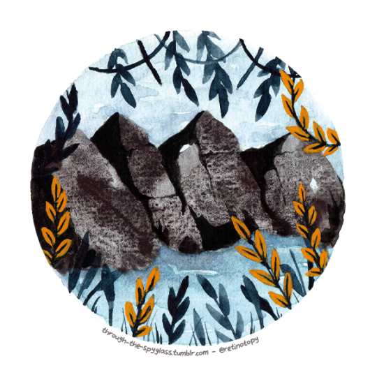
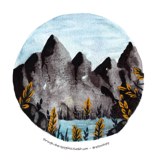
Photo
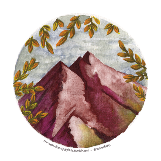
24
Through the spyglass on tumblr
@retinotopy on instagram
#through the spyglass#Daniel Smith#winsor and newton#watercolor#background#background concept art#sennelier#holbein watercolor#mountains#sky#retinotopy#traditonal art#spyglass#spyglass series#leaves#autumn#fall#landscape#watercolor art
6 notes
·
View notes
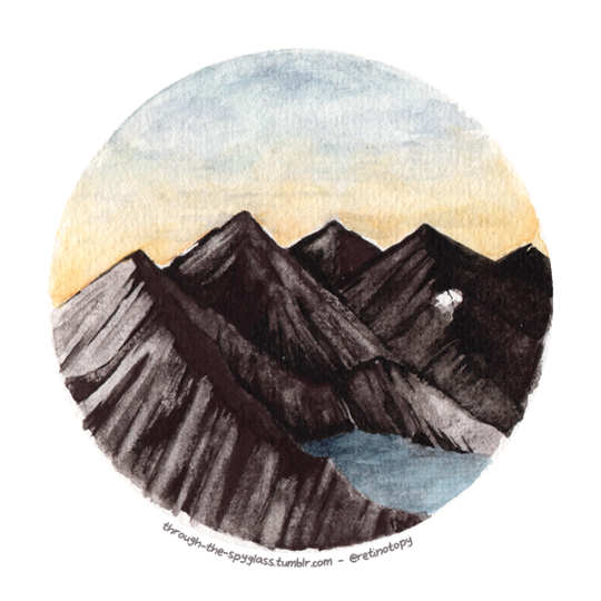
Photo
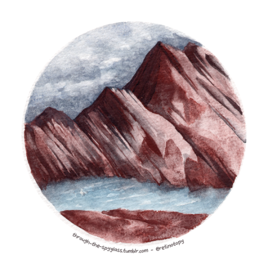
27
Through the spyglass on tumblr
@retinotopy on instagram
#through the spyglass#Daniel Smith#watercolor#background#background concept art#concept art#sennelier#holbein watercolor#mountains#scenery#sky#retinotopy#spyglass#spyglass series#landscape#river#water
2 notes
·
View notes
Photo
18
Through the spyglass on tumblr
@retinotopy on instagram
#through the spyglass#watercolor#watercolour#leaves#mountains#winsor and newton#Daniel Smith#background concept art#background#traditonal art#concept art#sennelier#water#lake#retinotopy#sky#gouache#acryla gouache#holbein#landscape#watercolor art
17 notes
·
View notes
Photo
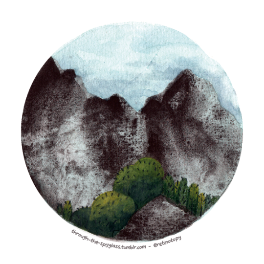
16
Through the spyglass on tumblr
@retinotopy on instagram
#through the spyglass#watercolor#watercolour#trees#mountains#winsor and newton#Daniel Smith#concept art#background concept art#traditonal art#artist on tumblr#sennelier#watercolor pencils#Polychromos#fabercastell#albrechtdurer#cacti#cactus#retinotopy#landscape#watercolor art
17 notes
·
View notes
Photo
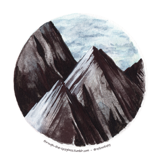
23
Through the spyglass on tumblr
@retinotopy on instagram
#through the spyglass#Daniel Smith#winsor and newton#watercolor#background#background concept art#sennelier#Holbein#mountains#sharp#watercolour#sky#clouds#retinotopy#traditonal art#spyglass#landscape#watercolor art
2 notes
·
View notes
Photo
15
Through the spyglass on tumblr
@retinotopy on instagram
#through the spyglass#watercolor#watercolour#trees#mountains#winsor and newton#daniel smith#concept art#background#background concept art#traditonal art#artist on tumblr#sennelier#watercolor pencils#polychromos#fabercastell#albrechtdurer#sky#retinotopy#landscape#watercolor art
13 notes
·
View notes
Photo
17
Through the spyglass on tumblr
@retinotopy on instagram
#through the spyglass#watercolor#watercolour#leaves#mountains#winsor and newton#Daniel Smith#background concept art#traditonal art#artist on tumblr#sennelier#water#lake#rocks#sky#retinotopy#gouache#holbein#acryla gouache#landscape#watercolor art
8 notes
·
View notes
Photo
21
Through the spyglass on tumblr
@retinotopy on instagram
#through the spyglass#Daniel Smith#winsor and newton#watercolor#watercolour#background#background concept art#sennelier#Holbein#holbein watercolor#mountains#sky#clouds#watercolor painting#retinotopy#traditonal art#spyglass#landscape#watercolor art
3 notes
·
View notes
Photo

7
Through the spyglass on tumblr
@retinotopy on instagram
#through the spyglass#watercolor#watercolour#trees#rocks#winsor and newton#danielsmith#concept art#background concept art#traditonal art#artist on tumblr#leaves#sennelier#watercolor painting#Polychromos#fabercastell#albrechtdurer#water#river#retinotopy#landscape#watercolor art
79 notes
·
View notes
Last Seen Blogs

homeforthebirdsofprey
She stole my fucking car!

brascatikompletadilat
çatı ustası

marcelosommer
sommer13
joshbriefs1428
Joshbriefs1428
joshbriefs1428
Joshbriefs1428