#tritc
Explore tagged Tumblr posts
Visit Tumblr Blog
Explore Tumblr blogs with no restrictions, modern design and the best experience.
Last Seen Tumblr Blogs


Fun Fact
1,644 Tumblr posts in 1 second.
Note
tritc or trent

your treat! half-life
3 notes
·
View notes
Text
Albumin, Tetramethylrhodamine isothiocyanate Bovine
Albumin, Tetramethylrhodamine isothiocyanate Bovine Catalog number: B2023610 Lot number: Batch Dependent Expiration Date: Batch dependent Amount: 10 mg Molecular Weight or Concentration: N/A Supplied as: Lyophilized Powder Applications: a molecular tool for various biochemical applications Storage: 2-8°C Keywords: TRITC-albumin Grade: Biotechnology grade. All products are highly pure. All…
0 notes
Text
Classification and application of fluorescent dyes
Classification and application of fluorescent dyes
August 16, 2023 Posted by Axispharm
Fluorescent dyes refer to substances that absorb light waves of a certain wavelength and emit light waves with a wavelength greater than that of the absorbed light. Most of them are compounds containing a benzene ring or a heterocyclic ring with a conjugated double bond. Fluorescent dyes can be used alone or combined into composite fluorescent dyes. Under the microscope, it functions by absorbing light at a given wavelength and re-emitting it at a longer wavelength. This fluorescence produces different colors that can be visualized and analyzed.
The composite fluorescent dye is a fluorescent dye synthesized by fluorescence resonance energy transfer technology, which is composed of a donor and an acceptor fluorescent substance molecules that are very close in distance and can transfer energy between each other. The complex dye is excited at the excitation wavelength of the acceptor molecule and emits a photon at the emission wavelength of the donor molecule. The development of fluorescent dyes is very rapid, and the fluorescent dyes developed for scientific research and clinical applications have basically covered the entire spectral range from ultraviolet to visible light and infrared.
Fluorescent dyes are commonly used for fluorescent labeling of various biomolecules (antibodies, peptides, various proteins, etc.) for monitoring drug delivery to target tissues, imaging, and other processes.
Fluorescent dye classification:
1. Fluorescein dyes, including fluorescein isothiocyanate (FITC), hydroxyfluorescein (FAM), tetrachlorofluorescein (TET), etc. and their analogs. This is a class of compounds with more benzene rings. The most widely used is FITC (as shown in the picture of FITC-labeled tissue fluorescence), which is excited by an argon ion laser at 488 nm and emits blue-green fluorescence at 525 nm. FITC can bind to various antibody proteins and stably exhibits blue-green fluorescence in alkaline solution.
2. Rhodamine dyes, including red rhodamine (RBITC), tetramethylrhodamine (TAMRA), rhodamine B (TRITC), etc. TRITC emits yellow fluorescence at 570 nm when excited at 550 nm.
3. Cy series cyanine dyes, cyanine dyes usually consist of two heterocyclic ring systems, including Cy2, Cy3, Cy3B, Cy3.5, Cy5, Cy5.5, Cy7, cy7 nhs ester , cy3b nhs ester and their analogs.
4. Alexa series dyes, which are series of fluorescent dyes developed by Molecular Probes. Its excitation light and emission light spectrum cover most of the visible light and part of the infrared spectral region, and it is widely used. Features high brightness, stability, instrument compatibility, multiple colors, pH insensitivity, and water solubility. Includes Alexa Fluor 350, 405, 430, alexa fluor 488 maleimide, 532, 546, 555, 568, 594, 610, 633, alexa 647 nhs ester, 680, 700, 750. At present, Alexa series dyes are widely used in the market and gradually replace traditional fluorescent dyes. For example, Alexa Fluor 488 can replace FITC and Cy2; Alexa Fluor 555 can replace Cy3 and TAMRA; Alexa Fluor 633 can replace APC, Cy5, etc.
5. APDye Fluors, APDye Fluors is the equivalent fluorescent dye of alexa fluors developed by axispharm. APDye Fluors series fluorescent dyes are bright in color, optically stable, very good in light stability of dyeing brightness, and very competitive in price. It can be applied to the labeling and localization of tissues, cells and biomolecules in biomedical research. Its excitation and emission spectra cover most of the visible and part of the infrared spectral region and are suitable for most fluorescence microscopes.
6. Protein dyes, including phycoerythrin (PE), phycocyanin (PC), allophycocyanin (APC), polydinoxanthin-chlorophyll protein (preCP), etc. Most of them are proteins found in cyanobacteria. Such fluorescent dyes can be coupled with Cy series cyanine dyes to form complex dyes for antibody labeling. For example, PE-Cy3/Cy5/Cy7, APC-Cy7, PerCP-Cy5.5, etc. are commonly used in the market.
Scientific research application
The earliest application of dyes in biochemistry was to directly stain sections and then visualize them. With the continuous development of biotechnology, computer technology and fluorescence spectrometry technology, many dyes, especially fluorescent dyes, have been widely used in cell detection, tumor gene protein analysis, toxic analysis, clinical medical diagnosis and so on.
Due to its high sensitivity and convenient operation, fluorescent dyes have gradually replaced radioisotopes as detection markers, and are widely used in fluorescent immunology, fluorescent probes, and cell staining. Including specific DNA staining, for chromosome analysis, cell cycle, apoptosis and other related research. Numerous nucleic acid dyes are also useful counterstains in multicolor staining systems as background controls, labeling nuclei so that the spatial relationships of intracellular structures can be seen at a glance.
Immunoassay
Fluorescence-labeled monoclonal antibody technology expands infinite application space for flow cytometry in the study of cell membranes and various functional antigens in cells, tumor gene proteins and other fields. Fluorescent probes can be covalently bound to monoclonal antibodies via protein cross-linkers. The most commonly used dyes for immunofluorescence labeling are fluorescein isothiocyanate (FITC), phycoerythrin (PE) and AlexaFluor series dyes.
Nucleic acid amplification testing
Nucleic acid fluorescent dyes stain the nucleus and quantitatively measure the fluorescence intensity of the cell, so that the content of DNA and RNA in the nucleus can be determined, and the cell cycle and cell proliferation can be analyzed. There are a variety of fluorescent dyes that can stain DNA or RNA in cells. Commonly used DNA dyes include propidium iodide (PI), DAPI, Hoechst 33342, etc., and RNA dyes include thiazole orange, acridine orange, etc.
Ideal fluorescent dyes generally have the following characteristics:
1. It has high photon yield and high signal intensity;
2. Strong absorption of excitation light, reducing background signal;
3. The distance between the excitation spectrum and the emission spectrum is large to reduce the interference of background signals;
4. It is easy to combine with the labeled antigen, antibody or other biological substances without affecting the specificity of the labeled substance;
5. Good stability, not easily affected by light, temperature, pH, specimen anticoagulant and fixative.

0 notes
Photo


In December 2019, there was a cluster of pneumonia cases in China.Investigations found that it was caused by a previously unknown virus – now named the 2019 Novel Coronavirus.
In this artical, we'll take a quick look at what's currently known about the virus.Keep in mind that this is a new virus and what's known about the virus now might change in the future.
Coronaviruses are a large group of viruses.They consist of a core of genetic material surrounded by an envelope with protein spikes.This gives it the appearance of a crown.Crown in Latin is called "corona" and that's how these viruses get their name.There are different types of coronaviruses that cause respiratory and sometimes gastrointestinal symptoms.Respiratory disease can range from the common cold to pneumonia.And in most people, the symptoms tend to be mild.
However, there are some types of coronaviruses that can cause severe disease.These include the Severe Acute Respiratory Syndrome coronavirus first identified in China in 2003 and the Middle East Respiratory Syndrome coronavirus that was first identified in Saudi Arabia in 2012.The 2019 Novel Coronavirus was first identified in China.It initially occurred in a group of people with pneumonia who'd been associated with a seafood and live animal market in the city of Wuhan.The disease has since spread from those who were sick to others, including family members and health care staff.There are many cases at present and the disease has spread within China and also to a number of other countries.
Q1:Where did the virus come from?
It's known that coronaviruses circulate in a range of animals.Sometimes these viruses can make the jump from animals to humans.This is called a spillover and could be due to a range of factors such as mutations in the virus or increased contact between humans and animals.
For example, MERS-CoV is known to be transmitted from camels and SARS-CoV, from civet cats.The animal reservoir of the 2019 Novel Coronavirus is not known yet.
Q2:How is it transmitted?
The exact dynamics of how the virus is transmitted is yet to be determined.In general, respiratory viruses are usually transmitted through droplets created when an infected person coughs or sneezes, or through something that has been contaminated with the virus.People most at risk of infection from the Novel Coronavirus are those in close contact with animals, such as live animal market workers, and those who are caring for people infected with the virus, such as family members or healthcare workers.
Q3:How does the disease present?
Well, from what is known so far, there can be a number of symptoms ranging from mild to severe.There can be fever and respiratory symptoms such as cough and shortness of breath.In more severe cases, there's been pneumonia, kidney failure, and death.The mortality rate is not known yet.
Q4:How can we tell whether someone is infected?
The infection can be diagnosed by a test called PCR or Polymerase Chain Reaction.This test identifies the virus based on its genetic fingerprint.There is currently no specific medication for the virus and treatment is supportive care.There's currently no vaccine to protect against the virus.Treatment and vaccines are in development.
Q5:How do we prevent transmission of the virus?
This new virus currently has a limited geographic spread.However, there are a number of standard hygiene practices that have been recommended to protect against infection and further spread.These include covering your mouth and nose when coughing or sneezing with a medical mask, tissue or flexed elbow; avoiding close contact with those who are unwell; the appropriate use of masks and personal protective equipment, especially in a healthcare setting; washing hands regularly with soap and water, or alcohol-based hand rub.
Q5:How does the virus looks like?
Images of Novel Coronavirus sames were captured using the OPTOLONG@fluorescence filter set:FITC,DAPI,TexaRed,Fish Orange filter set(EX,EM,DM),An Nikon microscope with a 100x objective were used.
Single-band Sets:FITC,DAPI,TexaRed,CY5,TRITC Individual Filter:Longpass,shortpass,bandpass filter,AR,IR,IRCUT... Dichroic Mirror:416nm,455nm,492nm,555nm,655nm.... FISH Set:Orange,Qqua,Green,Red. Cubes or Holders: U-MF2,U-FF,Zeiss CUBE. By Application:Machine Vision,Life Science Instrument,Biomedical Imaging System, Industrial Detection,Imging or Photonics...
Actions that can be taken to prevent infection from an animal source include: avoiding unnecessary unprotected contact with animals;washing hands after contact with animals or animal products; and ensuring that animal products are cooked thoroughly before they're consumed.It's important to stay home if you're feeling unwell.But if you have a fever, cough, and difficulty breathing, seek medical care early and share your previous travel history with your healthcare provider.
That's a quick look at this emerging infectious disease.
#FITCDAPITexaRed,CY5,TRITC#FITC#DAPI#TexaRed#cy5#tritc#416nm#455nm#FISH Set#U-MF2#U-FF#Zeiss CUBE#Industrial Detection#Fish Orange filter#Nikon microscope#100x objective
0 notes
Text

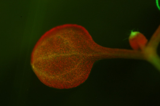
I am so glad you've mentioned absorption/emission spectras because it doesn't just apply to the elements in stars; it's literally everything! this here is a fluorescent microscope photo i took of a slice of rat brain where the blue (the DAPI) is a stain for the nuclei, the red (the TRITC) is a stain for the densest bundles of nerves, and the green (the FITC) isn't stained at all.
that's right: you are naturally green. most every living critter composed of mostly water and carbon averages out an emission spectrum that puts it squarely in the green portion of the visible light spectrum. this is known as autofluorescence. it's so faint you can only pick it up with a very sensitive microscope lens in a darkroom, though.
Okay fuck it if this post reaches 666k notes by the end of 2023 I'll practise basic self care
Why 666k? Because it's funny and impossible so good fucking luck
742K notes
·
View notes
Text
How to choose a right camera for your Fluorescence Microscope ?
It’s a challenge to choose a right camera for your research. There are various cameras in the market . So, what should you focus on when choosing a digital camera for fluorescence imaging .
At first , you need to know about your imaging requirements . Secondly, it’s also important to know the technical characteristics of each camera .
Here are some options to considering :
>Imaging cells and tissues
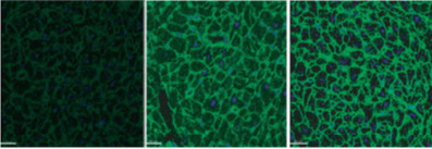
For a wide-field fluorescence microscope, the camera will be used to image stationary cells and tissues . In the below picture,two different cameras were used to image the Alexa Fluor 488-labeled actin filaments immobilized in endothelial cells . There are some key technical difference between the two cameras that make for a four-fold difference in cost . Using standard imaging conditions, both cameras produce sharp , clear images ,and the noise and background seem to make no difference . Well, you may ask what is the difference between the two cameras and is it necessary to buy the more expensive one ?

The main difference between the two images is that the one on the left was captured with an uncooled camera , while the one on the right was captured with a cooled camera . In the past ,the fluorescent imaging has to be cooled, however, advances in camera sensor technology over the last 25 years have meant that cooling is no longer required for conventional fluorescent imaging . For challenging fluorescent samples that require sophisticated microsopes(such as confocal microscopes) ,cooling cameras are ideal choice .But for most fluorescent imaging ,cooling is not required . Uncooled cameras also produce great images at a lower cost .
>Nosie
For fluorescence microscopes, noise affects signal-to-noise ratio and dynamic range .
A high signal-to-noise ratio(SNR) results in sharp contrast and good sharpness ,while as SNR decreases, background signals increase and image sharpness and contrast decrease . Pixel merging can be used to reduce noise and improve SNR , but, it will affect space resolution. The main source of noise in a fluorescence microscopes is the read noise, which is much higher than the dark current noise .
The dark current noise is independent of temperature of the sensor . Cooling cameras reduce dark current noise ,which can be considered for use in situations requiring long exposures or very low light intensity . In general,dark currents have no significant effect on standard fluorescent imaging. So, unless you are imaging in a situation that requires a long exposure or low light intensity, the effect of dark current on the image is minimal .
>Other considerations
Of course, other imaging variables also affect image quality . To maximize the image quality,we should use a uniform light sourc,a high NA objective and a good optical filter combination .
Combining the right camera technology with your microscope system will ensure you get excellent images .
In addition , some challenging applications, such as confocal imaging, calcium signal imaging and near-infrared fluorescence, have more stringent requirements for the camera to achieve the desired results . However, for general fluorescent imaging ,a high quality , uncooled camera can also produce sensitive images . That way , you can spend your precious money on other equipments .
You may also need fluorescence filter set for your fluorescence microscope .
Optolong optics(www.optolong.com) supply filter set DAPI , FITC , TRITC,TexasRed, Cy5 , dual-band green/orange , which you may interesting .

0 notes
Text
New Post has been published on Abbkine - Antibodies, proteins, biochemicals, assay kits for life science research
The third generation of fluorescent dye AbFluor™/highly water-soluble fluorescent dye

AbFluor™ dyes are a series of highly water-soluble fluorescent dyes introduced by Abbkine after many years of research, covering the ultraviolet, visible and near-infrared spectrum, suitable for labeling biological macromolecules, especially proteins and nucleic acids. Their excellent hydrophilicity enables protein-conjugated labeling to be performed easily in aqueous media, while minimizing the need for organic solvents. AbFluor™ dyes not only have better fluorescence performance than other fluorescent dyes (such as FITC, TRITC, Alexa Fluor and Dylight dyes), but also the coupling products are more stable.

Why recommend AbFluor™ fluorescent dyes?
With innovative modifications to the core structure of the dye, AbFluor™ dyes have more brilliant properties than other commercial dyes, including stronger fluorescent brightness, better light stability, wider pH tolerance range, and better penetration and lower background. As a third-generation fluorescent dye, AbFluor™ dyes not only perform better than traditional dyes (such as FITC, TRITC, and Cy dyes), but also perform better than other second-generation commercial dyes (such as Alexa Fluor, Dylight and IRDye).
1. AbFluor™ dyes have excellent core structure, stable reactive groups, and good light stability, ensuring high stability and high labeling efficiency of bioconjugation.
2. High water solubility (>100 mg/ml), which minimizes fluorescence quenching; it is also easily soluble in other polar solvents, such as DMSO, DMF, methanol and ethanol.
3. A wide range of fluorescence spectrum, covering ultraviolet, visible, and near-infrared, to meet most of the experimental needs of researchers.
4. Good pH tolerance, stable in pH 2-11.
Taking AbFluor™ 488 as an example, the following is an IF comparison chart of different dye effects:
Different dye : FITC Alexa Fluor 488 AbFluor™ 488
So, are you excited, and what are you waiting for???
Abbkine AbFluor™ product recommendation:
Product name #Cat Szie Price ($) LinKine™ AbFluor™ 405 Labeling Kit KTL0510 3*20μg/100μg/ 3*100μg/1mg 1200/1400/ 1600/2500 LinKine™ AbFluor™ 488 Labeling Kit KTL0520 3*20μg/100μg/ 3*100μg/1mg 1200/1400/ 1600/2500 LinKine™ AbFluor™ 555 Labeling Kit KTL0530 3*20μg/100μg/ 3*100μg/1mg 1200/1400/ 1600/2500 LinKine™ AbFluor™ 594 Labeling Kit KTL0540 3*20μg/100μg/ 3*100μg/1mg 1200/1400/ 1600/2500 LinKine™ AbFluor™ 647 Labeling Kit KTL0560 3*20μg/100μg/ 3*100μg/1mg 1200/1400/ 1600/2500 LinKine™ AbFluor™ 680 Labeling Kit KTL0580 3*20μg/100μg/ 3*100μg/1mg 1200/1400/ 1600/2500 LinKine™ AbFluor™ 770 Labeling Kit KTL0590 3*20μg/100μg/ 3*100μg/1mg 1200/1400/ 1600/2500
0 notes
Text
A picture of cells from the first time I did cell culture and, subsequently, immunohistochemistry in FITC and TRITC
you can tell a lot about someone based on their phone background. it shows what’s most important to them
738K notes
·
View notes
Text
Bioadvisers shared on Biotech Advisers
Essential guidance| Let apoptotic cells show the glory of life
nconsciously, we have entered the hot summer solstice, and the high temperature has accelerated the metabolism of living organisms. The vigorous increase of nascent cells gradually replaced apoptotic cells that completed their mission. Fallen flowers are not heartless things, into Chunni more quadrangle. Apoptotic cells use their own fall to complete the life cycle of the organism.
Apoptosis refers to the autonomous and orderly death of cells controlled by genes in order to maintain the stability of the internal environment. Apoptosis is different from cell necrosis. Apoptosis is not a passive process, but an active process. It involves the activation, expression and regulation of a series of genes; it is not a kind of auto-injury under pathological conditions. Phenomenon, but a process of death that is actively striving for better adaptation to the living environment. When a cell undergoes apoptosis, it is like the natural fall of a leaf or flower. For this biological observation, borrowed from the Greek “Apoptosis”, it means that the natural fall of a leaf or flower can be translated as apoptosis. So what is the existence of apoptosis?
Apoptosis is the only way in the development of animals. Looking at the following examples, the phenomenon of apoptosis is everywhere in the development of life:
Abnormal development from tadpoles to frogs
Metamorphosis of insects
The disappearance of webs between the fingers of higher mammals. For example, during the development of human embryos, flat disc-shaped limbs sprouting when the embryo grows to the fifth week, and the webs between the fingers and toes disappear, so that the limbs can form their final shape smoothly.
The cell death of the vitreous and lens in the eye is an important step for the eye to penetrate light.
Even in mature individuals, apoptosis is essential:
Control of cell number and tissue size
Renewal of tissues (e.g. replacement of nasal olfactory epidermis and skin epidermal cells)
The immune system selects and removes cells that are either non-functional or potentially dangerous, such as the non-specific cell killing effect of natural killer cells (NK) on cancer cells.
Remove degeneration cells
Give plasticity to the central nervous system. The researchers tried to activate the mouse’s bcl-2 gene during the embryonic stage. This gene prevents the apoptosis of nerve cells. The number of neurons in the brain of the born mouse is therefore larger than that of the normal mouse, and the volume becomes larger. But in the test, it seems slower than normal mice.
Selection of germ cells (about 95% of germ cells will be cleared by apoptosis before they mature)
Abbkine scientists’ research on apoptosis has reached the cellular, protein and molecular levels:
Protein level Annexin V series cell early apoptosis kit to detect valgus phosphatidylserine (PS) eversion:
Product name Cat# Abs/Em Alternative Annexin V-AbFluor™ 405 Apoptosis Detection kit KTA0001 Annexin V- AbFluor™ 405: Abs/Em = 404/431 nm; PI: Abs/Em = 535/617 nm (with DNA) Pacific Blue, Alexa Fluor 405, Dylight 405 Annexin V-AbFluor™ 488 Apoptosis Detection kit KTA0002 Annexin V- AbFluor™ 488: Abs/Em = 491/517 nm; PI: Abs/Em = 535/617 nm (with DNA) FITC, Alexa Fluor 488, Dylight 488 Annexin V-AbFluor™ 555 Apoptosis Detection kit KTA0003 Annexin V- AbFluor™ 555: Abs/Em = 555/565 nm Cy3, TRITC, Alexa Fluor 546, Dylight 549 Annexin V-AbFluor™ 647 Apoptosis Detection kit KTA0004 Annexin V- AbFluor™ 647: Abs/Em = 650/665 nm; PI: Abs/Em = 535/617 nm (with DNA) Cy5, Alexa Fluor 647, Dylight 649
Cell-level Caspase series kits to detect the expression of apoptosis-related protein Caspas and mid-term apoptosis:
Product NO Test purpose Product Name KTA3020 Apoptosis
(Colorimetry)
Caspase-1 Assay Kit (Colorimetric) KTA3021 Caspase-2 Assay Kit (Colorimetric) KTA3022 Caspase-3 Assay Kit (Colorimetric) KTA3023 Caspase-4 Assay Kit (Colorimetric) KTA3024 Caspase-6 Assay Kit (Colorimetric) KTA3025 Caspase-8 Assay Kit (Colorimetric)
The cascade reaction initiated by the Caspase cysteine protease family is the central link in the process of apoptosis. Its activation mainly includes mitochondrial-dependent pathways and death receptor-mediated signal transduction pathways. The activated downstream Caspase, through cleavage specificity Substrate, leading to apoptosis.
Molecular level TUNEL kit, detection of late apoptosis kit
Product Name Product NO TUNEL Apoptosis Detection Kit (Green Fluorescence) KTA2010 TUNEL Apoptosis Detection Kit (Orange Fluorescence) KTA2011
The apoptotic cells stained by the TUNEL fluorescence kit have a very bright fluorescent color, which is favored by Abbkine customers.
There are many studies on the relationship between apoptosis and the occurrence of cancer and various autoimmune diseases. People hope to stimulate the apoptosis of cancer cells in order to eliminate cancer. In addition, in order to maintain the size of the human brain, the genes that cause apoptosis are suppressed in humans.
The role of apoptosis in neurodegenerative diseases (such as Alzheimer’s disease (one type of dementia), Huntington’s disease, Parkinson’s disease, muscle Atrophic lateral sclerosis (ALS) is currently a hot research topic, and there are many studies in this area.
In order to provide readers with a platform for learning biological knowledge, Abbkine’s technical experts have worked hard to sort out the dry goods of various high-end biological experiments. Later, they will be published in the WeChat public account, and readers who love science will continue to pay attention to the Abbkine public account. .Abbkine specializes in the fields of proteinology and cytology, and is committed to innovating and developing various antibodies, proteins, analytical reagents and kits, with a view to becoming a key promoter in the fields of life science research and development, drug research and development. We provide you with favorite products of protein and immunological research users, from basic immunological products, such as protein extraction and quantification, to internal reference labeling antibodies, primary antibodies and secondary antibodies of immunological experiments, etc.; favorite products of cell research users, from Dyes and kits for detecting the state of cells, kits for extracting organelles, staining and tracking of cell substructures and cell metabolism detection products, to cytokine and protein detection kits for cell culture, only to help your research career !About AbbkineOur position: to serve global users of cell and protein research, to provide users with economical and technical product solutions in the form of application processes and product portfolios.Our mission: to stimulate our inner creativity, provide competitive biomedical products and services, and continue to create maximum value for customers.Our vision: to be a respected, world-class supplier of biomedical products and services.
0 notes
Text
Polysucrose 400-Tetramethylrhodamine Isothiocyanate Conjugate
Polysucrose 400-Tetramethylrhodamine Isothiocyanate Conjugate Catalog number: B2013035 Lot number: Batch Dependent Expiration Date: Batch dependent Amount: 100 mg Molecular Weight or Concentration: 350-450 kDa Supplied as: Lyophilized Powder Applications: molecular tool for various biochemical applications Storage: -20°C Keywords: TRITC-Polysucrose 400, Tetramethylrhodamine…

View On WordPress
0 notes
Text
New Post has been published on Abbkine - Antibodies, proteins, biochemicals, assay kits for life science research
New Post has been published on https://www.abbkine.com/ifkine-orange-donkey-anti-mouse-igg-becomes-new-addition-abbkine-family/
IFKine™ Orange Donkey Anti-Mouse IgG becomes the new addition to the Abbkine family
Abbkine Scientific Limited has recently announced the launch of its new product - IFKine™ Orange Donkey Anti-Mouse IgG. The Orange dye Donkey Anti-Mouse IgG conjugated antibodies are made to absorb light maximally at 555 nm and fluoresce with a peak around 570 nm. The antibody is brighter than its counterparties like TRITC, Cy3 antibody.
Hosted in donkeys and with mouse reactivity, the antibody’s immunogen is Mouse IgG whole molecule, with applications such as FCM, ICC, and IF. The antibody is polyclonal and is affinity purified using solid phase Mouse IgG (H&L) with finally > 95% purity based on SDS-PAGE.
Available in liquid solution, the antibody reacts with whole molecule mouse IgG. The antibody also reacts with light chains of all other mouse immunoglobulins. The minimization of the antibody’s cross-reaction with human, rabbit, goat, sheep and guinea pig serum proteins helps to reduce non-specific hybrization.
Abbkine IFKine™ is series of unique fluorence staining secondary antibodies with improved brightness, photostability and less nonspecific hybridization and background. The latest generation of IFKine fluorescent dyes ensure the best fluorescent performance, while it’s donkey host and other species of serum/IgG absorbed make IFKine™ secondary antibodies the ideal for use in fluorence staining, especially in fluorence multiple labeling.
About Abbkine Scientific Co. Ltd
Abbkine Scientific Co. Ltd is scientific research company founded in 2012 by number of scientists and marketing experts in the field of life science in California, USA. The company is headquartered in China and has effectively combined cutting edge technology from United States with China's manufacturing engineering and cost advantages to provide innovative, high quality assay kits, recombinant proteins, antibodies and other research tools to accelerate life science fundamental research and drug discovery.
0 notes
Text
New Post has been published on Abbkine - Your life science partner
Interests on http://www.abbkine.com/ifkine-orange-donkey-anti-mouse-igg-released/
IFKine™ Orange Donkey Anti-Mouse IgG Released
IFKine™ Orange conjugated antibodies absorb light maximally at 555 nm and fluoresce with a peak around 570 nm. They are brighter than TRITC, Cy3. IFKine™ Orange conjugates are recommended for for all immunofluorescence procedures requiring a orange -fluorescing dye. IFKine™ Orange Donkey Anti-.... Click for moreIFKine™ Orange Donkey Anti-Mouse IgG Released.
0 notes
Photo


What is fluorescence filter ?
Fluorescence filter is a key component used in biomedical and life science instruments.
It’s main function is to separate and select the characteristic band spectrum of the substances’ excitation light and emitted fluorescence in the biomedical fluorescence inspection analysis system .
Generally speaking , a fluorescence filter set consists of three different filters.
①Excitation Filter (Size φ25×5mm)
②Emission Filter (Size φ25×3.5mm)
③Dichroic Mirror (Size 25.7×36×1mm)
0 notes
Video
tumblr
In life science instrument, for the sake of quality image analyze and accurate diagnose, optical filters must separate desired wavelength from whole spectrum. Meanwhile to keep high standard attunation to the unwanted area.
Fluorescence Filters are widely used in varies kinds of life science and bio tech instrument. It’s known as high transmission to bright the subject and deep blocking to dark background. Also to make the scatter light to be least.
OPTOLONG has been offered fluorescence filters for many instrument manufacturer and also dealer. We could offer right solution to client according to client’s application. Standard blocking dept for fluorescence filters can be OD4, OD5 or above. Standard transmission could be from 70% to 95%. OEM for size from Dia4mm to Dia70mm, transmission, slop are acceptable and with reasonable and economic price.
Below is the application we involved in:
Microplate reader
Realtime PCR
Fluorometer
Cell counter
Flow cytometry
Fluorescence quantitative analyzer
Fluorescent Cell Analysis System
DNA sequence and analyzer
Microscope imaging system
Cell imaging system
Fluorometer and Absorbance Reader
Biochemical analyzer
Longpass filter set ( B,G,UV,V,R standard for fluorescence microscope) or other longpass set.
Shortpass filter set.
Single band filters set :Regular DAPI, FITC, Texas Red, TRITC, CFP, YFP, GFP, Cy3.5, Cy5.5.
FISH Application:FISH Aqua, FISH Red, FISH Orange, FISH Green, more coming soon.
Dual band filters set:DAPI-FITC, DAPI-Texas Red, FITC-Texas Red.
Triple band filter set:DAPI-FITC-Texas, DAPI-FITC-TRITC.
Individual filters:Individual exciter, emitter, dichroic mirror.
#coating film#fish aqua#FITCDAPITexaRed,CY5,TRITC#Dual band filter#Fluorescence filters#hard coating film#Longpass filter#Longpass filter set#Shortpass filter set#dichroic mirror
0 notes
Text
Polysucrose 70-Tetramethylrhodamine Isothiocyanate Conjugate
Polysucrose 70-Tetramethylrhodamine Isothiocyanate Conjugate Catalog number: B2012989 Lot number: Batch Dependent Expiration Date: Batch dependent Amount: 500 mg Molecular Weight or Concentration: 60-80 kDa Supplied as: Lyophilized Powder Applications: molecular tool for various biochemical applications Storage: -20°C Keywords: TRITC-Polysucrose 70, Tetramethylrhodamine isothiocyanate-Polysucrose…

View On WordPress
0 notes
Text
Rabbit anti-Hamster IgG (H&L) Conjugated to TRITC
Rabbit anti-Hamster IgG (H&L) Conjugated to TRITC
Rabbit anti-Hamster IgG (H&L) Conjugated to TRITC Catalog number: B2011218 Lot number: Batch Dependent Expiration Date: Batch dependent Amount: 1.0 mg Molecular Weight or Concentration: 1.0 mg/mL Supplied as: Lyophilized Applications: molecular tool for various biochemical applications Storage: 2-8 °C Keywords: anti-Hamster IgG (H+L) TRITC Grade: Biotechnology grade. All products are highly pure.…

View On WordPress
0 notes