Don't wanna be here? Send us removal request.
Statistics
We looked inside some of the posts by fitcfilter and here's what we found interesting.
Average Info
Notes Per Post
195
Likes Per Post
160
Reblog Per Post
29
Reply Per Post
6
Time Between Posts
21 days
Number of Posts By Type
Text
8
Photo
6
Video
1
Link
1
Last Seen Tumblr Blogs


Fun Fact
Tumblr Inc. is funded by 13 investors.
Text
Can anyone help with a fluorescence microscopy choose
Most of the applications of the FluidFM systems can be easily followed through fluorescence microscopy: from spotting a fluorescent solution in a glass surface to the injection of specific complexes into single adherent cells.
Fluorescence and fluorophores
Fluorescence refers to the physical property of an object absorbing light at one specific wavelength and then remitting it at another one. Then, when a molecule absorbs light (or it is excited) of one wavelength and emits in another one, that molecule is called fluorophore.
Each fluorophore has a characteristic excitation and emission spectra. Maximum values are the peaks of excitation and emission, respectively. However, most fluorophores absorb and emit in a range of wavelengths; for imaging, it is useful to think not just about their peaks, but also about the full spectrum of absorption (or excitation) and emission:
Excitation (blue) and emission (orange) spectra for Lucifer Yellow; peaks for excitation and emission are 428 and 544 nm, respectively.
The properties in the excitation and emission spectra can also determine the visualization under the fluorescence microscopy: the closer the spectra are, the more difficult it will be to see the emitted light from the labeled object as a separate from the one used for excitation.
Fluorescence filters
optical filters bandpass In order to collect the light form the specific fluorophore, a set of filters is introduced in the microscope: exciter filter, dichroic mirror and emission filter (or barrier filter). In our system, the three filters are located into a cube (figure 2).

Scheme of a fluorescence filter set (cube). (b) Available filters in the FluidFM BIO Series. Images adapted from
www.optolongfilter.com
website
The exciter filter has generally a defined band of wavelengths that it allows through. After going through this filter, it reflects onto the sample. Fluorophores in the sample will then become excited and, thus, emit light at a different wavelength. The dichroic mirror and, then, the emission filter will filter this emitted light.
In the FluidFM BIO Series, the filter sets available are the following ones:
Filter
Excitation Filter
Emission Filter
Corresponding color in the visible spectrum
U-FUNA
360-370 nm
420-460 nm
Blue
U-F39002
480 nm
535 nm
Green
U-F39004
540 nm
605 nm
Red
What should be considered when selecting a fluorophore and a filter set?
In order to obtain a better resolution of our samples and adapt the fluorescence intensity to our needs, it is important to take into account the following concepts when selecting the fluorescence presets and fluorophores for the experiment: fluorescent light filter
1. Selectivity of the fluorescent label. The fluorophore must be specific for a target: a molecule, a biological activity or a cellular location, without nonspecific background signal.
2. Photostability. The fluorophore must be able to maintain its properties after repeated exposures to illumination light.
3. Excitation and emission properties of the fluorophore. The filter set should be compatible with the range of wavelengths of the fluorophore (and vice versa).
4. Environmental stability. Some fluorophores can be sensitive to air, light or temperature.
Fluorophore
Excitation wavelength
Emission wavelength
Fluorescence filter
Lucifer Yellow
428 nm
544 nm
U-F39002 (green)
DAPI
359 nm
461 nm
U-FUNA (blue)
Hoechst
352 nm
455 nm
U-FUNA (blue)
Propidium Iodide
300 nm
610 nm
U-F39004 (red)
Alexa Fluor 555 dextran
555 nm
585 nm
U-F39004 (red)
Alexa Fluor 568
580 nm
600 nm
U-F39004 (red)
mCherry
587 nm
610 nm
U-F39004 (red)
GFP
480 nm
500 nm
U-F39002 (green)
Here, there is a list of the most common fluorescence filters used in FluidFM experiments, together with the filter sets used for its observation:
0 notes
Text
The filters is the key to successful fluorescence microscopy
The terminology applied to fluorescence filters has become a jumble as a result of various initials utilized by different manufacturers to identify their filters. In this discussion, we attempt to make some order of this confusing terminology. Basically there are three categories of filters to be sorted out: exciter filters, barrier filters and dichromatic beamsplitters (dichroic mirrors) that are usually combined to produce a filter cube similar to the one illustrated in Figure 1. Proper selection of filters is the key to successful fluorescence microscopy Red Excitation Filter Sets.
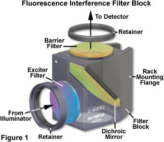
Exciter filters permit only selected wavelengths from the illuminator to pass through on the way toward the specimen. Barrier filters are filters which are designed to suppress or block (absorb) the excitation wavelengths and permit only selected emission wavelengths to pass toward the eye or other detector. Dichromatic beamsplitters (dichroic mirrors) are specialized filters which are designed to efficiently reflect excitation wavelengths and pass emission wavelengths. They are used in reflected light fluorescence illuminators and are positioned in the light path after the exciter filter but before the barrier filter. Dichromatic beamsplitters are oriented at a 45 degree angle to the light passing through the excitation filter and at a 45 degree angle to the barrier filter as illustrated in Figure 1. Filter curves (spectra) show the percentage of transmission (or the logarithm of percentage) as the vertical axis and the wavelengths as the horizontal axis.
Fluorescence filters were formerly almost exclusively made of colored glass or colored gelatin sandwiched between glass. As a result of more sophisticated filter technology, interference filters have been developed that consist of dielectric coatings
dichroic beamsplitter
(of varied refractive indices and reflectivity) on glass. These filters are designed to pass or reject wavelengths of light with great selectivity and high transmission. Most of today's exciter filters are the interference type; some barrier filters are also, for special needs, the interference type. Dichromatic beamsplitters are specialized interference filters. Sometimes short pass filters (SP) and long pass (LP) filters are combined to narrow the band of wavelengths passing through such a combination.

OPTOLONG has been offered fluorescent light filter for many instrument manufacturer and also dealer. We could offer right solution to client according to client’s application. Standard blocking dept for fluorescence filters can be OD4, OD5 or above. Standard transmission could be from 70% to 95%. OEM for size from Dia4mm to Dia70mm, transmission, slop are acceptable and with reasonable and economic price.Optical Filter Sets for Fluorescence
Below is the application we involved in:
· Microplate reader
· Realtime PCR
· Fluorometer
· Cell counter
· Flow cytometry
· Fluorescence quantitative analyzer
· Fluorescent Cell Analysis System
· DNA sequence and analyzer
· Microscope imaging system
· Cell imaging system
· Fluorometer and Absorbance Reader
· Biochemical analyzer
#non polarizing beamsplitter#beam splitter for cameras#Optical Filter Sets for Fluorescence#dichroic beamsplitter#Red Excitation Filter Sets#beam splitter for microscope#fiber beam splitter#dichroic glass mirror#short pass dichroic mirror#long pass dichroic mirror
0 notes
Text
Clearly imaging single fluorescent molecules.
Epifluorescence microscopes are a commonly used tool for studying specimens. Improved specificity and contrast provided by the application of fluorescence to the field of microscopy has stimulated the advancement of various discoveries in the biosciences.
In 1843, George Gabriel Stokes described fluorescence as being characterized by the wavelength of an emitted light that is longer than the wavelength of the exciting light.
This discovery led to the invention of the epifluorescence microscope, now a ubiquitous instrument in biological and medical laboratories.
The high contrast and specificity of the images produced have allowed for the increased understanding of cell structures, the location, and dynamics of gene expression and distinct interactions at a molecular level within the cell.
Epifluorescence illumination or epi-illumination
Epifluorescence illumination or epi-illumination is the specific arrangement of these optical components that allow for the illumination of the specimen from above. The objective lens acts as both the source of light which excites the fluorophore and the element of the microscope that collects the emission filter in fluorescence microscopy .
In comparison to other forms of fluorescence microscopy, epifluorescence illumination has the advantage of only requiring a small amount of emitted light to be blocked.
Though this approach means that the excitation light and emission light overlap, the dichroic mirror efficiently separates the excitation from the emission.
The combination of an excitation filter, dichroic mirror and barrier filter in the filter cubes means that only one photon in 10,000 of the wrong excitation wavelength to the specimen passes, with a similar amount for the emission light. This high ratio is important for clearly imaging single fluorescent molecules.
Terminology:
· Fluorophore: A chemical compound that fluoresces by emitting light upon excitation.
· Photon: A fundamental particle of visible light.
· Ground state: The normal, non-excited state of a molecule.
· Excited state: Electrons can move to a higher energy level by absorbing light, therefore achieving an excited state emission filter fluorescence .
OPTOLONG has been offered fluorescent light filter for many instrument manufacturer and also dealer. We could offer right solution to client according to client’s application. Standard blocking dept for fluorescence filters can be OD4, OD5 or above. Standard transmission could be from 70% to 95%. OEM for size from Dia4mm to Dia70mm, transmission, slop are acceptable and with reasonable and economic price. Optical Filter Sets for Fluorescence
Below is the application we involved in:
· Microplate reader
· Realtime PCR
· Fluorometer
· Cell counter
· Flow cytometry
· Fluorescence quantitative analyzer
· Fluorescent Cell Analysis System
· DNA sequence and analyzer
· Microscope imaging system
· Cell imaging system
· Fluorometer and Absorbance Reader
· Biochemical analyzer
#beam splitter quantum#beamsplitter deck#light beam splitter#uv beam splitter#non polarizing beamsplitter#Red Excitation Filter Sets#dichroic beamsplitter#Optical Filter Sets for Fluorescence
1 note
·
View note
Text
How do Epifluorescence Microscopes Work?
Principles of fluorescence
Fluorescence microscopy is a particular form of light microscopy in which, instead of utilizing visible light to illuminate specimens, a higher intensity light source excites a fluorescent molecule called a fluorophore.
The fluorophore absorbs photons leading to electrons moving to a higher energy state. When the electrons return to the ground state by losing energy, the fluorophore emits light of a longer wavelength.
The emitted light is then separated from the original excitation light via a filter and this produces a magnified image of the specimen being studied.
The use of filters for fluorescence microscopy allows for the object of interest to be specifically targeted. As the excitation light is filtered out, the emitted fluorescence allows only the fluorescent objects of interest to remain visible.Here are some of filter sets that mostly used in the market:
1) Single band pass: DAPI, FITC, TRITC,TexasRed,Cy5
2) Double Band Pass: FITC /TRITC
3) FISH app: FISHAqua, FISHGreen, FISHOrange, FISHRed. (Plan to add FISHGold)
4) Long Wave pass series: UV-DAPI, B,G,V

OPTOLONG has been offered fluorescence filters for many instrument manufacturer and also dealer. We could offer right solution to client according to client’s application. Standard blocking dept for fluorescence filters can be OD4, OD5 or above. Standard transmission could be from 70% to 95%. OEM for size from Dia4mm to Dia70mm, transmission, slop are acceptable and with reasonable and economic price.
Components of an epifluorescence microscope
An epifluorescence microscope requires particular components to separate the excitation and emission lights, acquire the image and allow for the target object to be observed. fluorescent camera filter The following are important components of an epifluorescence microscope:
1. The light source: A source of high-intensity light that can emit a broad spectrum of wavelengths is required. This is usually in the form of a xenon arc lamp or a high-pressure short arc mercury lamp.
2. Filter cubes: They allow for the selection of specific excitation and emission wavelengths and consist of the excitation filter, dichroic mirror and barrier filter. The transmission of the excitation light is selected via the excitation filter and the transmission of the emission light is selected via the barrier filter, by blocking the excitation light. The dichroic mirror separates the excitation light from the emission light further by reflecting the shorter wavelengths of the excitation light and transmitting the longer wavelengths of the emission light.
3. Objective lens: The objective lens transmits light to the sample to form the image. The emission light passes down through the dichroic mirror before reaching the objective lens.
4. Camera system: Modern epifluorescence microscopes can record the images of the specimen with high-resolution. Electron multiplying CCD cameras are often used as they are capable of imaging single-photon events without requiring an image intensifier. This means that images can be captured at high speed without loss of sensitivity digital photo filters .

#digital photo filters#fluorescent camera filter#filters for fluorescence microscopy#bandpass optical filters#optical filters bandpass#machine vision systems#machine vision lighting#beamsplitter mage deck
0 notes
Photo

OPTOLONG has been offered fluorescence filters for many instrument manufacturer and also dealer. We could offer right solution to client according to client’s application. Standard blocking dept for fluorescence filters can be OD4, OD5 or above. Standard transmission could be from 70% to 95%. OEM for size from Dia4mm to Dia70mm, transmission, slop are acceptable and with reasonable and economic price.
Below is the application we involved in:
Microplate reader
Realtime PCR
Fluorometer
Cell counter
Flow cytometry
Fluorescence quantitative analyzer
Fluorescent Cell Analysis System
DNA sequence and analyzer
Microscope imaging system
Cell imaging system
Fluorometer and Absorbance Reader
Biochemical analyzer
#Microplate reader#pcr#fluorometer#flow cytometry#cell analysis#dna sequence#dna analysis#Cell imaging system#Absorbance Reader#biochemical analyzer#biochemical
0 notes
Text
Olympus fluorescence filter set VS Optolong FITC TexasRed
Most precision optics, whether used in scientific, medical, industrial, military,life science applications, incorporate some type of thin film coating to enhance their capabilities. This Sherry Yan from the Optolong Optics company and let discussion into some of the more popular types of fluorescence coatings filter , what they do, how they're applied,what’s they working compare with famous brand and optolong fluorescence filter set. Here are image topics that will be discussed by this platform :The challenges in multilayer coatings will be examined as well as innovations in improving thin-film coating accuracy, with focus on achieving greater accuracy in matching theoretical thin-film designs.Optolong is a professional fluorescence filters manufactures who is dedicated on single band filter set, individual filters and multiband filter sets. Optical fluorescence filters(fluorescent light filters) are the essential optical components for most of the biotech instrument and analytical instrument. Most of the fluorescence imaging system consists of three kinds filters: exciter( or exitation filters), emitter (or emission filters) and dichroic filter(or dichroic mirror) to work properly. Fluorescence ModuleThis module is designed to work with monochrome cameras for fluorescence imaging. It adds:Control of camera gain, gamma and histogram settings for maximum sensitivity in low light conditions.Allows the user to add colour (blue, green and red) to both live and captured images.Each colour channel has individual camera settings (e.g. exposure on the red channel may be longer than the exposure in blue).Capture images in mono or with added colour.Create a composite image of two or three channels.Here is the image of Olympus TexaRed and Optolong TexaRed filter set: Color Combine OLYMPUS FITC TexaRed Color Combine OPTOLONG FITC TexaRed Filter application on high quality biotech instrument imaging requirement:· High transmission to ensure the brightness of fluorophore.· Dark background to enhance contrast of final image.· Deep blocking depth for the overlap of exciter and emitter. · Elimate auto fluorescence of interest fluorophore and all the other scattering light.· High signal tense. With 20 years of mature hard coating experience which makes a stable quality of filters and their prices are half or half lower than Chroma, Semrock, Omega.Do you wanna to try samples at first?Looking on Optolong company website or contact salesman for more image.
#COATINGS FILTER#EMISSION FILTERS#EXITATION FILTERS#FLUORESCENCE COATINGS FILTER#LIGHT FILTERS#MEDICAL FILTERTHIN#FILM COATING#OLYMPUS FITC#SCIENTIFIC FILTER
0 notes
Text
Why choose OPTOLONG Optics For Optical Coating Filter?
ith the foundation history since 1999, OPTOLONG has been working and supporting our clients’ optical filter needs professionally. We keep satisfying our client’s requirement on high volume OEM requirement or small quantity prototype. With the company additional expansion, we moved to a new 2000 square warehouse and imported one new coating machine OTFC-1300 from OPTORUN Japan. Together with Mars-1300 from Korea, they enables us to manufacture and export complex and high requirement optical filters for fluorescence life science instrument, astronomy planet nubela and machine vision industrial manufacturing related. From the beginning of company, we have served more than 2000 clients so far in optical design and manufacturing. To meet the increasing change requirement of technology or time, OPTOLONG would be your optical partner all the time.
Coating technology we applied
We design and apply the coating technology based on the equipment and the application or specification on client offer. The coating technology include IAD evaporation, PVD e-beam thermal evaporation. All the optical film belongs to hard coating. All is the latest metallic and dielectric film. We would apply the coating technology according to different applications.
Bandpass filter: Broad bandpass filter, narrow bandpass filters, high pass filter, low pass filter.
Filters types we offer
Bandpass filter: Broad bandpass filter, narrow bandpass filters, high pass filter, low pass filter.
Pass filter: longpass filter which is known as edge filters, shortpass filter which is also known as cut off filters, UV filter, Multibandpass filter.
Others:neutral density filters, hot mirror, cold mirror, beam splitter, dichroic mirror, Anti-reflection filter, Waterproof anti-reflection filter, protective windows, half mirrors.
Core competition advantage
Company internal high scratch dig standard and Optical component quality inspection US military standard.
Optolong internal standard for environmental and physical durability totally according to and exceed MIL-PRF-13830B(MIL-O-13830A). With high internal standard and microscope tools, operators inspect scratch dig of filters with highest qualities.
Ability to assist prototype design.
Optolong has experienced technician who worked in optical industry for years. They well know and understand the needs of client about application and spectrum design. We do not only manufacturers but also designers and inventors.
Waterproof thin film layers.
Some application requires to coat thin film layers to be waterproof. Without waterproof coating, water would fall apart and hard to clean up. Optolong has advanced coating method to coat waterproof thin film layers. Nothing left on waterproof filters when facing water.
Carving & Silk printing & Black anodized metal rings.
We have carving machine, silk printing machine to meet client’s relative requirement to make printing on filters and carving different words for better recognize. Nearly most of the filters size can be mounted with black anodized metal ring.
Tunable filters
We could offer wide angle of incidence change filters. Normal and standard AOI is 0 degree. We also could offer 45 degrees and 60 degrees.
Processing advantage
Strict substrate selection process
Substrate material can be selected among optical glass, sapphire, quartz, fused silica, color glass according to client requirement and different application.
Double side of substrate are polished to ensure the high base material parallel.
High uniformity of planetary wheel coating fixture improve the uniformity of filter wavelength.
Mature production management control
We have a mature system of production control. Every process processing with a strict standard and clear in every worker’s control.
Pure water ultrasonic cleaning clean clear any mineral dust on clear glasses. The cleanliness of glass substrate surface is equal to the pure water.
Experienced coating technicians improve qualified rate of filters. Higher qualified rate reduce the cost of filters.
Accurate thin film thickness calculation and thin film deposition make complex filter with minimal wavelength shift. Uniformity and repeated filters consistent from lot to lot.
Quality control of spectrum
Spectrum of each filters which would be delivered to client are measured strictly with photometer UV3150 and UV2600. Measuring range of photometer is from 190nm to 3200nm. Measuring accuracy 0.1nm and the optical density values to OD5. None compromise for 100% conform contract agreement.
1 note
·
View note
Video
tumblr
In life science instrument, for the sake of quality image analyze and accurate diagnose, optical filters must separate desired wavelength from whole spectrum. Meanwhile to keep high standard attunation to the unwanted area.
Fluorescence Filters are widely used in varies kinds of life science and bio tech instrument. It’s known as high transmission to bright the subject and deep blocking to dark background. Also to make the scatter light to be least.
OPTOLONG has been offered fluorescence filters for many instrument manufacturer and also dealer. We could offer right solution to client according to client’s application. Standard blocking dept for fluorescence filters can be OD4, OD5 or above. Standard transmission could be from 70% to 95%. OEM for size from Dia4mm to Dia70mm, transmission, slop are acceptable and with reasonable and economic price.
Below is the application we involved in:
Microplate reader
Realtime PCR
Fluorometer
Cell counter
Flow cytometry
Fluorescence quantitative analyzer
Fluorescent Cell Analysis System
DNA sequence and analyzer
Microscope imaging system
Cell imaging system
Fluorometer and Absorbance Reader
Biochemical analyzer
Longpass filter set ( B,G,UV,V,R standard for fluorescence microscope) or other longpass set.
Shortpass filter set.
Single band filters set :Regular DAPI, FITC, Texas Red, TRITC, CFP, YFP, GFP, Cy3.5, Cy5.5.
FISH Application:FISH Aqua, FISH Red, FISH Orange, FISH Green, more coming soon.
Dual band filters set:DAPI-FITC, DAPI-Texas Red, FITC-Texas Red.
Triple band filter set:DAPI-FITC-Texas, DAPI-FITC-TRITC.
Individual filters:Individual exciter, emitter, dichroic mirror.
#coating film#fish aqua#FITCDAPITexaRed,CY5,TRITC#Dual band filter#Fluorescence filters#hard coating film#Longpass filter#Longpass filter set#Shortpass filter set#dichroic mirror
0 notes
Photo


In December 2019, there was a cluster of pneumonia cases in China.Investigations found that it was caused by a previously unknown virus – now named the 2019 Novel Coronavirus.
In this artical, we'll take a quick look at what's currently known about the virus.Keep in mind that this is a new virus and what's known about the virus now might change in the future.
Coronaviruses are a large group of viruses.They consist of a core of genetic material surrounded by an envelope with protein spikes.This gives it the appearance of a crown.Crown in Latin is called "corona" and that's how these viruses get their name.There are different types of coronaviruses that cause respiratory and sometimes gastrointestinal symptoms.Respiratory disease can range from the common cold to pneumonia.And in most people, the symptoms tend to be mild.
However, there are some types of coronaviruses that can cause severe disease.These include the Severe Acute Respiratory Syndrome coronavirus first identified in China in 2003 and the Middle East Respiratory Syndrome coronavirus that was first identified in Saudi Arabia in 2012.The 2019 Novel Coronavirus was first identified in China.It initially occurred in a group of people with pneumonia who'd been associated with a seafood and live animal market in the city of Wuhan.The disease has since spread from those who were sick to others, including family members and health care staff.There are many cases at present and the disease has spread within China and also to a number of other countries.
Q1:Where did the virus come from?
It's known that coronaviruses circulate in a range of animals.Sometimes these viruses can make the jump from animals to humans.This is called a spillover and could be due to a range of factors such as mutations in the virus or increased contact between humans and animals.
For example, MERS-CoV is known to be transmitted from camels and SARS-CoV, from civet cats.The animal reservoir of the 2019 Novel Coronavirus is not known yet.
Q2:How is it transmitted?
The exact dynamics of how the virus is transmitted is yet to be determined.In general, respiratory viruses are usually transmitted through droplets created when an infected person coughs or sneezes, or through something that has been contaminated with the virus.People most at risk of infection from the Novel Coronavirus are those in close contact with animals, such as live animal market workers, and those who are caring for people infected with the virus, such as family members or healthcare workers.
Q3:How does the disease present?
Well, from what is known so far, there can be a number of symptoms ranging from mild to severe.There can be fever and respiratory symptoms such as cough and shortness of breath.In more severe cases, there's been pneumonia, kidney failure, and death.The mortality rate is not known yet.
Q4:How can we tell whether someone is infected?
The infection can be diagnosed by a test called PCR or Polymerase Chain Reaction.This test identifies the virus based on its genetic fingerprint.There is currently no specific medication for the virus and treatment is supportive care.There's currently no vaccine to protect against the virus.Treatment and vaccines are in development.
Q5:How do we prevent transmission of the virus?
This new virus currently has a limited geographic spread.However, there are a number of standard hygiene practices that have been recommended to protect against infection and further spread.These include covering your mouth and nose when coughing or sneezing with a medical mask, tissue or flexed elbow; avoiding close contact with those who are unwell; the appropriate use of masks and personal protective equipment, especially in a healthcare setting; washing hands regularly with soap and water, or alcohol-based hand rub.
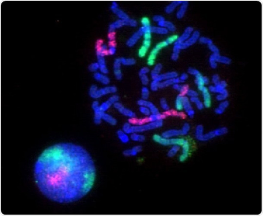
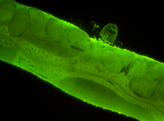
Q5:How does the virus looks like?
Images of Novel Coronavirus sames were captured using the OPTOLONG@fluorescence filter set:FITC,DAPI,TexaRed,Fish Orange filter set(EX,EM,DM),An Nikon microscope with a 100x objective were used.
Single-band Sets:FITC,DAPI,TexaRed,CY5,TRITC Individual Filter:Longpass,shortpass,bandpass filter,AR,IR,IRCUT... Dichroic Mirror:416nm,455nm,492nm,555nm,655nm.... FISH Set:Orange,Qqua,Green,Red. Cubes or Holders: U-MF2,U-FF,Zeiss CUBE. By Application:Machine Vision,Life Science Instrument,Biomedical Imaging System, Industrial Detection,Imging or Photonics...
Actions that can be taken to prevent infection from an animal source include: avoiding unnecessary unprotected contact with animals;washing hands after contact with animals or animal products; and ensuring that animal products are cooked thoroughly before they're consumed.It's important to stay home if you're feeling unwell.But if you have a fever, cough, and difficulty breathing, seek medical care early and share your previous travel history with your healthcare provider.
That's a quick look at this emerging infectious disease.
#FITCDAPITexaRed,CY5,TRITC#FITC#DAPI#TexaRed#cy5#tritc#416nm#455nm#FISH Set#U-MF2#U-FF#Zeiss CUBE#Industrial Detection#Fish Orange filter#Nikon microscope#100x objective
0 notes
Photo

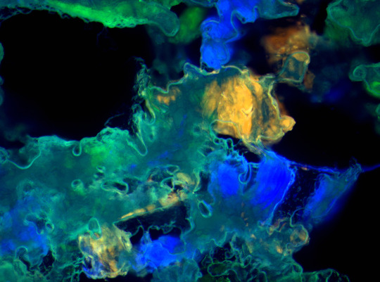
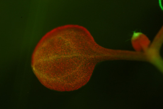
fluorescence spectroscopy has had a profound effect on medical diagnostics, both directly and indirectly. In terms of direct effects, advances in fluorescence endoscopy in the past few years have produced important diagnostic procedures, such as autofluorescence broncoscopy, LIFE (lung imaging fluorescence microscopy), and infrared fluorescence endoscopy, for detection of diverse malignancies, including bladder cancers. Autofluorescence of skin is also proving useful for detection of pre-malignant and malignant lesions. Indirectly fluorescence has been absolutely critical in the development of techniques such as high throughput screening for drug discovery and FISH (fluorescence in situ hybridization) for cytogenetics. And of course many of the diagnostics protocols used in clinical chemistry, including new generation of biosensors, are based on fluorescence methods. The Human Genome Project, which is proving to be a boon to medicine, was also, of course, totally dependent on fluorescence methods.
Fluorescence spectroscopy has affected medical diagnostics by enabling assays for specific molecules in complex samples like tissues and cells from clinical samples. Advances in fluorescence spectroscopy have enabled the use of multiple probes at the same time in the analysis of biomarkers in clinical medicine. Stains for use in anatomic pathology in surgical tissue specimens and biopsy specimens are deployed widely in clinical pathology labs. These stains are also used in tissue micro arrays in research settings.
Fluorescence imaging is also beginning to see use in clinical screening applications. For example, in bronchoscopy, a system for using autofluorescence to localize lesions, which are not visible using traditional, brightfield reflection imaging, has received FDA approval.
Optolong Optics Ltd is a china manufacturer of astronomy filters, fluorescence filters with the development experience for 20 years since 1999, varies wavelength of longpass, shortpass, edge filters, exitation filter, emission filters are widely used. For all those part, OPTOLONG could provide the proper thin film design and deposition according to the laser source of system.
For bandpass filter from 200nm to 1100nm, we could offer design and advice for bandwidth from 3nm to 20nm. Transmission would be as high as 90% above. Steepness and blocking requirement are also easy to handle. Contact us for your needs.
0 notes
Link
Optolong optics Ltd,your optical filter partner online here
0 notes
Photo
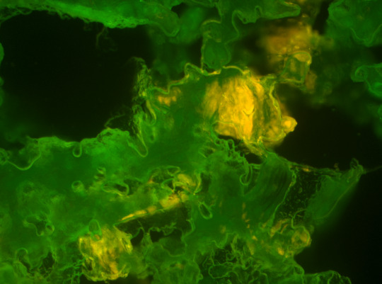
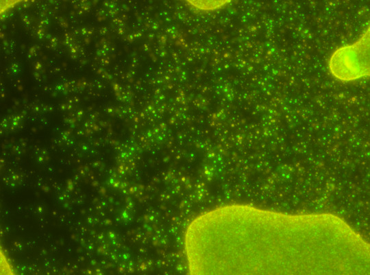
Fluorescence spectroscopy has had a profound effect on medical diagnostics, both directly and indirectly. In terms of direct effects, advances in fluorescence endoscopy in the past few years have produced important diagnostic procedures, such as autofluorescence broncoscopy, LIFE (lung imaging fluorescence microscopy), and infrared fluorescence endoscopy, for detection of diverse malignancies, including bladder cancers. Autofluorescence of skin is also proving useful for detection of pre-malignant and malignant lesions. Indirectly fluorescence has been absolutely critical in the development of techniques such as high throughput screening for drug discovery and FISH (fluorescence in situ hybridization) for cytogenetics. And of course many of the diagnostics protocols used in clinical chemistry, including new generation of biosensors, are based on fluorescence methods. The Human Genome Project, which is proving to be a boon to medicine, was also, of course, totally dependent on fluorescence methods.
Fluorescence spectroscopy has affected medical diagnostics by enabling assays for specific molecules in complex samples like tissues and cells from clinical samples. Advances in fluorescence spectroscopy have enabled the use of multiple probes at the same time in the analysis of biomarkers in clinical medicine. Stains for use in anatomic pathology in surgical tissue specimens and biopsy specimens are deployed widely in clinical pathology labs. These stains are also used in tissue micro arrays in research settings.
Fluorescence imaging is also beginning to see use in clinical screening applications. For example, in bronchoscopy, a system for using autofluorescence to localize lesions, which are not visible using traditional, brightfield reflection imaging, has received FDA approval.
Optolong Optics Ltd is a china manufacturer of astronomy filters, fluorescence filters with the development experience for 20 years since 1999, varies wavelength of longpass, shortpass, edge filters, exitation filter, emission filters are widely used. For all those part, OPTOLONG could provide the proper thin film design and deposition according to the laser source of system.
For bandpass filter from 200nm to 1100nm, we could offer design and advice for bandwidth from 3nm to 20nm. Transmission would be as high as 90% above. Steepness and blocking requirement are also easy to handle. Contact us for your needs.
#longpass#shorpass#fitc test#fitc fluorescence#fluorescence spectroscopy#fluorescence#spectroscopy#spectroscopyfitc#exitation#exitation filter#emission#emission filter#longpass filter#uvpass#uv ir cut#uv ir cut filter#uv it
0 notes
Text
How do you differentiate UV wavelengths
According to the different biological effects, the ultraviolet ray is divided into four wave bands by wavelength: long wave uv-a, medium wave uv-b, short wave uv-c, and vacuum wave uv-d. The longer the wavelength, the greater the penetration. uv-a Long wave uv-a, wavelength between 320 ~ 400 nm, also known as long wave black spot effect ultraviolet. Strong penetration, able to penetrate glass, even 9 feet of water; And throughout the year, whether rain or shine, day and night exist. Harm degree to human body: the ultraviolet ray that daily skin contacts 95% above is uv-a, because this its harm to the skin is greatest. Uv-a can penetrate through the epidermis and attack the dermis, severely damaging the collagen and elastin in the skin. And dermal cell self-protection ability is poor, very little uv-a can cause great damage. As time passes, skin produces loose, furrow, microvasculature appears wait for a problem. At the same time, it can also activate tyrosinase, resulting in immediate melanin deposition and the formation of new melanin, making the skin dark, lack of luster. Uv-a is also known as aging rays because it causes long-term, chronic, and long-lasting damage and causes premature aging of the skin. Application: UV-A at 360nm conforms to the phototropic response curve of insects and can be used to make insect trap lamp. The uv-a wavelength of 300-420nm can pass through the special colored glass lamp tube which is completely cut off from visible light. It can only radiate the near-ultraviolet light which is centered on 365nm. It can be used in the fields of ink glue curing, ore identification, stage decoration and bankbill inspection.
UV - B Medium wave uv-b, wavelength between 275 and 320 nm, also known as medium wave erythema effect ultraviolet. Medium penetration, the shorter wavelengths of which are absorbed by the clear glass. The uvb rays in sunlight are mostly absorbed by the ozone layer, and less than 2% of them reach the earth's surface.Damage to human body: it will make the epidermal protective lipid layer oxidation, dry skin; Further make the nucleic acid inside epidermal cell and protein denaturation, produce acute dermatitis (namely sunburn) wait for a symptom, the skin can become red, hair is painful. When serious, for example long time insolate, still cause skin cancer easily change. In addition, the long-term damage of uv-b can also cause melanocytes to mutate, resulting in hard-to-remove solar spots. Application: ultraviolet health lamp and plant growth lamp are made of special permeable glass (no light below 254nm) and phosphors with peaks near 300nm.
UV - C Short-wave uv-c, wavelength between 200 ~ 275 nm, also known as short-wave sterilization ultraviolet. It has the weakest penetration, unable to penetrate most clear glass and plastic. The short-wave ultraviolet light contained in sunlight is absorbed almost completely by the ozone layer and is absorbed by the ozone layer before it reaches the ground. Harm degree to human body: the uv-c in nature because did not reach the ground to be absorbed by the ozone layer, the effect on the skin can be ignored, but in fact the harm of short-wave ultraviolet ray to the human body is very big, cannot direct illuminate the human body. If direct irradiation, short time irradiation can burn the skin, long-term or high intensity irradiation can also cause skin cancer. Application: ultraviolet sterilization lamp is the UV-C short - wave ultraviolet. It is widely used in the fields of hospitals, air conditioning systems, disinfection cabinets, water treatment equipment, water dispensers, sewage treatment plants, swimming pools, food and beverage processing and packaging equipment, food factories, cosmetics factories, dairy factories, breweries, beverage factories, bakeries and freezers.
#uv filter#led lights#led devices#UVbandpass#uv ir#uvnm#uv 222nm#uv254nm#254nm filter#bandpass filter#bandpass#uv filter glass#uv filter film#uv light#uv filtering#uv ir cut filter#uv/ir cut filters
0 notes
Text
How do you differentiate UV wavelengths
#uv filter#uv coating filter#uv glass#uv wavelengths#uv ir#uv coating#filter#bandpass#bandpass filter#254nm#222nm filter#365nm fllter
0 notes
Photo


What is fluorescence filter ?
Fluorescence filter is a key component used in biomedical and life science instruments.
It’s main function is to separate and select the characteristic band spectrum of the substances’ excitation light and emitted fluorescence in the biomedical fluorescence inspection analysis system .
Generally speaking , a fluorescence filter set consists of three different filters.
①Excitation Filter (Size φ25×5mm)
②Emission Filter (Size φ25×3.5mm)
③Dichroic Mirror (Size 25.7×36×1mm)
0 notes