#umbilical cord tissue allograft therapy
Text
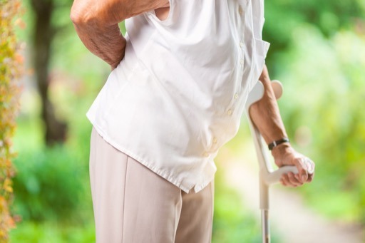
Standard post published to Joint Repair Clinic of Montana at May 18, 2021 13:45
The procedure can be divided into two types: Stem cell injection containing stem cells and Allograft injection containing stem cells. In Stem cell therapy, the stem cells are injected directly into the target area where the problem is. But this doesn't mean that it would take you only one year to experience good pain relief after receiving this treatment.
Source: https://www.hindawi.com/journals/sci/2016/6901286/
Joint Repair Clinic of Montana is a stem cell therapy center nearby Three Forks. Joint Repair Clinic of Montana offers human umbilical cord tissue stem cell treatment plan. Joint Repair Clinic of Montana is centrally located near Ackermann Mary (Bozeman, MT 59715).
Driving directions to Joint Repair Clinic of Montana in Bozeman:
https://www.google.com/maps/dir/?api=1&origin=45.6806631531643,-112.304788318867&destination=Joint%20Repair%20Clinic%20of%20Montana
See the last article:
https://bozeman-stem-cells-clinic.business.site/posts/1574834895853696627
https://local.google.com/place?id=5497948111560534332&use=posts&lpsid=1574834895853696627
source https://local.google.com/place?id=5497948111560534332&use=posts&lpsid=8843248831276547363
0 notes
Photo

What is umbilical cord allograft? Umbilical cord allograft is a dehydrated, minimally manipulated, non-viable cellular epicord for homologous use. The Epicord is a thick, unique #membrane derived from the umbilical cord, the structure of which protects the #veins and arteries. It is derived from the #umbilicalcord blood. Umbilical allograft therapy is one of the most amazing advancements in medicine. Umbilical allograft therapy is a major innovation in #regenerative medicine. What are the benefits of umbilical allograft?. . ✅Gives an immune environment for the healing process. ✅Provides a connective tissue matrix to supplement or replace damaged tissue. ✅Contains essential factors required for wound healing. ✅Can be stored in a suitable setting for up to 5 years. Welcome to COLORADO MEDICAL SOLUTIONS Please feel free to Give us a call and book an appointment: Denver Office: (720) 550-6907 Colorado Springs Office: (719) 308-2314 Or please visit: www.coloradomedicalsolutions.com #hormoneoptimization #wellnesslifestyle #testosterone #healthylifestyle #supplements #maxim #miami #weightloss #wellness #optimize #vitality #ageless #colorado #Denver #preventativemedicine (en Colorado Springs, Colorado) https://www.instagram.com/p/B7WK6P4l0QP/?igshid=4b3t7acr5z5l
#membrane#veins#umbilicalcord#regenerative#hormoneoptimization#wellnesslifestyle#testosterone#healthylifestyle#supplements#maxim#miami#weightloss#wellness#optimize#vitality#ageless#colorado#denver#preventativemedicine
0 notes
Text
Stem Cells to Improve the Wound Healing-Juniper publishers
Abstract
The cellular therapy using Mesenchymal Stem Cells, specially its two subtypes - Bone-marrow Mesenchymal Stem Cells and Adipose Derived Stem Cells - can benefit by virtue of the possibility of differentiating in specialized cells that secrete and suppress growing factors and cytokines necessary in the lesion niche. When attracted by the pro-inflammatory sinalization of the lesion, they act using the paracrine signaling, decreasing the inflammation, increasing the angiogenesis and the cell migration and proliferation. The development in the researches regarding the association of the application of MSCs, with reconstructive surgery practices, leads to effective future results that can bring more benefits to the clinic practice of this field. This paper has the objective of briefly reviewing the literature about the usage of MSCs and its subtypes, the ADSCs, to improve the skin cicatrization.
Keywords: Mesenchymal stem cells; Adipose derived stem cells; Bone-Marrow Mesenchymal Stem cells; Skin flaps; Skin grafts; Wound contraction; Paracrine signaling; Pro-inflammatory Interleukins; Anti-inflammatory Interleukins.
Abbreviations: MSC: Mesenchymal Stem Cells; HLA-DR: Human Leukocyte Antigen; ADSC - Adipose derived stem cells; BM-MSC: Bone-Marrow Mesenchymal Stem Cells; SDF1: Stromal Cell-Derived Factor; PDGF: Platelet-Derived Growth Factor; IL: interleukin; TNFα: Tumor Necrosis Factor-α; IFNγ: Interferon-γ; VEGF: Vascular Endothelial Growth Factor; BFGF: Basic Fibroblast Growth Factor; EGF: Epidermal Growth Factor; KGF: Keratinocyte Growth Factor; HGF: Hepatocyte Growth Factor; PDGF: Platelet-Derived Growth Factor; TGF-β: Transforming Growth Factor-β; FGF: Fibroblast Growth Factor; α-SMA: Alpha-smooth muscle actin's; IRI: Ischemia-Reperfusion Injury
Introduction
Mesenchymal stem cells (MSCs) were first described by Friedenstein et al. [1,2]. MSCs are heterogeneous cell populations, called multipotent adult cells or somatic stem cells [3,4], capable of differentiation in most cell types in order to maintain and repair the organism. They are resident in different organs and tissues such as adipose tissue [4,5], bone marrow, umbilical cord, amniotic membrane [2,4], kidneys, liver, spleen, lungs, pancreas, tendons, synovial membranes, placenta, amniotic fluid and dental pulp [4]. The International Society for Cellular Therapy proposed three minimal criteria for defining MSCs:
a) Plastic adherence;
b) Positive expression (greater than 95% of the cell population) for CD105, CD73 and CD90 markers and negative, (maximum 2% of the population) for CD45, CD34, CD14, CD11b, CD79a, CD19 and Human leukocyte antigen (HLA-DR) surface molecules, and
c) Ability to differentiate into osteoblasts, adipocytes and chondroblasts under standard in vitro differentiating conditions [2,6].
Later, others researchers complemented this characterization with new markers: CD13, CD44, CD54, 106 and Stro-1 [4] and the presentation of fibroblast cellular morphology [3,4].
MSCs have a great potential to the tissue repairment and regeneration in reconstructive plastic surgery, preventing ischemic lesions in skin flaps [7-14] and in skin grafts [15,16]. MSCs are capable of reducing severe atrophy, retraction, fibrosis and ulceration skin signals, caused by radiotherapy [17,18]. Combined with the lipoinjection in face and body defects and in rejuvenating esthetic treatments, MSCs raise in 35% the survival rate and the microvasculature from the injected fat [18]. Also, diabetic patients with ischemic wounds show greater survival rates and lower number of amputations in affected limbs, when treated with Adipose derived stem cells (ADSCs) [19]. The MSCs improved the skin healing at induced burns in rats [20,21]. In dermatology, the MSCs are being clinically used for inflammatory diseases [22-28], Graft-Versus-Host Disease [23,25], Chrown Disease [29], Systemic Sclerosis, Lupus or dermatomyositis [24,28,30].
When compared with the usage of Growing Factors, that like any drug has a limited half-life, the cellular therapy using MSCs, specially in its two subtypes - the Bone-Marrow Mesenchymal Stem Cells (BM-MSCs) and ADSCs - can bring greater benefits because of: their expansion capacity in cellular culture; stable phenotypic expression; and possibility of differentiating in specialized cells, that secrete and suppress cytokines and growing factors in the injury [13,31-33]. However, long expansion periods of the cellular culture, to achieve effective therapeutic doses, raise the possibility of infection from the host. Because of that, the ADSCs still have to be heavily studied to the application in the clinic routine [2]. This way, this study has as objective briefly review the literature about the usage of MSCs and its subtypes, the ADSCs, to improve the skin healing.
Discussion
The MSCs are capable to answer and adjust their functions, when exposed to cells or typical factors of the lesion environment [33,34]. They have an attraction capacity to the inflammation areas [33-35] or tumors, because of the molecules CCL21 or HMGB1, known as homing. In case of lacking of this signalization, they are attracted in order of preference to the lungs, liver and spleen [35]. The MSCs show chemotaxis in vitro by the Stromal Cell-Derived Factor (SDF1), Platelet-Derived Growth Factor (PDGF), Insulin-Like Growth Factor-1, Interleukin-8 (1L-8) and Tumor Necrosis Factor-α (TNFα). In murine models, the MSCs used in the systemic administration are capable of getting into the damaged tissues [14,34,36].
The MSCs act, in many levels, in the three healing phases: inflammatory, proliferative and remodeling phase. Current studies indicate that the differentiation of the MSCs, that contributed to the tissue regeneration, is limited by the small survival rate of these cells in the damaged area. However, the paracrine signaling to the cells from the injury is the main mechanism of the MSCs, reducing the inflammation, stimulating the angiogenesis and inducing the cell migration and proliferation [33,37]. Nevertheless, other authors suggest that the differentiation of the MSCs in keratinocytes and endothelial cells have the same function as the paracrine signaling to accelerate the neovascularization and the reepithelialization of wounds [32] (Figure 1 & 2).
Some studies suggest that immunosuppressive potential is the same of both types of MSCs, ADSCs and BM-MSCs, in an inflammatory environment. Their local administration inhibited the T cells recruitment and proliferation [37-41] and decrease Interferon-γ (lFNγ),Tumor Necrosis Factor-α (TNFα), 1L-6 and IL-1β expression in the skin grafts [39-41]. In the inflammatory phase (one to three days), the expression of anti-inflammatory cytokines 1L-4 and 1L-10 is increased while 1L-2 pro- inflammatory cytokines, TNF-α and 1FN-γ are decreased, causing less local inflammatory reaction [37,42]. MSCs can also regulate macrophage activation and convert phenotype expression of M1 macrophages to M2 anti-inflammatory macrophages [39-41], which accelerates the wound healing. Additionally, MSCs act over the innate immunity to prevent the rejection of transplanted allografts, inhibiting the complement system activation [39]. The decrease of TFN-α also helps the reepithelialization after the proliferation phase [43]. The antimicrobial action from the MSCs are also important to limit the infection occurrence [37].
With the utilization of cell therapy in the proliferative phase (14 days) the production of important growth factors is increased: Vascular Endothelial Growth Factor (VEGF), Basic Fibroblast Growth Factor (bFGF) [32,37,42,43], Epidermal Growth Factor (EGF), Keratinocyte Growth Factor (KGF), Hepatocyte Growth Factor (HGF), Platelet-Derived Growth Factor (PDGF) and Transforming Growth Factor-beta (TGF-β). Through their paracrine action, the MSCs raise the migration and proliferation of keratinocytes, endothelial and epithelial cells. The fibroblasts proliferation is also enhanced, as well as the blood vessels' formation. This way, there is a better growing of a more vigorous granulation tissue [33,37,42,43]. The angiogenesis is a very important factor to the wound's healing, and the VEGF and the Fibroblast Growth Factor (FGF) are very important angiogenic factors [32].
Through the paracrine signaling, from the MSCs in the remodeling phase (21 days to one year), the fibroblasts produce collagen fibers in greater quantity and density, generating an increase in wound tensile strength [37,42,43]. At the same time, there is a smaller wound contraction and increased matrix-metalloproteinases production, that control the collagen deposition exacerbation, improving the aesthetic result and keeping the skin's function [33,42,43]. MSCs also contribute to the scar's appropriate remodeling with the increased secretion of VEGF and HGF, the adequate balance between TGF-β1 and TGF-β3 [37] and the increase of bFGF and TGF-β which inhibit the Alpha-Smooth Muscle Actin (α-SMA) expression, responsible for myofibroblast phenotype [32].
Studies indicate that there is also the possibility of using BM-MSCs and ADSCs to protect the damaged tissues caused by the 1schemia-Reperfusion 1njury (1R1) and to decrease the dysfunction of organs undergoing ischemia. The ADSCs are able to protect axial flaps from the 1R1 with results as satisfactory as the ones obtained in procedures without ischemia, because of the improvement in the angiogenic response and the blood perfusion elevation [10]. Many studies use the ADSCs to prevent ischemic injuries in skin flaps [7-14], as others [15,16] used them to improve the quality of skin grafts.
Conclusion
The MSCs - both BM-MSCs and ADSCs - are capable to answer and module their functions, when exposed to cells or typical factors of the lesion environment, attracted by the cell signaling to the inflammation areas. The researches developed till the moament point the paracrine signaling as the main mechanism from the MSCs, decreasing the inflammation, benefiting the angiogenesis and inducing the cell migration and proliferation in the damaged area. The progress on the researches about the association of MSCs with reconstructive surgery practices indicates good and productive results, which can benefit the clinical practice in this field.
To know more about Open Access Journal of Head Neck & Spine Surgery please click on:
https://juniperpublishers.com/jhnss/index.php
To know more about Open access Journals Publishers please click on : Juniper Publishers
To know more about juniper publishers: https://juniperpublishers.business.site/
#Head and Neck Surgery Journal#Journal of Head Neck & Spine Surgery#Journal of Spine Surgery#Juniper Publishers#Open Access Journals
0 notes
Text
Biomed Grid | Use of Regulatory T Cells as a Cellular Therapy for Transplant Rejection/Autoimmunity
Introduction
Foxp3+ regulatory T cells (Treg) are a crucial sub-population of T cells in the control of immunity and the maintenance of immunological tolerance aiding the prevention of autoimmunity. As such Treg have been proposed to be a candidate cell that could be used as cellular therapy to suppress unwanted immune responses. Indeed, there is an expanding literature on the potential use of Treg to induce or re-establish tolerance in both autoimmune diseases such as type I diabetes, arthritis and chronic inflammatory diseases as well as in preventing allograft rejection and graft versus host disease (GVHD). Furthermore, the finding that antigen specific Treg are superior to polyclonal Treg in a range of disease settings has raised the possibility that Treg based therapies may be refined and improved by genetic manipulation.
The first description of Treg was by Sakaguchi and his colleagues in 1995, where they showed the depletion of a CD4+CD25+ T cell subset led to the development of autoimmune disease e.g. gastritis, thyroiditis, adrenalitis and insulitis [1]. However, CD25 is expressed by activated lymphocytes so it was the discovery that Treg express the master transcription, forkhead box protein 3 (Foxp3) that defined Treg as being a separate T cell lineage from conventional T cells [2,3]. In addition, mice that lack Foxp3 expression (scurfy mice) develop T cell dependent autoimmune diseases [4]. Importantly, a spontaneous inflammatory disease called Immune dysregulation polyendocrinopathy enteropathy X linked syndrome (IPEX) is the result of a mutation in the foxp3 gene which links Treg and foxp3 to the development of autoimmunity in humans as well as in mice [4]. The mechanisms employed by Treg to suppress immunity can be mediated by cell contact (e.g. via CTLA-4) or may be cell contactindependent (e.g. IL-10 and TGFβ) [5].
Treg can be isolated from a number of different sources including peripheral blood, umbilical cord blood and discarded paediatric thymuses, however, the most common source for Treg thus far used in clinical trials is peripheral blood. In order to take advantage of the suppressive capacity of such cells and due to the low frequency of Treg in peripheral blood, ex-vivo expansion of the cells using non-specific stimulation with anti-CD3\CD28 in the presence of a high concentration of IL-2 is generally required. This type of expansion protocol has proved to be able to successfully generate sufficient numbers of Treg to be infused in clinical trials [6].
Indeed, Treg ex-vivo expanded in this way have been introduced as an immunotherapeutic agent in autoimmune diseases such as type 1 diabetes [7,8]. It is worth noting that in all studies to date the administration of ex-vivo expanded Treg to patients has not resulted in any serious complications or side-effects. A recent study reported that the infused Tregs can be detected in the circulation for at least 3 months post infusion and that such cells largely retain their original Treg phenotype [9]. Interestingly, it has been shown that culturing Treg together with certain cytokine combinations during ex-vivo expansion may result in Treg expressing chemokine receptors that may facilitate Treg migration to sites of inflammation and/or peripheral tissues [10].
Ex-vivo expanded Treg have now been infused into patients in a number of different disease settings. For example, such Treg have been transferred to patients at risk of acute and chronic graft versus host disease (GVHD); a condition that can occur following allogeneic HSC transplantation due to alloreactive T lymphocytes present in the donor HSC product. These studies demonstrated that it is feasible and safe to inject Treg into patients receiving HSC transplantation and that their infusion correlated with the suppression of GVHD and enhanced immune recovery [11,12]. Ex-vivo expanded Tregs have also been administered to liver and kidney transplant patients [13,14]. Indeed, the ONE study intensively investigated the safety and feasibility of injecting ex vivo expanded Treg into kidney and liver transplant patients and has reported no side-effects associated with the infused Treg [13,15]. The contribution, if any, of the administered Treg to the suppression of rejection has yet to be confirmed as both sets of patients received conventional immunosuppressive agents in addition to the Treg.
The Tregs that have been given to patients are largely the result of polyclonal expansion such that only a small percentage of the Treg are likely to recognise the target disease associated antigen/alloantigen. However, advances in gene-engineering may have opened an avenue to re-direct Treg specificity toward a particular antigen thereby potentially increasing the suppressive capacity of the Treg whilst lowering the possibility of off-target immunosuppression [16]. Indeed, T cell receptor (TCR) or chimeric antigen receptor (CAR) gene engineering of conventional T cells has been widely trailed to treat solid and haematological tumours and such clinical trials have shown encouraging and often remarkable results [17-119]. The successful outcomes from re-directing conventional T cells toward tumour antigens through either TCR or CAR gene engineering raises the question of whether similar approaches could be applied to regulatory T cells in order to suppress autoimmunity and allograft rejection.
A recent study highlighted the potential of CAR-Treg showing that lung epithelium-reactive CAR-Tregs demonstrated an enhanced ability to suppress allergic airway inflammation in a mouse model compared to their polyclonal counterparts [20]. Moreover, directing Treg specificity to ta issue-specific antigen or disease related antigen revealed a robust suppression of Type I diabetes and rheumatoid arthritis [21,22]. In terms of transplantation, several recent studies have shown that HLA-A2-specific CAR Tregs displayed an enhanced ability to suppress HLA-A2+ skin allograft rejection and xenogeneic GVHD in humanised mouse models of transplantation [23-25]. Another promising approach in the field is the use of an engineered B cell antibody receptor (BAR) in conventional T cells or Treg to ameliorate B cell mediated autoimmunity and alloimmunity [26]. Recently, BAR transduced Treg have been shown to potently prevent the secretion of the neutralising antibodies that develop against the therapeutic factor VIII in haemophilia [27]. This raises the question whether using BAR Treg would be advantageous to suppress pathogenic antibodies in autoimmunity and transplantation.
Taken together there is now convincing evidence that Treg can be isolated and expanded from patients under cGMP conditions to enable their re-infusion as part of a therapeutic strategy. Phase 1 clinical trials have shown that these cells are able to survive for prolonged periods of time post infusion and that the generation of location-specific or antigen-specific Treg is likely to increase the potency of such cells. However, it remains to be seen whether Treg can be used as a cellular therapy for autoimmune disease and allograft rejection/GVHD in isolation or perhaps more likely will be used as part of an adjunctive strategy combined with more traditional immunosuppressive agents. The other important point is whether Tregs when confronted with certain inflammatory stimuli could lose their suppressive phenotype and convert to T helper subsets such as IFN or IL-17 secreting CD4+ T cells that may exacerbate rather than suppress immune-mediated disease [28]. Addressing these potential confounding factors will be a necessary next step in the potential implementation of Treg as a cellular therapy for autoimmunity, transplant rejection and GVHD.
Read More About this Article: https://biomedgrid.com/fulltext/volume5/use-of-regulatory-t-cells-as-a-cellular-therapy-for-transplant-rejection-autoimmunity.000882.php
For more about: Journals on Biomedical Science :Biomed Grid | Current Issue
#biomedgrid#american journal of biomedical science & research#Open access Journals of Biomedical Science#Biomedical Research Journals
0 notes
Text
TISSUE GLUE AND BIO-ADHESIVE SEALANTS MARKET ANALYSIS
Tissue glue is used as an alternative method for wound closure that is painless, fast, and does not require a follow-up visit for removal. Tissue glue is made from cyanoacrylate compounds. Tissue glue finds application in superficial lacerations, low wound tension, and after placement of deep sutures to close skin.
Bio-adhesive sealants are developed from proteins, collagen, thrombin, fibrin, gelatin, and polysaccharides such as chitosan are used for acute thoracic aortic dissection repair, controlling intraoperative bleeding in vascular procedures, and for soft tissue sealing.
The global tissue glue and bio-adhesive sealants market is estimated to account for US$ 1,509.6 Mn in terms of value in 2019 is expected to reach US$ 2,876.9 Mn by the end of 2027.
Global Tissue Glue and Bio-adhesive Sealants Market: Drivers
Increasing prevalence of bone-related disorders is expected to propel growth of the global tissue glue and bio-adhesive sealants market over the forecast period. For instance, according to the paper ‘New Osteoporotic/Vertebral Compression Fractures’, published in 2018 by National Center for Biotechnology Information (NCBI), the U.S. records 700,000 cases of vertebral compression fractures, annually.
Increasing number of arthroplasty procedures is also expected to aid in growth of the market. For instance, in 2018, the American Academy of Orthopaedic Surgeons (AAOS) and the American Joint Replacement Registry (AJRR) recorded over 1 million patients and over 1.4 million hip and knee arthroplasty procedures.
North America region held dominant position in the global tissue glue and bio-adhesive sealants market in 2018, accounting for 51.5% share in terms of value, followed by Europe.
Global Tissue Glue and Bio-adhesive Sealants Market: Restraints
There are several complications in cemented arthroplasty such as inflammation of surrounding soft tissue and breakdown of cement, which may prompt the need for second arthroplasty surgery. Such risks decrease the demand for cemented arthroplasty, which in turn restrains growth of the global tissue glue and bio-adhesive sealants market to a certain extent.
Moreover, lack of adoption of tissue glue and bio-adhesive sealants in emerging economies is also expected to hamper growth of the market. This can be attributed to lack of awareness regarding the benefits, clinical efficacy, safety, and cost-effectiveness of tissue glue and bio-adhesive sealants compared to substitute products or procedures and tissue repair therapies.
Global Tissue Glue and Bio-adhesive Sealants Market: Opportunities
Increasing number of surgeries and amputations is expected to offer lucrative growth opportunities for players in the global tissue glue and bio-adhesive sealants market. For instance, according to the study, ‘Estimating the Prevalence of Limb Loss in the United States: 2005 to 2050", published in the Archives of Physical Medicine and Rehabilitation, there will be an estimated 3.6 million people in the U.S. living with limb loss by 2050.
Moreover, increasing R&D in tissue glue and bio-adhesive sealants is also expected to aid in growth of the market. For instance, in December 2018, researchers from Uppsala University and Stockholm University reported development of new class of bioinspired calcium phosphate cements that can glue tissues together and bond tissues to metallic and polymeric biomaterials.
Market Trends/Key Takeaways
The market is witnessing increasing adoption of inorganic activities by market players. For instance, in December 2019, CryoLife, Inc. partnered with Misonix for commercialization of CryoLife's NeoPatch product for the treatment of various indications outside of cardiac and vascular surgery.
Key players in the market are focused on adopting tissue glue and bio-adhesive sealants in reconstruction of peripheral nerves. For instance, in January 2018, Gecko Biomedical received a funding of US$6 million under the Investments for the Future Program for R&D in reconstruction of peripheral nerves using 3D-printed micro-conducts secured by one of the company’s exclusive adhesives.
Regulations
U.S.
U.S. FDA Regulations for Human Cells, Tissues, and Cellular and Tissue-based Products (HCT/Ps)
Human cells, tissues, and cellular and tissue-based products (HCT/P's) are regulated by the Center for Devices and Radiological Health (CDRH)
Products are included under Section 361 of PHS Act
Premarket review and approval not required; Product regulated solely under Tissue Regulations to control spread of communicable disease (21 CRF 1271)
Establishment registration and product listing required (21 CRF 1271 -Subpart B)
These HCT/P's are regulated solely as "361 products" when they meet all of the criteria in 21 CFR 1271.10(a):
Bone (including demineralized bone)
Ligaments
Tendons
Fascia
Cartilage
Ocular tissues (corneas & sclera)
Skin
Vascular grafts (veins & arteries), except preserved umbilical cord veins
Pericardium
Amniotic membrane (when used alone (-without added cells-) for ocular repair)
Dura mater
Heart valve allografts
Hematopoietic stem cells derived from peripheral or umbilical cord blood
Semen
Oocytes
Embryos
Global Tissue Glue and Bio-adhesive Sealants Market: Competitive Landscape
Major players operating in the global tissue glue and bio-adhesive sealants market include, Cryolife, Baxter International Inc., Luna Innovations Incorporated, B. Braun Melsungen AG, Johnson and Johnson, Dentsply Sirona Inc., Cohera Medical, Inc., Chemence Medical, Inc., Tissuemed Ltd., C.R. Bard Inc., and Integra LifeSciences Corporation.
Global Tissue Glue and Bio-adhesive Sealants Market: Key Developments
Key players in the market are focused on product development and approval to expand their product portfolio. For instance, in January 2020, Polyganics received CE marking for LIQOSEAL, its easy-to-use and innovative dural sealant patch.
Key players in the market are focused on adopting MA&A strategies to expand their product portfolio. For instance, in December 2019, Advanced Medical Solutions Group plc., a provider of tissue adhesives, acquired Biomatlante SA, a developer and manufacturer of surgical biomaterial technologies.
About Us-
Coherent Market Insights is a global market intelligence and consulting organization focused on assisting our plethora of clients achieve transformational growth by helping them make critical business decisions.
What we provide:
Customized Market Research Services
Industry Analysis Services
Business Consulting Services
Market Intelligence Services
Long term Engagement Model
Country Specific Analysis
Mr. Shah
Coherent Market Insights Pvt.Ltd.
Address: 1001 4th Ave, #3200 Seattle, WA 98154, U.S.
Phone: +1–206–701–6702
Email: [email protected]
0 notes
Text
COVID-19 Impact and Recovery Analysis | Global Tissue Scaffolds Market 2020-2024 | Evolving Opportunities with Bioventus LLC and Integra LifeSciences Holdings Corp. | Technavio
The global tissue scaffolds market is expected to grow by USD 1.38 billion as per Technavio. This marks a significant market slow down compared to the 2019 growth estimates due to the impact of the COVID-19 pandemic in the first half of 2020. However, steady growth is expected to continue throughout the forecast period, and the market is expected to grow at a CAGR of 12%. Request free sample pages

Read the 120-page report with TOC on "Tissue Scaffolds Market Analysis Report by Product (Natural tissue scaffolds and Synthetic tissue scaffolds) Geographic Landscape (APAC, Europe, MEA, North America, and South America), and the Segment Forecasts, 2020-2024".
https://www.technavio.com/report/tissue-scaffolds-market-industry-analysis
The market is driven by the increasing adoption of tissue scaffolds. In addition, the increasing technological advances are anticipated to boost the growth of the tissue scaffolds market.
The demand for organ and stem cell transplantations has increased due to the rising incidences of organ failure, and growth in the geriatric population. However, the shortage of donor organs is a major challenge. Despite the advances in immunosuppression therapies, organ rejection also remains another huge concern. In addition, patients need to be kept on immunosuppressive medications following transplant procedures which may involve complications. Tissue-engineered scaffolds are gaining prominence in the healthcare sector as an effective therapeutic approach for replacing tissue loss as it can help in overcoming limitations associated with stem cell and organ transplantations and supply shortage problems. Consequently, the adoption of tissue scaffolds is increasing, which in turn, is driving the growth of the market.

Buy 1 Technavio report and get the second for 50% off. Buy 2 Technavio reports and get the third for free.
View market snapshot before purchasing
Major Five Tissue Scaffolds Companies:
Bioventus LLC
Bioventus LLC operates the business under various segments such as Ultrasound Bone Healing, Joint Therapies, and Bioventus Surgical. The company offers a synthetic bone void filler device, SIGNAFUSE. Surgeons use this device to support the new bone formation. The company also offers a wide range of therapies for joint pain such as DUROLANE, and SUPARTZ FX.
Integra LifeSciences Holdings Corp.
Integra LifeSciences Holdings Corp. offers products through the following business units: Codman Specialty Surgical and Orthopedics and Tissue Technologies. The company offers a dermal repair scaffold, PriMatrix. It also provides specialty surgical implants and instrumentation and soft tissue repair and tissue regeneration products.
Medtronic Plc
Medtronic Plc operates under various business segments, namely Cardiac and Vascular Group, Minimally Invasive Therapies Group, Restorative Therapies Group, and Diabetes Group. The company offers an osteoconductive, resorbable scaffold, Mastergraft. It also provides hernia mechanical devices, vessel sealing instruments, mesh implants, electrosurgery products, and surgical stapling devices.
Merck KGaA
Merck KGaA offers products through the following business segments: Healthcare, Life Science, and Performance Materials. The company provides varied kind of scaffolds such as Polycaprolactone Scaffolds, Gelatin Scaffolds, Nanofiber Scaffolds, Collagen Scaffolds, Polystyrene Scaffolds, and Hydrogel Scaffolds.
MiMedx Group Inc.
MiMedx Group Inc. offers products through the following business segments: Amniotic Membrane, Umbilical Cord, and Placental ECM. The company offers AmnioFix, EpiFix, and AmnioFill. AmnioFill is a non-viable cellular tissue matrix allograft. AmnioCord and EpiCord are non-viable, dehydrated umbilical cord allograft.
Tissue Scaffolds Market Product Outlook (Revenue, USD Billion, 2020-2024)
Natural tissue scaffolds
Synthetic tissue scaffolds
Tissue Scaffolds Market Geographic Landscape Outlook (Revenue, USD Billion, 2020-2024)
APAC
Europe
MEA
North America
South America
Key leading countries
Technavio’s sample reports are free of charge and contain multiple sections of the report, such as the market size and forecast, drivers, challenges, trends, and more. Request a free sample report
0 notes
Text
High-Dose Intravenous Vitamin C Therapy at Riordan Clinic Offers New Hope to Young Cancer Patient After Chemotherapy Fails
New Post has been published on https://vizrelease.com/press-release/3236506/
High-Dose Intravenous Vitamin C Therapy at Riordan Clinic Offers New Hope to Young Cancer Patient After Chemotherapy Fails
Wichita, Kansas (PRWEB) November 15, 2016
At just three months of age, doctors diagnosed Hoyt Lee with Neurofibromatosis 1 (NF-1). At 16 months old, he started chemotherapy for a brain tumor. After completing a grueling year of drug cocktails that wracked his young body, Hoyt’s mother, Shawna Overbey, received the news that she’d feared the most. Hoyt’s tumors were not responding to the chemo. They were growing.
Magnetic Resonance Imaging (MRI) showed accelerated tumor growth in his optic nerve chiasm. The optic chiasm is an X-shaped structure formed by the crossing of the optic nerves in the brain. The tumor was affecting his right and left eyes, his pituitary gland, and his hypothalamus.
The only available treatment alternatives were radiation or a different chemotherapy cocktail that can cause nerve damage and affect motor skills like walking and hand movement.
Decades after the Riordans’ pioneering research, ascorbic acid treatment for cancer is entering the mainstream, with clinical trials being conducted at John’s Hopkins, University of Iowa, Jefferson University and Cornell. A clinical trial on vitamin C and prostate cancer was recently completed at Copenhagen University Hospital at Herlev, Denmark and in a study published November 5th in Science, a team of researchers from Weill Cornell Medicine, Cold Spring Harbor Laboratory, Tufts Medical Center, Harvard Medical School and The Johns Hopkins Kimmel Cancer Center found that high doses of vitamin C – roughly equivalent to the levels found in 300 oranges – impaired the growth of KRAS mutant and BRAF mutant colorectal tumors in cultured cells and mice.
Since undergoing IV high-dose vitamin C therapy, Hoyt Lee’s progress has been miraculous. According to his mother, MRIs have shown tumor stability or shrinkage over the past two and a half years. The tumor is no longer affecting his right eye, pituitary gland or hypothalamus. As of February 2016, there is almost no sign of a tumor in Hoyt’s optic nerve chiasm. Hoyt is doing so well that he won’t have to return to the hospital for another year.
“As Schopenhauer said, ‘All truth passes through three stages. First, it is ridiculed. Second, it is violently opposed. Third, it is accepted as being self-evident.’,” said Dr. Riordan. “As far as high-dose vitamin C goes, I think that, fortunately, we are finally entering the third stage,’ he added.
“We are delighted with Hoyt’s progress and equally proud that through generous charitable contributions, we’ve been able to do it without the crushing costs that can be associated with conventional treatments like chemotherapy,” commented Donna Kramme, CEO of Riordan Clinic. “It can cost as much as $15,000 per year to treat a child like Hoyt. We ask that everyone who is passionate about helping children like Hoyt please contact the Riordan Clinic to donate today. Without your help we cannot continue vital research that will make Hugh Riordan’s dream four decades ago, a reality today and into the future,” she concluded.
About Riordan Clinic
Riordan Clinic is a not-for-profit 501(c)(3), nutrition-based health facility in Wichita, Kansas. We have integrated lifestyle and nutrition to help you find the underlying causes of your illness. Since our inception in 1975, our mission has been clear and unwavering. Our functional medicine providers “stimulate an epidemic of health.”
People turn to the Riordan Clinic to restore, improve, and maintain health. Our integrative health practitioners listen to the needs of patients. Then we test and measure to map out a research-based, nutrition-fueled path to well being. Together, our professionals move beyond simply treating symptoms to address illness at its cause. Your Way to Well communicates a positive, hope-filled message. It stresses our individualized approach and achieves the best possible outcomes.
About Riordan-McKenna Institute (RMI)
RMI specializes in non-surgical treatment of acute and chronic orthopedic conditions using *amniotic tissue allograft and bone marrow aspirate concentrate (BMAC) that is harvested using the patented BioMAC bone marrow aspiration cannula. Common conditions treated include meniscal tears, ACL injuries, rotator cuff injuries, runner’s knee, tennis elbow, and joint pain due to degenerative conditions like osteoarthritis.
Additionally, RMI augments orthopedic surgeries with BMAC and amniotic tissue allograft to promote better post-surgical outcomes and uses amniotic membranes as part of a complete wound care treatment regimen.
BMAC contains a patient’s own mesenchymal stem cells (MSC,) hematopoietic stem cells (CD34+), growth factors and other progenitor cells. Amniotic tissue allograft is composed of collagens and other structural proteins, which provide a biologic matrix that supports angiogenesis, tissue growth and new collagen during tissue regeneration and repair.
*Amniotic tissue is donated after normal healthy births.
About Stem Cell Institute Panama
Founded in 2007 on the principles of providing unbiased, scientifically sound treatment options; the Stem Cell Institute (SCI) has matured into the world’s leading adult stem cell therapy and research center. In close collaboration with universities and physicians world-wide, our comprehensive stem cell treatment protocols employ well-targeted combinations of autologous bone marrow stem cells, autologous adipose stem cells, and donor human umbilical cord stem cells to treat: autism, cerebral palsy, multiple sclerosis, spinal cord injury, osteoarthritis, rheumatoid arthritis, heart disease, and autoimmune diseases.
For more information on stem cell therapy:
*Tissue is donated after normal, healthy births.
0 notes
Text
Tissue Banking Market Growth, Market Insights and Forecast to 2025
Precision Business Insights (PBI) in its report titled “Global Tissue Banking Market” Market Estimation, Dynamics, Regional Share, Trends, Competitor Analysis 2013-2017 and Forecast 2018-2024” assesses the market performance over seven years forecast period over 2018-2024. The report analyses the market value forecast and provides the strategic insights into the market driving factors, challenges that are hindering the market revenue growth over forecast period.
The global tissue banking market was valued at US$ XX Mn in 2017 and increasing at a significant CAGR over the forecast timeframe owing to Rise in demand for regenerative medicines, the prevalence of cancer, diabetes, cardiovascular diseases, and other diseases related to change in life style, technological advancements in the tissue storage technologies, increasing healthcare expenditure, enhanced awareness regarding tissue donation, and establishment of various tissue bank are expected to drive the global pain treatment market. However, the stringent regulatory requirement due to the needles are anticipated to hinder the revenue share of global pain treatment market.
Global tissue banking market segmented on the basis of tissue type, equipment, end user and region
For request sample : https://www.precisionbusinessinsights.com/request-sample?product_id=37355
Eye Tissue Dominates the Global Tissue Banking Market
Based on the type of tissue, global tissue banking market segmented into cardiac tissues, liver tissues, lung tissues, eye tissues, umbilical cords, and other tissues. Among them eye tissues segment dominated the global tissue banking market in 2018 and estimated to dominate over the forecast period owing to the increase in number of eye banks, enhanced awareness regarding the eye donation, increased number of patient suffering from various eye related diseases, technological advancements in ophthalmology, innovation of newer operational methods, corneal tissue storage methods are driving the market over the forecast years. However, a significant growth rate observed for the cardiac tissues, among them majorly for the heart valves due to increase in prevalence of cardiovascular diseases and obesity.
North America Leads the Global Tissue Banking Market
PBI’s global tissue banking market report analyses the market in different regions such as North America, Europe, Asia Pacific, Latin America, and Middle East and Africa. According to regional analysis, North America accounted for larger revenue share through 2013-2017 and expected to dominate over 2018-2024. Increase in prevalence of cardiovascular diseases, increase in healthcare expenditure, increased awareness regarding the tissue replacement therapies, availability of advanced healthcare services and facilities are anticipated to propel the market. Asia pacific Tissue Banking market projected to exhibit significant growth due to rise in awareness regarding the tissue donation, increase in prevalence of various metabolic diseases, new product launches, increasing number of tissue banks in China and India are likely to boost the market growth in this region.
Launch of newer types of therapies, treatment centers, and strategic alliances are the key strategies adopted by market players
Global tissue banking market further reveals that the key players increasingly adopting strategies such as launch of newer types of therapies, treatment centers, and long term alliance to improve market revenue share and gaining significant geographic presence across the region.
Key player’s profiles in the report are
· Thermo Fisher Scientific Inc. (US)
· Brooks Automation, Inc. (US)
· BioLife Solutions (US)
· PHC Corporation
· LifeNet Health, Inc. Virginia
· Chernobyl Tissue Bank (UK)
· Novabone Products LLC (US)
· Bone Bank Allografts (US)
· Cureline Inc. (US)
· Bluechiip Limited (Australia)
Detailed Segmentation
Based on Equipment
o Freezer
o Thawing equipment
o Labeling & coding equipment
o Storage system
o Alarming & monitoring system
o Others
Based on Tissue Type
o Cardiovascular tissue
o Lung tissue
o Umbilical Cord
o Liver tissue
o Eye tissue
o Other tissues
Based on End User
o Bio banks
o Hospitals and clinics
o Research and academic institutes
o Biotech companies
Geography
o North America
· U.S
· Canada
o Europe
· Germany
· France
· U.K
· Italy
· Spain
· Russia
· Poland
· Rest of Europe
o Asia-Pacific
· Japan
· China
· India
· Australia & New Zealand
· ASEAN (Includes Indonesia, Thailand, Vietnam, Philippines, Malaysia, and Others)
· South Korea
· Rest of Asia-Pacific
o Latin America
· Brazil
· Mexico
· Argentina
· Venezuela
· Rest of Latin America
o Middle East and Africa (MEA)
· Gulf Cooperation Council (GCC) Countries
· Israel
· South Africa
· Rest of MEA
For more information: https://www.precisionbusinessinsights.com/market-reports/tissue-banking-market/
Precision Business Insights is one of the leading market research and business consulting firm, which follow a holistic approach to solve needs of the clients. We adopt and implement proven research methodologies to achieve better results. We help our clients by providing actionable insights and strategies to make better decisions. We provide consulting, syndicated and customized market research services based on our client needs.
Kemp House,
152 – 160 City Road,
London EC1V 2NX
Email: [email protected]
Toll Free (US): +1-866-598-1553
Website @ https://www.precisionbusinessinsights.com
0 notes
Text
Cartilage Degeneration Market | Industry Outlook, Growth Prospects and Key Opportunities
Cartilage Degeneration Market: Overview
Cartilage is the tough, complex but flexible living tissue that covers the bony surface of joints. Articular cartilage, fibro cartilage and elastic cartilage are the three different types of cartilage present in the body. Articular cartilage is the one which covers joint surfaces, elastic cartilage is found in the outer ear and fibro cartilage is present in the vertebral disc and knee meniscus. All these three cartilages in the body are distinguished by elasticity, structure, and strength. It is responsible for providing shock absorption, and thus enables the joints to bear the weight associated with the daily activities as well as in various kinds of sports.
The most common type of cartilage damage is that of articular cartilage, and may be caused due to injury or degeneration. Articular cartilage cells may heal depending on the extent of degeneration, and location of injury, however only in cases where an injury penetrates into the bone beneath the cartilage. Self healing capacity of the cartilage reduces since the cartilage has no direct blood supply, so little or no capacity to repair itself.
Cartilage is also responsible for providing shape and support to other parts of the body, such as nose, ears, and windpipe. Cartilage also provides protection to the bones by preventing them from friction against each other. Pain and restricted movement is observed in cases of injured, inflamed or damaged cartilage including joint deformity in some cases. There are several causes for cartilage problems which include genetic, trauma (injuries), nutritional, mal alignment and meniscus tear. According to a medical center in the U.S., more than 12 million people visit doctors due to knee pain each year, 50 % of them are diagnosed with cartilage damage.
View Report-
http://www.transparencymarketresearch.com/cartilage-degeneration-market.html
Cartilage Degeneration Market: Treatment Trends
Osteoarthritis is a leading cause of cartilage damage. Osteoarthritis, which is also known as degenerative joint disease is characterized by a loss of cartilage, the body’s attempt to repair the cartilage and, ultimately the destruction of the bone underneath the articular cartilage. Since the cartilage tissue does not contain blood vessels or nerves, treating this degeneration pose a challenge and have a limited ability to re-grow. Various treatment options for cartilage degeneration, such as, arthroscopy, drug therapy and joint replacement, have yielded mixed results of regenerating the damaged cartilage.
Medical researchers are also conducting various clinical studies in the U.S. of innovative stem cell therapy for repairing knee cartilage degenerated due to causes such as trauma, aging, or even arthritis. Cartistem is one such example by Rush University Medical Center which is developed from mesenchymal stem cells derived from allogenic umbilical cord blood. This Cartistem is administered surgically into the area of cartilage damage following an arthroscopic surgery. Studies and research on exploring various biological solutions to treat cartilage degeneration is currently one of the fastest growing area of research and development.
Cartilage Degeneration Market: Segmentation
The global cartilage degeneration market can be segmented based on therapy type and pipeline assessment on the basis of target site, molecule type, mechanism of action and route of administration. Therapy type can be classified as monotherapy or combined therapeutics. Various drug molecules in pipeline for cartilage degeneration include sprifermin, chondrogen, and MSB-CAR-001 amongst various others. The alternative for the drug treatment includes minimal invasive procedures such as arthroscopy. The other regeneration procedures include autologus chonrocyte transplantation, cell based cartilage resurfacing, meniscus transplant, micro fracture, osteochondral allograft, and mosaicplasty.
Calzada Limited, Merck KGaA, Mesoblast Limited, Sanofi and Thrasos, Inc., are some of the key players contributing to the global cartilage degeneration market.
The report offers a comprehensive evaluation of the market. It does so via in-depth insights, understanding market evolution by tracking historical developments, and analyzing the present scenario and future projections based on optimistic and likely scenarios. Each research report serves as a repository of analysis and information for every facet of the market, including but not limited to: Regional markets, technology developments, types, applications, and the competitive landscape.
Request a brochure of this report to know what opportunities will emerge in the rapidly evolving Cartilage Degeneration Market during 2016- 2024
http://www.transparencymarketresearch.com/sample/sample.php?flag=B&rep_id=13229
About Us
Transparency Market Research (TMR) is a market intelligence company, providing global business information reports and services. Our exclusive blend of quantitative forecasting and trends analysis provides forward-looking insight for thousands of decision makers. TMR’s experienced team of analysts, researchers, and consultants, use proprietary data sources and various tools and techniques to gather, and analyze information. Our business offerings represent the latest and the most reliable information indispensable for businesses to sustain a competitive edge.
Each TMR syndicated research report covers a different sector – such as pharmaceuticals, chemicals, energy, food & beverages, semiconductors, med-devices, consumer goods and technology. These reports provide in-depth analysis and deep segmentation to possible micro levels. With wider scope and stratified research methodology, TMR’s syndicated reports strive to provide clients to serve their overall research requirement.
US Office Contact
90 State Street, Suite 700
Albany, NY 12207
Tel: +1-518-618-1030
USA – Canada Toll Free: 866-552-3453
Email: [email protected]
Website: http://www.transparencymarketresearch.com
0 notes
Photo

What is umbilical cord allograft? Umbilical cord allograft is a dehydrated, minimally manipulated, non-viable cellular epicord for homologous use. The Epicord is a thick, unique #membrane derived from the umbilical cord, the structure of which protects the #veins and arteries. It is derived from the #umbilicalcord blood. Umbilical allograft therapy is one of the most amazing advancements in medicine. Umbilical allograft therapy is a major innovation in #regenerative medicine. What are the benefits of umbilical allograft?. . ✅Gives an immune environment for the healing process. ✅Provides a connective tissue matrix to supplement or replace damaged tissue. ✅Contains essential factors required for wound healing. ✅Can be stored in a suitable setting for up to 5 years. Welcome to COLORADO MEDICAL SOLUTIONS Please feel free to Give us a call and book an appointment: Denver Office: (720) 550-6907 Colorado Springs Odfice: (719) 308-2314 Or please visit: www.coloradomedicalsolutions.com #hormoneoptimization #wellnesslifestyle #testosterone #healthylifestyle #supplements #maxim #miami #weightloss #wellness #optimize #vitality #ageless #colorado #Denver #preventativemedicine (en Colorado Springs, Colorado) https://www.instagram.com/p/B5qhMicA1vc/?igshid=1vzbs2y8udc23
#membrane#veins#umbilicalcord#regenerative#hormoneoptimization#wellnesslifestyle#testosterone#healthylifestyle#supplements#maxim#miami#weightloss#wellness#optimize#vitality#ageless#colorado#denver#preventativemedicine
0 notes
Photo

What is umbilical cord allograft? Umbilical cord allograft is a dehydrated, minimally manipulated, non-viable cellular epicord for homologous use. The Epicord is a thick, unique #membrane derived from the umbilical cord, the structure of which protects the #veins and arteries. It is derived from the #umbilicalcord blood. Umbilical allograft therapy is one of the most amazing advancements in medicine. Umbilical allograft therapy is a major innovation in #regenerative medicine. What are the benefits of umbilical allograft?. . ✅Gives an immune environment for the healing process. ✅Provides a connective tissue matrix to supplement or replace damaged tissue. ✅Contains essential factors required for wound healing. ✅Can be stored in a suitable setting for up to 5 years. Welcome to COLORADO MEDICAL SOLUTIONS Please feel free to Give us a call and book an appointment: Denver Office: (720) 550-6907 Colorado Springs Odfice: (719) 308-2314 Or please visit: www.coloradomedicalsolutions.com #hormoneoptimization #wellnesslifestyle #testosterone #healthylifestyle #supplements #maxim #miami #weightloss #wellness #optimize #vitality #ageless #colorado #Denver #preventativemedicine (en Colorado Springs, Colorado) https://www.instagram.com/p/B5GqmJfgHxu/?igshid=8d2y8m610umn
#membrane#veins#umbilicalcord#regenerative#hormoneoptimization#wellnesslifestyle#testosterone#healthylifestyle#supplements#maxim#miami#weightloss#wellness#optimize#vitality#ageless#colorado#denver#preventativemedicine
0 notes
Photo

What is umbilical cord allograft? Umbilical cord allograft is a dehydrated, minimally manipulated, non-viable cellular epicord for homologous use. The Epicord is a thick, unique #membrane derived from the umbilical cord, the structure of which protects the #veins and arteries. It is derived from the #umbilicalcord blood. Umbilical allograft therapy is one of the most amazing advancements in medicine. Umbilical allograft therapy is a major innovation in #regenerative medicine. What are the benefits of umbilical allograft?. . ✅Gives an immune environment for the healing process. ✅Provides a connective tissue matrix to supplement or replace damaged tissue. ✅Contains essential factors required for wound healing. ✅Can be stored in a suitable setting for up to 5 years. Welcome to COLORADO MEDICAL SOLUTIONS Please feel free to Give us a call and book an appointment: Denver Office: (720) 550-6907 Colorado Springs Odfice: (719) 308-2314 Or please visit: www.coloradomedicalsolutions.com #hormoneoptimization #wellnesslifestyle #testosterone #healthylifestyle #supplements #maxim #miami #weightloss #wellness #optimize #vitality #ageless #colorado #Denver #preventativemedicine (en Colorado Springs, Colorado) https://www.instagram.com/p/B4gefvzAlKV/?igshid=1lw28566e2vl5
#membrane#veins#umbilicalcord#regenerative#hormoneoptimization#wellnesslifestyle#testosterone#healthylifestyle#supplements#maxim#miami#weightloss#wellness#optimize#vitality#ageless#colorado#denver#preventativemedicine
0 notes
Photo

What is umbilical cord allograft? Umbilical cord allograft is a dehydrated, minimally manipulated, non-viable cellular epicord for homologous use. The Epicord is a thick, unique #membrane derived from the umbilical cord, the structure of which protects the #veins and arteries. It is derived from the #umbilicalcord blood. Umbilical allograft therapy is one of the most amazing advancements in medicine. Umbilical allograft therapy is a major innovation in #regenerative medicine. What are the benefits of umbilical allograft?. . ✅Gives an immune environment for the healing process. ✅Provides a connective tissue matrix to supplement or replace damaged tissue. ✅Contains essential factors required for wound healing. ✅Can be stored in a suitable setting for up to 5 years. Welcome to COLORADO MEDICAL SOLUTIONS Please feel free to Give us a call and book an appointment: Denver Office: (720) 550-6907 Colorado Springs Odfice: (719) 308-2314 Or please visit: www.coloradomedicalsolutions.com #hormoneoptimization #wellnesslifestyle #testosterone #healthylifestyle #supplements #maxim #miami #weightloss #wellness #optimize #vitality #ageless #colorado #Denver #preventativemedicine (en Denver, Colorado) https://www.instagram.com/p/B3vEfHKBe4j/?igshid=1wdbrtfg8gj5u
#membrane#veins#umbilicalcord#regenerative#hormoneoptimization#wellnesslifestyle#testosterone#healthylifestyle#supplements#maxim#miami#weightloss#wellness#optimize#vitality#ageless#colorado#denver#preventativemedicine
0 notes
Last Seen Blogs
cyannevdh
Cyanne van den Houten

sarcasmicdisaster
They Always Wanna Cum But They Never Wanna Leave

7ck
Sem título

sarcasticallyenabled
Viva la Noche

ailuruz
Oh well. Whatever. Nevermind.