#veterinarycytopathology
Photo
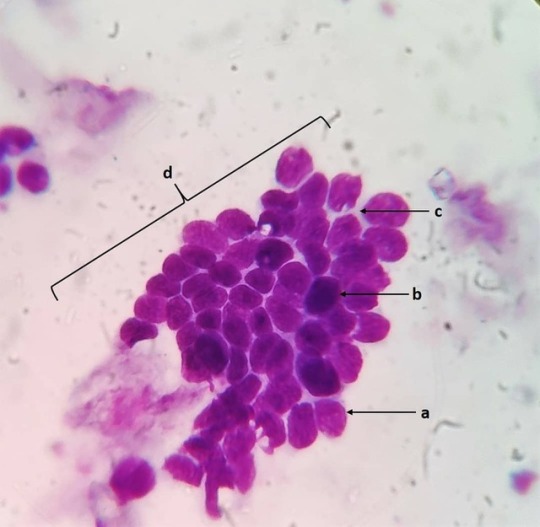
Basal cells: Location: Basal layer of all epithelia. Characteristics and function: Stem cell regenerative compartment for epithelial structures. Shape and size: Small round cells with a high N:C ratio. Nucleus: Round (a) to ovoid (b) centrally placed with compact chromatin. Cytoplasm: Scant with mild basophilia (c). Cytoarchitecture: Pavement (d) or columnar. Ddx: Matured lymphocytes. #vetpath #vetpathology #vetpathologist #vetcytology #vetcyto #veterinaryclinicalpathology #vetclinpath #veterinarycytology #veterinarycytopathology #veterinarymedicine #vetlabdiagnostic #vetcases #vetlife #normalcaninecell #basalcell #vetlablife #veterinary_medicine #vetmed https://www.instagram.com/drdashvetpath/p/BweUHo2Bi7J/?utm_source=ig_tumblr_share&igshid=1hu6g0s917um0
#vetpath#vetpathology#vetpathologist#vetcytology#vetcyto#veterinaryclinicalpathology#vetclinpath#veterinarycytology#veterinarycytopathology#veterinarymedicine#vetlabdiagnostic#vetcases#vetlife#normalcaninecell#basalcell#vetlablife#veterinary_medicine#vetmed
0 notes
Photo

Melanocytes Location: Pigmented areas of body. Characteristics and function: Melanin producing cells of the body. Associated with epidermis, mucosae and serosal surfaces. Shape and size: Medium sized with pleomorphic morphology and no defined cellular limits (a) Cytoplasm: Abundant, faint Basophilic (c), large numbers of dark fine melanin granules (d). Nucleus: Large, round, central with fine stippled chromatin (b). Background: Free melanin pigment. Differential: Melanophage. #vetpath #vetpath #veterinaryclinpath #veterinarypathology #veterinarycytopathology #veterinarycytology #vetcytology #cytology #cytologyvet #cytologystuff #normalcaninecell #normalcell #melanocytes #melaningranules https://www.instagram.com/drdashvetpath/p/BwRCJnNhCkM/?utm_source=ig_tumblr_share&igshid=v1hiuv0kzaws
#vetpath#veterinaryclinpath#veterinarypathology#veterinarycytopathology#veterinarycytology#vetcytology#cytology#cytologyvet#cytologystuff#normalcaninecell#normalcell#melanocytes#melaningranules
0 notes
Photo
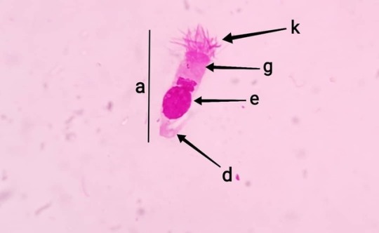
Ciliated epithelial cells: Location: Respiratory mucosa, conjunctiva Function: Lining of mucous membrane. Sweeping action of cilia helps in clearing out dust particles and debris. Shape: Elongated, columnar and wavy (a). Size: Medium sized cells seen sometimes with nucleus protuding out (b). Cytoplasm: Moderately Basophilic (g), apical ciliary apparatus (k), focal hyper basophilic area (h), superficial eosinophilic zone (i) and cilia (j). Nucleus: Round (e), oval (f), subterminal with fine stippled chromatin. Cytoarchitecture: Exfoliate as individual cells. Background: Mucinous matrix #vetpath #veterinarypathology #veterinarycytopathology #vetcytology #veterinarycytology #normalcell #cytopathology #normalcaninecell #respiratoryepithelialcells #ciliatedepithelium #ciliated #ciliatedcolumnarepithilium #hairycells https://www.instagram.com/drdashvetpath/p/BwHM6l0hvmM/?utm_source=ig_tumblr_share&igshid=1xzqjs1fbr0mv
#vetpath#veterinarypathology#veterinarycytopathology#vetcytology#veterinarycytology#normalcell#cytopathology#normalcaninecell#respiratoryepithelialcells#ciliatedepithelium#ciliated#ciliatedcolumnarepithilium#hairycells
0 notes
Photo

Conjunctival squamous: Location: Bulbar conjunctiva. Shape and size: Intermediate to superficial cells. Polygonal with a low N:C ratio. Characteristics: Epithelial cells of the bulbar conjunctiva. Nucleus: Centrally placed with compact fine chromatin (a) Cytoplasm: Clear, amphophilic with melanin pigments in perinuclear area (b). Cytoarchitecture: Single or in clusters. #vetclinicalpathology #vetpath #vetpathology #vetcytology #veterinarypathology #vetopthalmology #veterinaryclinicalpathology #veterinarycytology #veterinarycytopathology #normalcaninecell #normalcell #normalcytology #conjunctivalsquamouscell #eyecell https://www.instagram.com/drdashvetpath/p/BwB1X7OBjRi/?utm_source=ig_tumblr_share&igshid=lohfxwr2i8cg
#vetclinicalpathology#vetpath#vetpathology#vetcytology#veterinarypathology#vetopthalmology#veterinaryclinicalpathology#veterinarycytology#veterinarycytopathology#normalcaninecell#normalcell#normalcytology#conjunctivalsquamouscell#eyecell
0 notes
Photo
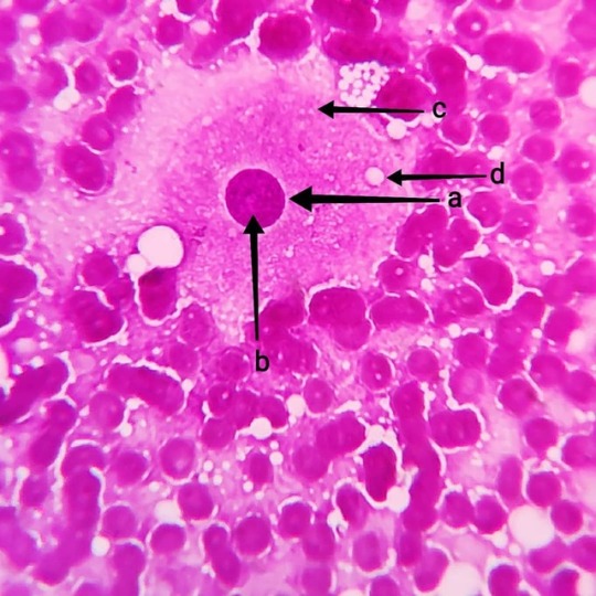
Luteal cell: Location- Female genital tract, #ovary #corpusluteum Characteristic and function - Production of #progesterone and formation of #corpusluteum Shape - Pear shaped Size - Vert large to large Cytoplasm - Basophilic (c) and abundant with empty vacuoles (d). Nucleus - Large (a), oval round, central to paracentral with finely stippled chromatin and prominent nucleolus (b). Cytoarchitecture - Pavement clusters or single cells. #vetpath #veterinarypathology #cytology #vetcytology #veterinarycytology #veterinarycytopathology #cytopathology #cytopath #normalcell #vetdiagnosis #impressionsmear #diffquick #diffquikstain #ovariancell https://www.instagram.com/drdashvetpath/p/Bv3Da3fhMdM/?utm_source=ig_tumblr_share&igshid=1pugr3qr10uxs
#ovary#corpusluteum#progesterone#vetpath#veterinarypathology#cytology#vetcytology#veterinarycytology#veterinarycytopathology#cytopathology#cytopath#normalcell#vetdiagnosis#impressionsmear#diffquick#diffquikstain#ovariancell
0 notes
Photo

And again!!!! Lipoma in a dog: Seen are plump adipocytes with optically empty cytoplasm due to the stored fats/lipids in the cells. The nucleus is pushed to the periphery to accomodate the maximum available space for storage of fats/lipids. Adipose tissue are usually found intertwined with collagenous fibres and blood vessels forming a 3D cytoarchitecture. #vetpath #vetpathology #cytopathology #vetdiagnosis #vetdiagnostics #fna #fnac #cytology #cytologyvet #veterinarycytopathology #vetlabdiagnostic #fineneedleaspirationcytology #fineneedleaspirate #lipoma #subcutaneous #veterinaryclinicalpathology #cytologicalexamination #aspirationcytology https://www.instagram.com/drdashvetpath/p/Bvenfy5hfrW/?utm_source=ig_tumblr_share&igshid=306j3bci9mbr
#vetpath#vetpathology#cytopathology#vetdiagnosis#vetdiagnostics#fna#fnac#cytology#cytologyvet#veterinarycytopathology#vetlabdiagnostic#fineneedleaspirationcytology#fineneedleaspirate#lipoma#subcutaneous#veterinaryclinicalpathology#cytologicalexamination#aspirationcytology
0 notes
Photo
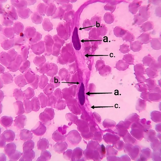
Smooth muscle cell: Location: Involuntary muscle tissue if many organs Function: Mesenchymal cells for involuntary muscle contraction. Shape: Elongated and thin Cytoplasm: Eosinophilic (c) Nucleus: Cigar shaped (b), elongated and thin (a) Ddx: Fibrocytes #vetpath #veterinarypathology #cellcytology #vetcytology #veterinarycytopathology #veterinarycytology #fna #fnac #fineneedleaspirationcytology #fineneedleaspiration #fineneedleaspirate #smoothmuscle #smoothmusclecells #mesenchymalcell https://www.instagram.com/drdashvetpath/p/BvZ-a3xBMYo/?utm_source=ig_tumblr_share&igshid=1codzb9tdhe3s
#vetpath#veterinarypathology#cellcytology#vetcytology#veterinarycytopathology#veterinarycytology#fna#fnac#fineneedleaspirationcytology#fineneedleaspiration#fineneedleaspirate#smoothmuscle#smoothmusclecells#mesenchymalcell
0 notes
Photo
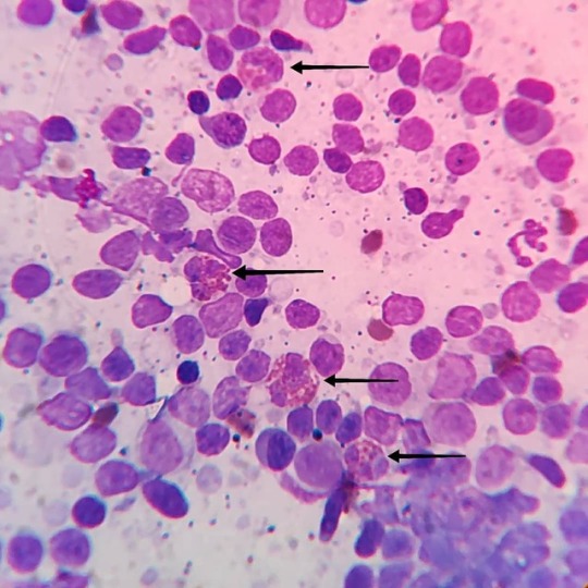
Eosinophilic lymphadenitis: Eosinophils are not commonly observed in the lymph nodes. Individually or seen in aggregates in cases like chronic skin infections, allergic and hypersensitivity reactions, paraneoplastic response, fungal and parasitic infections. Prognosis is good but also depends on the underlying cause (Inflammatory or paraneoplastic response). #vetpath #vetpathology #vetclinpath #veterinarypathology #veterinaryclinicalpathology #fineneedleaspirationcytology #fineneedleaspirate #fineneedleaspiration #cytology #cytologyvet #veterinarycytology #lymphnode #lymphadenitis #eosinophils #caninecytology #dogcytology #cytopathology #cytopath #veterinarycytopathology https://www.instagram.com/drdashvetpath/p/BvTETR4hc3A/?utm_source=ig_tumblr_share&igshid=k1qkf226vr57
#vetpath#vetpathology#vetclinpath#veterinarypathology#veterinaryclinicalpathology#fineneedleaspirationcytology#fineneedleaspirate#fineneedleaspiration#cytology#cytologyvet#veterinarycytology#lymphnode#lymphadenitis#eosinophils#caninecytology#dogcytology#cytopathology#cytopath#veterinarycytopathology
0 notes
Last Seen Blogs

musiclmaiden
Untitled

lg-art-200
Lg - Art
musehcwling
‒ moonlight musing

music-and-art-bean
Welcome to my lair

abacaxau
Abacaxau