#volk inview fundus camera
Explore tagged Tumblr posts
Visit Tumblr Blog
Explore Tumblr blogs with no restrictions, modern design and the best experience.
Last Seen Tumblr Blogs


Fun Fact
Total funding amounts to $125.3M.
Text
Revolutionizing Cataract Surgery Precision: Exploring the Zeiss IOL Master 500
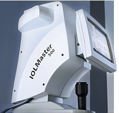
In the realm of cataract surgery, precision and accuracy are paramount for successful outcomes. The Zeiss IOL Master 500 has emerged as a game-changer in the field, revolutionizing the way surgeons measure and calculate intraocular lens (IOL) power. In this blog, we will delve into the features, benefits, and impact of the Zeiss IOL Master 500 on cataract surgery, showcasing its significance in ensuring optimal visual results for patients.
The Evolution of IOL Calculation:
Introduction of Optical Biometry:
1. Advancements in Measurement: Optical biometry marked a significant advancement, utilizing laser technology to measure axial length and corneal curvature for more accurate IOL power calculations.
2. Enhancing Predictability: Optical biometry improved the predictability of outcomes, but the quest for even greater accuracy led to the development of the Zeiss IOL Master 500.
Integration of Advanced Formulas:
Incorporating Latest IOL Calculation Formulas:
1. Customization for Patient Specifics: Surgeons can select the most appropriate formula based on individual patient factors, further refining the accuracy of IOL power predictions.
Consideration of Posterior Corneal Astigmatism:
1. Total Corneal Astigmatism: The device takes into account posterior corneal astigmatism, providing a more comprehensive assessment of the eye's total astigmatism for improved IOL power calculation accuracy.
Benefits for Surgeons and Patients:
Time Efficiency and Workflow:
1. Rapid Measurements: The Zeiss IOL Master 500 delivers quick and efficient measurements, streamlining the preoperative workflow for surgeons and optimizing clinic efficiency.
2. Real-Time Data Feedback: Surgeons benefit from real-time data feedback, allowing for immediate assessment and adjustment of IOL power calculations as needed.
Enhanced Predictability and Visual Outcomes:
1. Reduced Refractive Error: The precision offered by the Zeiss IOL Master 500 contributes to reduced refractive errors, enhancing the predictability of visual outcomes for patients.
2. Minimized Postoperative Surprises: Surgeons can confidently anticipate and minimize postoperative surprises, ensuring a higher degree of patient satisfaction with their visual results.
Clinical Validation and Research Findings:
Scientific Studies and Publications:
1. Validation in Clinical Settings: Numerous scientific studies and publications have validated the accuracy and reliability of the Zeiss IOL Master 500 in various clinical settings.
2. Positive Surgeon Feedback: The device has garnered positive feedback from surgeons worldwide, contributing to its widespread adoption as a standard tool in modern cataract surgery practices.
Conclusion: Elevating Cataract Surgery Precision with Zeiss IOL Master 500:
The Zeiss IOL Master 500 stands at the forefront of cataract surgery precision, offering surgeons an indispensable tool to achieve optimal visual outcomes for their patients. With advanced technology, precise measurements, and integration with cutting-edge formulas, this device has reshaped the landscape of IOL power calculation. As cataract surgery continues to evolve, the Zeiss IOL Master 500 remains a beacon of accuracy, reliability, and innovation, setting the standard for excellence in modern ophthalmic practices.
0 notes
Text
Unveiling the Marvel: Tomey TL-3000C Advanced – A Technological Triumph in Ophthalmic Instruments
In the ever-evolving field of ophthalmology, precision and innovation are paramount. One such groundbreaking advancement is the Tomey TL-3000C Advanced, a state-of-the-art ophthalmic instrument that is reshaping the landscape of eye care. Let's delve into the benefits of this technological marvel and how it is revolutionizing the way we approach eye examinations.
Crystal-Clear Imaging:
At the heart of the Tomey TL-3000C Advanced lies its unparalleled imaging capabilities. The device employs cutting-edge technology to provide crystal-clear images of the eye, allowing ophthalmologists to delve deep into the intricacies of ocular structures. With a high-resolution camera and advanced optics, this instrument delivers images that are not only detailed but also incredibly precise.
Ergonomic Design:
Ophthalmologists spend hours each day examining patients' eyes, making the ergonomics of their instruments crucial. The Tomey TL-3000C Advanced considers this with its ergonomic design, ensuring both comfort and efficiency for practitioners.The instrument features an adjustable chin rest and headrest, allowing for optimal patient positioning during examinations. This not only enhances the patient's comfort but also facilitates a more accurate assessment of the eye.
Streamlined Workflow:
In the fast-paced world of healthcare, efficiency is key. The Tomey TL-3000C Advanced is designed with a focus on streamlining the examination process, thereby optimizing the workflow in ophthalmic practices.The device integrates seamlessly with electronic health record (EHR) systems, allowing practitioners to store and retrieve patient data effortlessly. This not only reduces the risk of errors associated with manual record-keeping but also enhances the overall efficiency of the practice.
Patient Engagement:
In today's digital age, patient engagement is a crucial aspect of healthcare. The Tomey TL-3000C Advanced goes beyond traditional ophthalmic instruments by incorporating features that promote patient engagement and education.The device enables real-time sharing of images and diagnostic findings with patients, fostering a collaborative approach to eye care. This not only enhances patient understanding of their eye health but also strengthens the doctor-patient relationship.
Future-Proof Technology:
The field of ophthalmology is dynamic, with new challenges and technologies emerging regularly. The Tomey TL-3000C Advanced stands as a testament to its commitment to future-proof technology. Regular software updates ensure that the device stays abreast of the latest advancements in ophthalmic care, providing practitioners with the tools they need to deliver the best possible patient outcomes.
The Tomey TL-3000C Advanced is more than just an ophthalmic instrument – it's a technological triumph that is reshaping the way we approach eye care. With its crystal-clear imaging, ergonomic design, streamlined workflow, patient engagement features, and future-proof technology, this device is at the forefront of innovation in ophthalmology. As we embrace the future of healthcare, the Tomey TL-3000C Advanced leads the way in ensuring a clearer vision for both practitioners and patients alike.
0 notes
Text
Discover the Power of Clarity with the VOLK InView Fundus Camera
In the realm of ophthalmology, accurate imaging of the retina is essential for diagnosing and monitoring various eye conditions. ForesightIntl understands the importance of advanced technology in capturing detailed retinal images, which is why they proudly offer the VOLK InView Fundus Camera.
Unmatched Clarity and Detail: The VOLK InView Fundus Camera stands out for its ability to capture retinal images with unparalleled clarity and detail. Equipped with advanced optics and cutting-edge technology, this device provides high-resolution images of the retina. Eye care professionals can now visualize intricate retinal structures and pathologies with exceptional precision.
Efficiency and Ease of Use: The VOLK InView Fundus Camera is designed to optimize efficiency and streamline the examination process. Its user-friendly interface and intuitive controls make image capture a seamless experience. With its automated features and rapid capture capabilities, eye care professionals can efficiently obtain high-quality retinal images, saving valuable time and enhancing the overall patient experience.

Versatile Imaging Modes: The VOLK InView Fundus Camera offers a range of versatile imaging modes, catering to the diverse needs of eye care professionals. From standard color fundus photography to fluorescein angiography, this device enables practitioners to capture various types of retinal images. With the VOLK InView Fundus Camera, eye care professionals have the flexibility to tailor their imaging techniques based on individual patient requirements.
Enhanced Diagnosis and Collaboration: The clarity and detail captured by the VOLK InView Fundus Camera empower eye care professionals to make accurate and informed diagnoses. The high-quality retinal images serve as valuable references for monitoring disease progression and treatment efficacy over time.
Reliability and Durability: ForesightIntl is dedicated to providing high-quality ophthalmic equipment, and the VOLK InView Fundus Camera is no exception. Built with durability in mind, this device is designed to withstand the demands of a busy clinical environment. Its robust construction and precise engineering ensure long-term reliability, allowing eye care professionals to rely on the VOLK InView Fundus Camera for consistent and accurate retinal imaging.
Conclusion: The VOLK InView Fundus Camera from ForesightIntl empowers eye care professionals to achieve unparalleled clarity and detail in retinal imaging. With its advanced technology, ease of use, and versatile imaging modes, this device enhances efficiency and accuracy in examinations. Backed by ForesightIntl's commitment to reliability and durability, the VOLK InView Fundus Camera sets new standards in retinal imaging. Embrace the power of clarity with the VOLK InView Fundus Camera and unlock a new level of excellence in ophthalmic practice.
0 notes
Text
Key Features of Portable VOLK InView Fundus Camera
The VOLK InView Fundus Camera is a medical device used for capturing high-resolution images of the retina. It is primarily used by ophthalmologists and optometrists for diagnosing and monitoring various eye conditions, such as diabetic retinopathy, age-related macular degeneration, and glaucoma.
The VOLK InView Fundus Camera uses advanced imaging technology to capture detailed images of the retina, which is the light-sensitive tissue lining the back of the eye. These images provide valuable information about the health of the retina and can help in the early detection and management of eye diseases.
Some key features of the VOLK InView Fundus Camera may include:
High-resolution imaging: The camera is capable of capturing high-quality images with excellent detail, allowing for accurate diagnosis and monitoring of retinal conditions.
Non-mydriatic imaging: Non-mydriatic refers to the ability to capture images of the retina without the need for pupil dilation, making the procedure more comfortable for the patient.
Wide-field imaging: The camera may have the capability to capture wide-field images of the retina, allowing for a comprehensive view of the entire retina and peripheral areas.
User-friendly interface: The camera may feature an intuitive user interface, making it easy for healthcare professionals to operate and capture images efficiently.
Integration with software: The VOLK InView Fundus Camera may be compatible with image management software, enabling the storage, analysis, and comparison of images over time for better patient management.
The VOLK InView Fundus Camera may be used by an ophthalmologist for a number of reasons, including the following:
Early Detection and Diagnosis: The camera enables the early identification and diagnosis of a number of eye disorders, including glaucoma, diabetic retinopathy, and macular degeneration. Preventing eyesight loss and possibly blindness through early identification and treatment.
High-Quality Images: Ophthalmologists may view the retina in great detail and with accuracy thanks to the VOLK InView Fundus Camera, which also takes high-resolution pictures of the eye's interior tissues. Making more accurate diagnosis and treatment strategies can benefit from this.
Non-Invasive: Since the camera is a non-invasive diagnostic device, no contact with the eye or the administration of contrast agents are necessary. Because of this, patients may undergo the treatment in safety and comfort.
Quick and Efficient: The camera produces excellent photographs in a matter of seconds and is simple to operate. Ophthalmologists may be able to identify eye diseases more quickly and effectively as a result, keeping patients out of the waiting room.
Portable: Due to its portability, the VOLK InView Fundus Camera may be used in a variety of clinical situations. In isolated or underdeveloped places where access to medical equipment may be restricted, this can be extremely helpful.
Ophthalmologists can identify and diagnose a variety of eye diseases early on thanks to the VOLK InView Fundus Camera. It is a useful addition to any ophthalmologist's practise because to its high-quality imaging capabilities, simplicity of use, and mobility.
0 notes
Text
Pioneering Retinal Insights: Exploring the Potential of VOLK InView Fundus Camera
In the ever-evolving realm of ophthalmic technology, the VOLK InView Fundus Camera emerges as a transformative tool, unraveling the intricacies of retinal health with its cutting-edge capabilities and unrivaled precision. This advanced instrument stands as a testament to innovation, propelling eye care professionals into a new era of diagnostic excellence and retinal assessment.
As a fusion of technical prowess and optical ingenuity, the VOLK InView Fundus Camera takes a prominent place in modern ophthalmology, redefining the landscape of retinal imaging through its state-of-the-art design and visionary features.
Ophthalmic professionals around the globe are applauding the VOLK InView Fundus Camera for its remarkable ability to capture intricate retinal details with exceptional clarity. This instrument not only captures images; it provides invaluable insights into the health of the retina, setting a new standard for precision and diagnostic accuracy.
At the core of the VOLK InView Fundus Camera lies a commitment to seamless user experience, catering to both clinicians and patients. Its user-friendly interface and streamlined controls ensure that capturing retinal images is an effortless process, enhancing patient cooperation and facilitating efficient examinations.
The integration of advanced imaging technology within the VOLK InView Fundus Camera elevates its diagnostic potential. By offering high-resolution retinal images and seamless connectivity to imaging systems, it empowers clinicians to delve into comprehensive retinal analysis, enabling informed decision-making and personalized treatment plans.
In the fast-paced realm of modern healthcare, efficiency is paramount. The VOLK InView Fundus Camera addresses this demand through its swift image acquisition and seamless data management. This not only optimizes clinical workflows but also minimizes patient discomfort, ensuring a smooth and comfortable imaging experience.
Furthermore, the VOLK InView Fundus Camera emerges as a visionary tool for early detection and comprehensive retinal assessment. Its capabilities enable clinicians to identify subtle retinal changes and abnormalities, contributing to early interventions and improved patient outcomes.
In conclusion, the VOLK InView Fundus Camera transcends conventional retinal imaging, ushering in an era characterized by precision and diagnostic excellence. With its advanced technology and user-centric design, it has become an indispensable asset in the field of ophthalmology. As eye care professionals and patients embrace its transformative potential, the VOLK InView Fundus Camera stands as a beacon of progress, illuminating the path towards enhanced retinal assessment and proactive vision care.
0 notes
Text
Key Features of Portable VOLK InView Fundus Camera
The VOLK InView Fundus Camera is a medical device used for capturing high-resolution images of the retina. It is primarily used by ophthalmologists and optometrists for diagnosing and monitoring various eye conditions, such as diabetic retinopathy, age-related macular degeneration, and glaucoma.
The VOLK InView Fundus Camera uses advanced imaging technology to capture detailed images of the retina, which is the light-sensitive tissue lining the back of the eye. These images provide valuable information about the health of the retina and can help in the early detection and management of eye diseases.
Some key features of the VOLK InView Fundus Camera may include:
High-resolution imaging: The camera is capable of capturing high-quality images with excellent detail, allowing for accurate diagnosis and monitoring of retinal conditions.
Non-mydriatic imaging: Non-mydriatic refers to the ability to capture images of the retina without the need for pupil dilation, making the procedure more comfortable for the patient.
Wide-field imaging: The camera may have the capability to capture wide-field images of the retina, allowing for a comprehensive view of the entire retina and peripheral areas.
User-friendly interface: The camera may feature an intuitive user interface, making it easy for healthcare professionals to operate and capture images efficiently.
Integration with software: The VOLK InView Fundus Camera may be compatible with image management software, enabling the storage, analysis, and comparison of images over time for better patient management.
The VOLK InView Fundus Camera may be used by an ophthalmologist for a number of reasons, including the following:
Early Detection and Diagnosis: The camera enables the early identification and diagnosis of a number of eye disorders, including glaucoma, diabetic retinopathy, and macular degeneration. Preventing eyesight loss and possibly blindness through early identification and treatment.
High-Quality Images: Ophthalmologists may view the retina in great detail and with accuracy thanks to the VOLK InView Fundus Camera, which also takes high-resolution pictures of the eye's interior tissues. Making more accurate diagnosis and treatment strategies can benefit from this.
Non-Invasive: Since the camera is a non-invasive diagnostic device, no contact with the eye or the administration of contrast agents are necessary. Because of this, patients may undergo the treatment in safety and comfort.
Quick and Efficient: The camera produces excellent photographs in a matter of seconds and is simple to operate. Ophthalmologists may be able to identify eye diseases more quickly and effectively as a result, keeping patients out of the waiting room.

Portable: Due to its portability, the VOLK InView Fundus Camera may be used in a variety of clinical situations. In isolated or underdeveloped places where access to medical equipment may be restricted, this can be extremely helpful.
Ophthalmologists can identify and diagnose a variety of eye diseases early on thanks to the VOLK InView Fundus Camera. It is a useful addition to any ophthalmologist's practise because to its high-quality imaging capabilities, simplicity of use, and mobility.
0 notes
Text
VOLK InView Fundus Camera: Is it The Best Way to Monitor Your Eye Health?
A VOLK InView Fundus Camera is a medical device that is used to capture images of the retina and optic nerve. This camera is used to help diagnose and treat various eye conditions. The camera is a small, handheld device that is easy to use. It has a built-in flash and a digital display that allows the user to view the images that are captured. The images can be stored on a computer or printed out for further examination.
How does a VOLK InView Fundus Camera work?
The VOLK InView Fundus Camera is a handheld device that is used to take images of the fundus, or back of the eye. The device uses a bright light and a camera to capture images of the fundus. These images can be used to diagnose and treat eye diseases. The camera is held close to the eye and the light is shined into the eye. The camera captures images of the fundus as the light passes through the eye.
What are the benefits of using a VOLK InView Fundus Camera?
A VOLK InView Fundus Camera is a device used to capture images of the retina and optic nerve. This device is beneficial because it is portable, easy to use, and produces high-quality images.

The portability of the VOLK InView Fundus Camera makes it easy to move around the clinic or hospital. This camera is also easy to use, which makes it a great choice for clinics and hospitals with limited staff. The high-quality images produced by the VOLK InView Fundus Camera make it an ideal choice for clinics and hospitals that want to provide the best possible care for their patients.
How can a VOLK InView Fundus Camera help monitor your eye health?
A VOLK InView Fundus Camera is a device that can help monitor your eye health. It uses a special technology called fundus photography to take pictures of the inside of your eye. These pictures can help your doctor see any problems with your eye health.
How to take care of VOLK InView Fundus Camera?
If you have a VOLK InView Fundus Camera, it is important to take care of it so that it lasts a long time. Here are some tips on how to do that:
1. Store the camera in a cool, dry place.
2. Keep the camera clean and dust-free.
3. Be careful when handling the camera. Avoid dropping it or subjecting it to sudden impacts.
4. Bring the camera to a VOLK-authorized service center for regular maintenance.
By following these tips, you can be sure that your VOLK InView Fundus Camera will provide you with many years of trouble-free service.
0 notes
Text
Everything you ever need to know about the VOLK InView Fundus Camera
A high-resolution, simple-to-use fundus camera, the VOLK InView Fundus Camera gives doctors a better approach to recording and monitoring the development of their patients' retinal health. For general ophthalmology, glaucoma, and retina clinics, this camera is a superb tool.
A fundus camera is a medical tool used to capture images of the fundus, the eye's deepest layer. A light source, a lens, and a digital camera make up the fundus camera. The fundus is illuminated by the light source, which is then focused onto the digital camera by the lens.
VOLK InView Fundus Camera advantages
The InView Fundus Camera can be a good option if you are seeking a fundus camera that produces high-quality photos and is simple to use. This camera is made to be used in clinics and hospitals, and it has a number of advantages that make it a perfect option for many establishments.

The VOLK InView Fundus Camera's superior image quality is one of its main advantages. To take crisp, clear pictures of the retina, this camera employs a high-resolution sensor. For the detection and treatment of diseases like diabetic retinopathy, this is crucial.
The VOLK InView Camera's ease of use is another advantage. The camera is portable and light, making it simple to use. You may start using the camera right away because of the user interface's clarity and simplicity.
The VOLK Fundus Camera is a terrific choice if you are searching for a fundus camera that has high image quality and is simple to use. This camera is a fantastic option for many institutions and is a wonderful choice for both clinics and hospitals. It has a number of advantages that make it an amazing option.
Guidelines for maintaining the VOLK InView Fundus Camera
If you own an InView Fundus Camera, you are aware of its superior build quality. But in order for it to work at its best, it must be properly maintained, just like any other piece of equipment. The following are some suggestions for maintaining your VOLK InView Camera:
1. Maintain a clean camera. After each usage, clean the camera's body and lens with a gentle, dry cloth. Dry the camera off right away if it becomes wet.
2. When not in use, keep the camera in a secure, dry location. Steer clear of severe temperatures since they can harm the camera.
3. Frequently check the camera. Make sure all the controls and buttons are operational and look for any body or lens damage.
You can assist ensure that your VOLK InView Fundus Camera will provide years of trouble-free service by keeping in mind these easy suggestions.
0 notes
Text
Everything you ever need to know about the VOLK InView Fundus Camera
A high-resolution, simple-to-use fundus camera, the VOLK InView Fundus Camera gives doctors a better approach to recording and monitoring the development of their patients' retinal health. For general ophthalmology, glaucoma, and retina clinics, this camera is a superb tool.
A fundus camera is a medical tool used to capture images of the fundus, the eye's deepest layer. A light source, a lens, and a digital camera make up the fundus camera. The fundus is illuminated by the light source, which is then focused onto the digital camera by the lens.
VOLK InView Fundus Camera advantages
The InView Fundus Camera can be a good option if you are seeking a fundus camera that produces high-quality photos and is simple to use. This camera is made to be used in clinics and hospitals, and it has a number of advantages that make it a perfect option for many establishments.
The VOLK InView Fundus Camera's superior image quality is one of its main advantages. To take crisp, clear pictures of the retina, this camera employs a high-resolution sensor. For the detection and treatment of diseases like diabetic retinopathy, this is crucial.
The VOLK InView Camera's ease of use is another advantage. The camera is portable and light, making it simple to use. You may start using the camera right away because of the user interface's clarity and simplicity.
The VOLK Fundus Camera is a terrific choice if you are searching for a fundus camera that has high image quality and is simple to use. This camera is a fantastic option for many institutions and is a wonderful choice for both clinics and hospitals. It has a number of advantages that make it an amazing option.
Guidelines for maintaining the VOLK InView Fundus Camera
If you own an InView Fundus Camera, you are aware of its superior build quality. But in order for it to work at its best, it must be properly maintained, just like any other piece of equipment. The following are some suggestions for maintaining your VOLK InView Camera:

1. Maintain a clean camera. After each usage, clean the camera's body and lens with a gentle, dry cloth. Dry the camera off right away if it becomes wet.
2. When not in use, keep the camera in a secure, dry location. Steer clear of severe temperatures since they can harm the camera.
3. Frequently check the camera. Make sure all the controls and buttons are operational and look for any body or lens damage.
You can assist ensure that your VOLK InView Fundus Camera will provide years of trouble-free service by keeping in mind these easy suggestions.
0 notes
Link
The Volk iNview mobile app automatically allows the device to acquire retina photos during a patient session. The program automatically recognizes and chooses the best focused and defined photographs, allowing even inexperienced users to shoot images fast and effortlessly.
0 notes
Text
VOLK In View Fundus Camera
The VOLK In View Fundus Camera is an innovative new ophthalmic camera device able to capture wide-angle digital color images of the fundus using an Apple iPhone 6s, 6, 5s or 5E .The Volk In view mobile app authorizes the device to automatically capture images of the retina during a patient session. The app automatically detects and selects for the most focused & defined images permitting even first time users to capture images quickly and easily. Contact us directly for this product. https://www.foresightintl.com/product/volk-inview-fundus-camera
0 notes
Text
Portable VOLK InView Fundus Camera
The Volk iNview is an ophthalmic camera that uses an Apple iPhone or iPod to capture wide-angle digital colour photographs of the retina (fundus). Volk iNview is a hybrid product that combines a mobile app (Apple App Store) with an indirect ophthalmoscopic lens attachment for Apple iPhone/iPod.
The Volk iNview is designed to take images of the eye's retina for general visualisation and patient education. Combining the power of the Apple iPhone with Volk Optics' dependable quality.

The Volk Inview mobile app allows the device to take images of the retina automatically during a patient session. The software automatically analyses and selects the best focused and defined shots, allowing even inexperienced users to capture images quickly and easily. The Volk Inview programme may be securely secured with a password key, ensuring that patient data is kept safe and secure.
Features of VOLK InView Fundus Camera
· Facilitate communication between doctors and patients about illness progression and treatment options by rapidly and easily capturing fundus pictures for visualisation and patient education.
· A free mobile application is available in the Apple App Store.
· Make a secure purchase. Volk offers a 30-day money-back guarantee on all purchases. It's simple to use using Auto-capture Technology.
Consider buying this effective optometry equipment for best results.
0 notes