#physiologyfirst
Explore tagged Tumblr posts
Visit Tumblr Blog
Explore Tumblr blogs with no restrictions, modern design and the best experience.
Last Seen Tumblr Blogs


Fun Fact
In 2020, 27% of US Tumblr users had an annual household income of over $100,000.
Photo

Active Transport Intro · ‘Active transport is the process of transferring substances into, out of, and between cells, using energy. In some cases, the movement of substances can be accomplished by passive transport, which uses no energy. However, the cell often needs to transport materials against their concentration gradient.’ - https://biologydictionary.net/active-transport/ · Substances which cannot be diffused would be selected (e.g. too large solutes or they are incapable of dissolving in the lipid bilayer). · There are two types of active transport: o Primary o Secondary Primary Active Transport · ‘Primary active transport is also called direct active transport or uniport. It involves using energy (usually ATP) to directly pump a solute across a membrane against its electrochemical gradient.’ · Solute are transported across by the hydrolyses of ATP which changes the protein shape. This enables the solute to be pumped across. · An example of primary active transport is the Sodium-potassium pump (Na+ K+ pump). The pump drives Na+ out of the cell against a steep concentration gradient & pumps K+ back in. Secondary Active Transport · ‘In secondary active transport, also known as coupled transport or cotransport, energy is used to transport molecules across a..... Reference · https://biologydictionary.net/active-transport/ · https://microbenotes.com/active-transport/ · Marieb & Hoehn. Human Anatomy and Physiology. 10th ed. Chapter 3 . . . #discoverphysiotherapy #physiotherapists #physiotherapystudents #physioworld #physio #physiologyfirst #physiology_notes #physiologyfacts #physiologyofactivity #anatomyandphysiology #sportphysiology #humananatomyphysiology #musclephysiology #psychophysiology #transport #active #activetransport #activetransportation #new #daily #dailypost #tuesdayvibes #tuesday #tuesdaymotivation #tuesdaythoughts #cell #cells #biology🔬 #biologyislife #biologynotes https://www.instagram.com/p/CECLy_an2Tb/?igshid=1agegrhurfpn4
#discoverphysiotherapy#physiotherapists#physiotherapystudents#physioworld#physio#physiologyfirst#physiology_notes#physiologyfacts#physiologyofactivity#anatomyandphysiology#sportphysiology#humananatomyphysiology#musclephysiology#psychophysiology#transport#active#activetransport#activetransportation#new#daily#dailypost#tuesdayvibes#tuesday#tuesdaymotivation#tuesdaythoughts#cell#cells#biology🔬#biologyislife#biologynotes
0 notes
Photo

Passive Transport Intro · ‘Passive transport, also known as passive diffusion, is a process by which an ion or molecule passes through a cell wall via a concentration gradient, or from an area of high concentration to an area of low concentration. It’s like moving from the train to the platform of a subway station or stepping out of a crowded room. Basically, passive transport gives an ion or molecule “room to breathe.”’ - https://biologydictionary.net/passive-transport/ · There are different types of diffusion for passive transport: o Simple diffusion o Facilitated diffusion o Filtration Simple diffusion · ‘Simple diffusion is the process by which solutes are moved along a concentration gradient in a solution or across a semipermeable membrane. Simple diffusion is carried out by the actions of hydrogen bonds forming between water molecules and solutes’. · Lipid soluble, non-polar and small substances are diffused through the bilayer by simple diffusion. Facilitated diffusion · ‘Facilitated diffusion is a form of facilitated transport .... Reference�� · https://biologydictionary.net/passive-transport/ · https://biologydictionary.net/simple-diffusion/#:~:text=Simple%20diffusion%20is%20the%20process,between%20water%20molecules%20and%20solutes. · https://biologydictionary.net/facilitated-diffusion/ · https://courses.lumenlearning.com/boundless-biology/chapter/passive-transport/ . . . #discoverphysiotherapy #physioterapist #physiology_lab #physiologyfirst #physiology #physiology_notes #exercisephysiology #anatomyphysiology #sportphysiology #pathophysiology #animalphysiology #humananatomyandphysiology #clinicalphysiology #medicalphysiology #physiolab #physiotherapist #physiotherapists #physiotherapystudents #physio #physiologie #education #educationmatters #educating #educationfirst #educationpositive #tuesday #tuesdayvibes #physiotherapeut https://www.instagram.com/p/CDvQR7InkM2/?igshid=1io4dw3zno881
#discoverphysiotherapy#physioterapist#physiology_lab#physiologyfirst#physiology#physiology_notes#exercisephysiology#anatomyphysiology#sportphysiology#pathophysiology#animalphysiology#humananatomyandphysiology#clinicalphysiology#medicalphysiology#physiolab#physiotherapist#physiotherapists#physiotherapystudents#physio#physiologie#education#educationmatters#educating#educationfirst#educationpositive#tuesday#tuesdayvibes#physiotherapeut
0 notes
Photo

Membrane Transport Summary · The plasma membrane protects the cell from the external environment as well as permitting the transport of specific substances in or out of the cell. · Some substances(solutes) can move across a selectively permeable membrane. This means some substances are capable to pass through (e.g. oxygen, carbon dioxide, etc.) whilst blocking others (e.g. ions) · The plasma membrane has two processes to transport substances across: o Passive process - No energy is required (ATP) - Simple and facilitated diffusion takes place o Active process - ATP is required (directly or indirectly) - Primary, secondary or vesicular transport takes place Reference · Marieb & Hoehn. Human Anatomy and Physiology. 10th ed. Chapter 3 . . . #discoverphysiotherapy #physioterapist #physiotherapists #physio #physiologyfirst #physiorehab #physiology_lab #physiology #physiology_notes #humanphysiology #membrane #membranestructure #protein #process #active #passive #transport #permeable #carbon #carbondioxide #atp #simple #diffusion #energy #directly #primary #secondary #plasma #environment #cell . @physiology.s @physiology.anatomy @_physiology_facts_ @physiology_arts @physiologyofstudying @physiology_facts_exciting @physiology @easy_anatomy_physiology @frappeandphysiology @physio_logy_ @joselopezchicharro @effectexercisephysiology @anatomy.physiology.nursing @human_physiology2017 @physiology_facts_ @anatomy_physiology @vitruvianexercisephysiology @humanphysiologyonline @physiologyfacts20 @deckerapnotes @physiology_notes @exercise_training_physiology https://www.instagram.com/p/CDLtsb5HC1_/?igshid=1sbgddiqvu45g
#discoverphysiotherapy#physioterapist#physiotherapists#physio#physiologyfirst#physiorehab#physiology_lab#physiology#physiology_notes#humanphysiology#membrane#membranestructure#protein#process#active#passive#transport#permeable#carbon#carbondioxide#atp#simple#diffusion#energy#directly#primary#secondary#plasma#environment#cell
0 notes
Photo

Plasma membrane and its proteins Plasma membrane · ‘a microscopic membrane of lipids and proteins which forms the external boundary of the cytoplasm of a cell or encloses a vacuole and regulates the passage of molecules in and out of the cytoplasm.’ - https://www.lexico.com/definition/plasma_membrane · Plasma membrane: phospholipid bilayer · The structure of the plasma membrane (phospholipid bilayer) consists of two hydrophobic fatty acid “tails” and a hydrophilic “head “consisting of a phosphate group. o The hydrophobic fatty acid tails face inwards. It repels from water. o The hydrophilic head faces outwards. It faces the water. · The plasma membrane is joined together by the glycerol molecules. This glycerol molecule strengthens the membrane. Plasma membrane’s proteins Phospholipid bilayers contain different proteins: Integral proteins span the membranes with different domains on either side. Integral proteins hold strong association with the lipid bilayer and cannot easily..... References · https://www.lexico.com/definition/plasma_membrane · https://en.wikipedia.org/wiki/Phospholipid · https://en.wikipedia.org/wiki/Biological_membrane . . . #discoverphysiotherapy #physiotherapists #physioworld #physio #physioterapist #physiologyfirst #physiologyclass #physiology #physiology_notes #physiologylab #cell #membrane #protein #hydro #hydrophobic #hydrophilic #bilayer #plasmapen #plasma #biologynotes #biology #biologystudent #biology🔬 #biologylab #biologyclass #biologyislife #cellbiology #cellbiology #lipids #glycerol . @physiologyofstudying @physiology.anatomy @physio_logy_ @anatomy.physiology.nursing @physiology @human_physiology2017 @anatomy_physiology @aandp1 @thephysiologyofeverything @thephysiology_nb @soma_strength @cellulite_slayer @cells_.at._work @cellsofworld @pro_physiology @physiologyfit_ https://www.instagram.com/p/CC5u6A7n0ye/?igshid=mu69ir2y1zxq
#discoverphysiotherapy#physiotherapists#physioworld#physio#physioterapist#physiologyfirst#physiologyclass#physiology#physiology_notes#physiologylab#cell#membrane#protein#hydro#hydrophobic#hydrophilic#bilayer#plasmapen#plasma#biologynotes#biology#biologystudent#biology🔬#biologylab#biologyclass#biologyislife#cellbiology#lipids#glycerol
0 notes
Photo
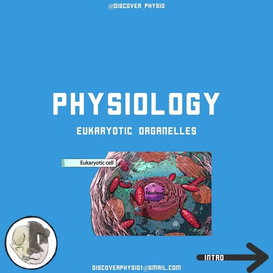
Eukaryotic Organelles Intro Eukaryotic cells contain: · Nucleus: it’s the control centre of the cell and contains gene/chromosomes · Ribosomes: where proteins are made · Endoplasmic reticulum: o Rough: it attaches sugar groups to proteins, also bounding vesicles for the transport to the Golgi apparatus o Smooth: it’s the site of lipid and steroid syntheses. Also, drug detoxification · Golgi apparatus: it modifies and packages proteins and lipids. It also puts them into vesicles for transport and makes lysosomes. · Lysosomes: contains digestive enzymes called lysosomal enzymes or lysozymes to breakdown biological tissues (e.g. hydrolyses pathogens and damaged cell organelles) · Mitochondria: it’s the site of aerobic respiration, where ATP is provided. Nucleus Structure: surrounded by a nuclear envelope. It contains fluid nucleoplasm, nucleoli, and chromatin. Function: controls the cell’s activity by transmitting genetic information and providing instruction for protein synthesis. Ribosomes Structure: it is a dense particle consisting of two subunits, ribosomal RNA and protein. Ribosomes are free or attached to rough endoplasmic reticulum. Function: where protein synthesis occurs. Endoplasmic reticulum Rough Structure: Sacs and tubules making a membrane system. Covered in ribosomes. Function: Makes ..... Reference · Marieb & Hoehn. Human Anatomy and Physiology. 10th ed. Chapter 3 · Marieb, E., Wilhelm, P. and Mallatt, J., 2017. Human Anatomy. 8th ed. London: Pearson Education Limited, p.61. . . . #discoverphysiotherapy #physiotherapists #physiolab #physiotherapystudents #physioworld #physiotutors #physiologyfirst #physiology_lab #physiology #humanphysiology #cell #cells #nucleus #pt #ptstudent #eukaryote #human #humanphysiology #biology #biologynotes #biology🔬 #biologystudent #cellbiology #humanbiology . @physiologyofstudying @_physiology_facts_ @physiology.anatomy @anatomy.physiology.nursing @physiology @thephysiologyofeverything @nucleushealth @physiology.s https://www.instagram.com/p/CCnkqE8H5zT/?igshid=1fies2gsqeviz
#discoverphysiotherapy#physiotherapists#physiolab#physiotherapystudents#physioworld#physiotutors#physiologyfirst#physiology_lab#physiology#humanphysiology#cell#cells#nucleus#pt#ptstudent#eukaryote#human#biology#biologynotes#biology🔬#biologystudent#cellbiology#humanbiology
0 notes
Photo
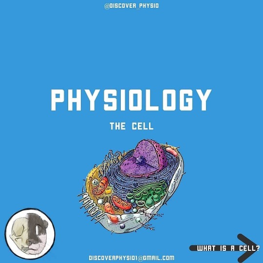
The cell What is a cell? · ‘The smallest structural and functional unit of an organism, typically microscopic and consisting of cytoplasm and a nucleus enclosed in a membrane. Microscopic organisms typically consist of a single cell, which is either eukaryotic or prokaryotic.’ - https://www.lexico.com/en/definition/cell · All living creatures are comprised of cells. · There are trillions of cells in the human body. Each cell varies in type, shape, size and function. · Human cells are eukaryotes. Eukaryotic cells have a nucleus whereas prokaryotes don’t. · The eukaryotic cell contains a: o Nucleus o Ribosomes o Endoplasmic reticulum - Rough - Smooth o Golgi apparatus o Lysosomes o Mitochondria References · https://www.lexico.com/en/definition/cell · https://ghr.nlm.nih.gov/primer/basics/cell#:~:text=The%20human%20body%20is%20composed,and%20carry%20out%20specialized%20functions.&text=Within%20cells%2C%20the%20cytoplasm%20is,structures%20that%20surround%20the%20nucleus. . . . #discoverphysiotherapy #physio #physiostudent #physiotherapy #physiotherapist #student #studentphysio #physiology #physiologynotes #physiology_notes #humanphysiology #ptstudent #pt #physiologyfirst #anatomyandphysiology #educated #education #uni #university #universitylife #study #studyinghard #work #cell #cells #cellbiology #human #humanphysiology #tuesdayvibes #tuesday #nucleus . @physiology.anatomy @human_physiology2017 @_physiology_facts_ @arbillaexercisephysiology @physiologyfacts20 @clinicalphysio @human._.anatomy_ @easy_anatomy_and_physiology @physiology.s @physiology_facts_exciting https://www.instagram.com/p/CCWEyEnHnoN/?igshid=16lfur8jvm9vl
#discoverphysiotherapy#physio#physiostudent#physiotherapy#physiotherapist#student#studentphysio#physiology#physiologynotes#physiology_notes#humanphysiology#ptstudent#pt#physiologyfirst#anatomyandphysiology#educated#education#uni#university#universitylife#study#studyinghard#work#cell#cells#cellbiology#human#tuesdayvibes#tuesday#nucleus
0 notes
Photo

The cell What is a cell? · ‘The smallest structural and functional unit of an organism, typically microscopic and consisting of cytoplasm and a nucleus enclosed in a membrane. Microscopic organisms typically consist of a single cell, which is either eukaryotic or prokaryotic.’ - https://www.lexico.com/en/definition/cell · All living creatures are comprised of cells. · There are trillions of cells in the human body. Each cell varies in type, shape, size and function. · Human cells are eukaryotes. Eukaryotic cells have a nucleus whereas prokaryotes don’t. · The eukaryotic cell contains a: o Nucleus o Ribosomes o Endoplasmic reticulum - Rough - Smooth o Golgi apparatus o Lysosomes o Mitochondria References · https://www.lexico.com/en/definition/cell · https://ghr.nlm.nih.gov/primer/basics/cell#:~:text=The%20human%20body%20is%20composed,and%20carry%20out%20specialized%20functions.&text=Within%20cells%2C%20the%20cytoplasm%20is,structures%20that%20surround%20the%20nucleus. . . . #discoverphysiotherapy #physio #physiostudent #physiotherapy #physiotherapist #student #studentphysio #physiology #physiologynotes #physiology_notes #humanphysiology #ptstudent #pt #physiologyfirst #anatomyandphysiology #educated #education #uni #university #universitylife #study #studyinghard #work #cell #cells #cellbiology #human #humanphysiology #tuesdayvibes #tuesday #nucleus . @physiology.anatomy @human_physiology2017 @_physiology_facts_ @arbillaexercisephysiology @physiologyfacts20 @clinicalphysio @human._.anatomy_ @easy_anatomy_and_physiology @physiology.s @physiology_facts_exciting https://www.instagram.com/p/CCWCvBinOPq/?igshid=1erstoztglj13
#discoverphysiotherapy#physio#physiostudent#physiotherapy#physiotherapist#student#studentphysio#physiology#physiologynotes#physiology_notes#humanphysiology#ptstudent#pt#physiologyfirst#anatomyandphysiology#educated#education#uni#university#universitylife#study#studyinghard#work#cell#cells#cellbiology#human#tuesdayvibes#tuesday#nucleus
0 notes
Photo

Negative & Positive Feedback Homeostatic Control Mechanisms · The three interdependent components of control mechanisms are: - Receptor – monitors the environment and responds to changes (stimuli) - Control center – determines the set point at which the variable is maintained - Effector – provides the means to respond to the stimulus · The variable produces a change in the body Negative feedback Negative feedback takes place to reduce the output or change. This result in the system to return to a stable condition. · Homeostatic regulatory mechanisms are mainly negative feedback responses. · An example of this is thermoregulation control o Control center: hypothalamus (set point approximately 37oC) o Receptors: skin & hypothalamus § If temp rises above 37 oC activity in the hypothalamus targets… o Effectors: smooth muscle in blood vessels supplying the skin dilate & sweat glands increase secretion · As temp returns to normal control center stops signaling the effectors Positive Feedback Positive feedback occurs to enhance or increase the output or change. This results in the reaction to be amplified to speed the process. · Positive feedback is less frequent than negative feedback. · Activated when an urgent response is required o i.e. blood loss & blood clotting and labor during childbirth · In positive feedback systems, the output enhances or exaggerates the original stimulus rather than opposing it. . . . #physiotheraphy #physiostudent #physiogram #physio #studentphysio #students #physiotherapyday #physiotherapist #physiolife #physiologyfirst #physiology #physiologynotes #physiology_notes #feedback #positive #negative #positivefeedback #negativefeedback #mechanism #control #receptor #effector #homeostasis #homeostasis #nerve #physiolab #humanphysiology #pathophysiology #anatomyandphysiology #physiotherapeut #education https://www.instagram.com/p/CBx9tXjJ5ER/?igshid=1fnf39v89mu76
#physiotheraphy#physiostudent#physiogram#physio#studentphysio#students#physiotherapyday#physiotherapist#physiolife#physiologyfirst#physiology#physiologynotes#physiology_notes#feedback#positive#negative#positivefeedback#negativefeedback#mechanism#control#receptor#effector#homeostasis#nerve#physiolab#humanphysiology#pathophysiology#anatomyandphysiology#physiotherapeut#education
0 notes
Photo

Homeostasis Intro · ability of the body to maintain a relatively stable internal environment despite the constantly changing external environment. · Chemical, thermal, and neural factors interact to maintain homeostasis · The internal environment of the body is in a dynamic state of equilibrium o Small fluctuations around a set point to maintain balance Autoregulation · Ability of a cell/tissue/organ to adjust automatically at a local level to environmental change o E.g. declining O2 levels in the tissue mediates the release of chemical factors that dilate local blood vessels, increasing flow to provide more O2 to the area Extrinsic Regulation · Impact of nervous & endocrine system activity on many other systems simultaneously. o E.g. during exercise the autonomic nervous system initiates an increase in heart rate, to provide adequate circulation, whist reducing blood flow to less active organs. o the hormone epinephrine (adrenalin) increases cardiac activity References • Marieb & Hoehn. Human Anatomy and Physiology. 10th ed. Chapter 3. . . . #ptstudent #pt #physiostudent #physiogram #physiotherapistlife #physiotherapists #physiorehab #physiologyofthecells #physiology #physiologynotes #physiology_notes #homeostasis #extrinsic #regulation #autoregulation #pathophysiology #physiologyclass #physiotutors #physio #physios #studentphysio #students #studentphysiotherapist #chemical #education #educational #educationmatters #educateyourself #physiologyfirst https://www.instagram.com/p/CBftuafFUZ6/?igshid=10hxhniq2nvzy
#ptstudent#pt#physiostudent#physiogram#physiotherapistlife#physiotherapists#physiorehab#physiologyofthecells#physiology#physiologynotes#physiology_notes#homeostasis#extrinsic#regulation#autoregulation#pathophysiology#physiologyclass#physiotutors#physio#physios#studentphysio#students#studentphysiotherapist#chemical#education#educational#educationmatters#educateyourself#physiologyfirst
0 notes