
Everything you need to know about x-ray imaging within the Veterinary world!
Don't wanna be here? Send us removal request.
Statistics
We looked inside some of the posts by bone-appetit and here's what we found interesting.
Average Info
Notes Per Post
5
Likes Per Post
3
Reblog Per Post
2
Reply Per Post
0
Time Between Posts
2 months
Number of Posts By Type
Text
6
Last Seen Tumblr Blogs


Fun Fact
The total number of visits Tumblr.com received during January 2021 is 327 million.
Text
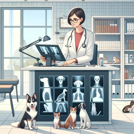
Understanding Vet X-Ray Markers: A Key Tool in Veterinary Radiography
When it comes to veterinary radiography, one component that often flies under the radar, yet plays a pivotal role, are vet x-ray markers. These small, but mighty tools are essential for any veterinary practice that employs x-ray technology to diagnose and treat our furry friends.
What Are X-Ray Markers?
Simply put, x-ray markers, also known as radiographic markers or lead markers, are used to mark x-ray images for identification and orientation purposes. In the veterinary field, these markers are indispensable for ensuring accuracy and clarity in radiographic images.
Why Vet X-Ray Markers are Crucial
Identification: Vet x-ray markers include information such as the date, the veterinarian's initials, and sometimes the patient's name or ID. This aids in maintaining accurate medical records.
Orientation: They help in determining the left or right side of the animal in the x-ray. This is crucial for accurate diagnosis and treatment planning.
Quality Assurance: They ensure that the x-ray is of optimal quality and that the image hasn't been flipped or altered.
Types of Vet X-Ray Markers
There are various types of x-ray markers available:
Lead Letters and Numbers: These are the most common type, providing clear, legible identification on x-ray films.
Custom Markers: Some practices opt for custom markers that might include specific shapes or symbols relevant to their clinic.
Digital Markers: With the advent of digital radiography, digital markers can be added to x-ray images using software.
How Xray4Vets Enhances Veterinary Radiography
Xray4Vets, a renowned UK-based manufacturer and supplier of veterinary x-ray equipment, understands the importance of high-quality vet x-ray markers. Their markers are designed with precision, ensuring that they are easily readable on radiographic images. Their range of markers is suitable for various types of veterinary practices, from small clinics to large hospitals.
Best Practices for Using X-Ray Markers in Veterinary Practices
Correct Placement: Always place the marker within the primary beam and on the same side as the labeled side of the animal.
Legibility: Ensure that the marker's information is clear and legible on the x-ray.
Maintenance: Regularly check markers for wear and tear and replace them as needed to maintain image quality.
The Evolution of X-Ray Markers
The evolution of x-ray markers has mirrored advancements in veterinary radiography. Traditional lead markers have evolved into more sophisticated forms, including digital options that integrate seamlessly with modern digital x-ray systems. This evolution signifies the constant strive for better diagnostic tools in veterinary medicine.
Xray4Vets: Your Partner in Veterinary Radiography
Xray4Vets stands at the forefront of this technological evolution. Their products are not just tools; they are part of a larger commitment to enhance veterinary healthcare, click here. Their expertise in manufacturing vet x-ray markers, combined with a comprehensive range of veterinary radiographic equipment, positions them as a trusted partner for veterinary professionals across the UK.
Vet x-ray markers might be small, but their impact on the accuracy and effectiveness of veterinary radiography is undeniable. They are a testament to the meticulous care and attention to detail that goes into veterinary diagnostics.
For those in the veterinary field looking to enhance their radiographic capabilities, Xray4Vets offers a range of top-notch veterinary x-ray equipment and markers. Their dedication to quality and innovation makes them a wise choice for any veterinary practice. Explore their offerings and take the first step towards elevated veterinary care at Xray4Vets.
1 note
·
View note
Text
The Essential Guide to Vet X-Ray Accessories
Embarking on a journey through the intricate world of veterinary radiology, we uncover the critical role played by vet x ray accessories. As a linchpin in animal healthcare, these tools not only enhance diagnostic precision but also redefine the capabilities of veterinary professionals, from seasoned practitioners to those newly entering this fascinating field.
The Importance of High-Quality X-Ray Accessories in Veterinary Practice
X-ray imaging is a cornerstone in veterinary diagnostics, offering invaluable insights into our furry friends' health. However, the quality of the images relies heavily on the x-ray accessories used. High-quality vet X-ray accessories ensure clearer images, leading to more accurate diagnoses and effective treatments.
Types of Vet X-Ray Accessories and Their Functions
Protective Gear: Safety first! Lead aprons, thyroid collars, and gloves are essential for protecting vets and technicians from radiation exposure.
Positioning Aids: Foam wedges, sandbags, and positioning sponges make it easier to position animals correctly, ensuring high-quality X-ray images.
Cassettes and Screens: These are crucial for capturing clear images. Digital cassettes have revolutionized veterinary X-rays, offering higher quality images and quicker processing times.
Grids: Grids are used to reduce scatter radiation, which can blur images. They are especially useful for imaging larger animals.
Viewing Equipment: Lightboxes and digital viewers aid in the detailed examination of X-ray films.

Choosing the Right Accessories for Your Practice
Selecting the right X-ray accessories can be daunting. Consider the following:
Compatibility: Ensure the accessories are compatible with your existing X-ray equipment.
Quality: Invest in high-quality accessories for longevity and better imaging.
Specialisation: Tailor your choices to the types of animals you treat.
Case Studies: Success Stories with the Right Accessories
Here, we can share anecdotes from veterinary practices that have seen remarkable improvements in diagnostics and treatment outcomes by using the right X-ray accessories. Real-world examples add a personal touch and illustrate the practical benefits.
Maintenance and Care of X-Ray Accessories
Maintaining your X-ray accessories is as crucial as selecting the right ones. Regular cleaning, proper storage, and routine checks ensure longevity and consistent performance.
Xray4Vets: A Reliable Source for Vet X-Ray Accessories
While discussing various suppliers, it's worth mentioning Xray4Vets. This UK-based company specializes in manufacturing and supplying top-notch veterinary X-ray equipment. Though we're just touching on them briefly here, their commitment to quality and innovation is noteworthy.
Conclusion: Enhancing Your Veterinary Practice with the Right Accessories
Investing in the right vet X-ray accessories is a game-changer for any veterinary practice. It not only enhances diagnostic accuracy but also improves the overall efficiency of your practice. For those in the UK, considering a supplier like Xray4Vets could be a smart move. They offer a wide range of quality accessories tailored to the needs of veterinary professionals.
For more information and to explore their extensive product range, head over to Xray4Vets. Enhance your veterinary practice today with the right X-ray accessories!
https://xray4vets.com/
0 notes
Text
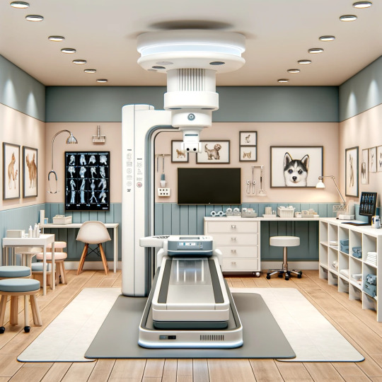
Digital XRay for Vets: Transforming Small Animal Healthcare
Hey there, fellow animal lovers and veterinary professionals! Today, let's dive into the world of veterinary diagnostics and explore how digital X-ray technology, specifically tailored for small animal Xray systems, is revolutionising our approach to animal healthcare. It's a topic close to my heart and one that's increasingly important in our digital age.
The Evolution of X-Ray Technology in Veterinary Care
Remember the days of traditional X-ray films? They were cumbersome, time-consuming, and not always the clearest. But the game has changed dramatically with the advent of digital Xray for vets. This technology offers unparalleled clarity and speed, transforming how we diagnose and treat our furry patients.
Why Digital X-Ray?
Speed and Efficiency: Digital X-ray systems provide immediate results. There's no more waiting for films to develop. This speed is crucial, especially in emergency situations where every second counts.
Image Quality: The clarity of digital X-rays is a game-changer. It allows for more accurate diagnoses, ensuring our small friends get the treatment they need without guesswork.
Ease of Use and Sharing: Digital images can be easily shared with specialists or pet owners. This collaborative approach is invaluable for comprehensive care.
Environmentally Friendly: Going digital means saying goodbye to chemical developers and storage films. It's a win for our planet!
Small Animal XRay Systems: A Closer Look
When it comes to small animals, precision is key. Digital X-ray systems designed for small animals are tailored to capture the minutest details, crucial for diagnosing conditions in smaller breeds or exotic pets. These systems also allow for various positions and angles, ensuring a comprehensive view of the animal's anatomy.
The Real-Life Impact
Let's talk about real-life scenarios. Imagine a small dog rushed in after an accident. With a digital X-ray system, within minutes, you have a clear image of the extent of its injuries. Treatment begins immediately, significantly improving the chances of a full recovery.
Or consider a cat with a chronic condition. Digital X-rays can be used regularly to monitor its progress without the stress of long vet visits.
Choosing the Right Digital X-Ray System
Selecting the right digital X-ray system is crucial. Consider factors like image quality, ease of use, and after-sales support. And if you're looking for a reliable provider, Xray4Vets is a fantastic resource, click here. They offer a range of digital X-ray solutions tailored for vets, backed by excellent customer service.
Xray4Vets: Your Partner in Veterinary Care
Xray4Vets stands out for its commitment to providing top-notch digital X-ray solutions for vets. Their expertise in small animal X-ray systems is particularly noteworthy, offering systems that cater to the specific needs of smaller breeds and exotic animals.
Embracing digital X-ray technology is not just about keeping up with the times; it's about providing the best care possible for our animal companions. Whether you're a vet looking to upgrade your practice or a pet owner interested in the latest in veterinary technology, digital X-ray systems, especially those designed for small animals, are worth exploring.
For more information on how digital X-ray can benefit your practice, head over to Xray4Vets (https://xray4vets.com/). They’re your go-to experts for everything related to digital X-ray for vets, offering solutions that make a real difference in animal healthcare.
Remember, in the world of veterinary medicine, staying ahead with technology like digital X-ray systems is not just an option; it's a necessity for providing the best care for our beloved pets.
2 notes
·
View notes
Text
The Advantages of Modern X-ray Imaging Systems for Vets

X-ray imaging systems are a type of medical imaging technology, that uses electromagnetic radiation to study the patient. Over the years, these technologies have progressed, and have further developed to keep up with the digitalised world. These imaging machines are used on a daily basis in veterinary clinics, to diagnose medical problems in a range of animals. The use of radiography in medical practices is a revolutionary process, as veterinarians are able to view the inside of an animal without surgery or making incisions. With a variety of modern x-ray imaging systems available on the market (click here), how can vets benefit from them?

Modern and digital veterinary x-ray imaging systems produce less radiation than traditional x-ray films, which ensures your pet, or any animal is safe. Although there is a slight exposure to the radiation to take the image, the strength of the waves is reduced, and the length of time the animal is exposed is much shorter too.
Digital x-rays are more accurate and clear, producing images which have a high resolution. This enables vets to clearly diagnose the issue, as the x-ray produces a more vivid image compared to conventional film.
Similarly, digital radiography allows vets to analyse x-ray images easily, as they can edit the image to view things more clearly. For example, the x-ray can be resized, enlarged, have added annotation, and can be changed in brightness/contrast.
Digital x-rays are very efficient, as the image is viewable within seconds of it being taken. Plus, once it has been captured, it is stored on the computer until it has been manually removed. This allows all veterinarians to have access to the file, resulting in a more productive workplace. Technicians will also have more time to spare, as there is no need to file and retrieve films for traditional x-rays.
Modern x-ray imaging systems are more cost-effective than traditional methods, as the x-ray is taken and stored on the computer. Although the initial price will be a lot higher, there will be fewer additional costs as time goes on. With traditional film, you will need to purchase processing chemicals, imaging processors, mailing jackets, and rent out a darkroom, every time you take an x-ray, which results in a lot of expenses!
If you are a vet or technician working in a veterinary clinic and still using conventional film, then you should make the change over to digital. Digital radiography offers you an abundance of benefits and will make your workforce a lot more productive.
I hope you have enjoyed this blog post, please feel free to check out my others!
https://xray4vets.com/
1 note
·
View note
Text
How Vet Xray Systems Have Advanced
Just like all technologies that advance over time, radiography in both human medicine and veterinary practices has developed too. Radiography and Xray systems are used to understand the insides of patients, and to look for any internal abnormalities such as broken bones, tumours, and even diseases. However, over the past decades, imaging technologies have improved more and more, with CT, MRI, and MR scans available for increased functionality. Particularly in the veterinary industry, there are three main types of vet Xray systems available: conventional film-screen radiography, computed radiography (CR), and direct digital radiography (DR).

So, how are these new technologies different to the older ones?
Film Screen or Digital
In this digital age, the majority of things that undergo digital advancements will offer more benefits than those that are more traditional. There are many companies that offer a wide range of modern imaging technologies, click here.
Traditional Film Screen Radiography
Uses an Xray cassette
Uses intensifying screens to convert Xrays into visible light
The radiographic film undergoes film processing, to convert the ‘latent’ image into a permanent one
Digital Radiography (including CR and DR)
Digital radiography uses the same generator as conventional film; however, the image is printed on a plate, which is converted to digital to be displayed on a monitor
CR – uses a portable imaging plate to record the raw image. The plate is then placed into a reader, where the interior screen is scanned and produced into a digital image
DR – doesn’t need a reader to convert the image, the digital image can be sent directly to the computer utilising sensors
Digital radiographs are saved as DICOM (digital imaging and communications in medicine), stored within a PACS (picture archiving and communication system) network for all staff members to see
The Benefits of Modern Vet Xray Systems
Faster imaging – digital radiographs can produce images instantly, offering a more productive and efficient working environment. Traditional film radiographs, need to undergo a long process of converting the image, which can slow down diagnosis time.
Less radiation – compared to traditional methods, digital radiographs don’t need as much radiation, which can offer a range of health benefits to the patient.
Cost-effective – for veterinary practices, digital radiographs are relatively low cost. Conventional Xray films can be rather expensive and cannot be reused, whereas sensor plates can be. Other than the initial upfront cost, CR and DR are cost-effective.
Computerisation benefits – as the images are digitalised, they can be edited (contrast and brightness) and altered (cropped, zoomed in, and notations), which allows veterinarians to interpret the images a lot easier. Plus, as they are stored in a PACS network, all members of staff have access to the images.
Although conventional radiographs are still used, modern technologies are taking over, as they offer a range of advantages that makes jobs easier!
https://xray4vets.com
0 notes
Text
When Should You Replace Your Lightweight Lead Apron?
If you work close to radiation in fluoroscopy, perhaps in a Veterinary clinic or medical facility, then you will more than likely own a lead apron. If you’ve owned your apron for a long time, there might be some wear and tear visible, but when does it need replacing? In this blog post, I have listed the top defects you should look out for, that may affect the functionality of your apron. Especially, if you have a lightweight lead apron, these tend to be a bit thinner and may need a little more care and attention. To help keep your aprons intact, I have also listed ways in which to look after your apron, so it can last you an even longer time!

Looking Out For Cracks, Tears, Holes, and Creases
Cracks within your lead apron can occur naturally over time, however, with incorrect storage they can appear a lot sooner than expected! Even the smallest of cracks can allow radiation to seep through, exposing you to levels that may impact your health. Similarly, any thinning or tearing of the material should also mean that your apron needs replacing. As lead aprons are designed to protect vital organs of the body, it is 100% necessary for your apron to be in top-notch condition. Additionally, the closures of the apron, such as the buckles, Velcro, or magnets should also be inspected for any flaws, in case they need repairing or replacing. If you need to replace your apron, click here.
How to Inspect Your Lightweight Lead Apron
Lead aprons should undergo routine inspections, to ensure they are safe to use. There are many ways in which you can inspect your apron including visual, tactile and radiographic.
A visual inspection involves you looking at your apron for any defects, specifically larger tears ad imperfections in the fabric. You should lay your apron down on a clean surface and examine all areas of the material.
A tactile inspection involves you feeling the apron, and looking for any thinning and creases within the material. You can search the apron whilst it is laid down or hung up, just make sure that your hands can run up and down the front and back surfaces of the apron.
Finally, a radiographic inspection uses a similar type of imaging that you use at work! By using an x-ray or fluoroscopy, unnoticed flaws from visual/tactile examinations can be evaluated. X-ray images will show cracks and tears in the lead, appearing as dark grey if there are any. Fluoroscopy will highlight any flaws in the seams and stitches of the apron.
Top 5 Lead Apron Caring Tip
1. Routine Cleaning
Aprons should always be cleaned after use, to maintain its appearance, as well as for hygiene reasons. To clean your apron, use cold water, mild soap, and a soft brush to clean the surface of the apron. Make sure to NEVER use bleach on the apron and don’t submerge it in water, as this may affect the level of protection the apron provides.
2. Store Correctly
Storing your apron correctly, is a vital step, to ensure that it remains in tip-top condition. Lead aprons should be hung on hangers, so they remain upright and flat. They should not be folded or draped over furniture, as this will cause the material to crease, which will eventually turn to cracks.
3. Wear the Right Size
This tip may not seem like an obvious one, however, wearing the correct sized apron for you and your body can impact how the apron lasts. Not only will the right-sized apron protect you better from radiation, but it will reduce the chances of creases, snags, and potential tears. An apron that is too large may be more susceptible to creases, and the fastenings will be loose, increasing the chances of other equipment getting caught on it. On the other hand, an apron that is too small will cause thinning as the materials will need to stretch more.
4. Always Inspect!
The key to maintaining your apron is inspection, inspection, inspection! By examining your apron frequently, any defects can be avoided. Plus, if you do find any, you know not to use the apron and find a new one.
5. Disposal
Finally, if you find that you need to replace your apron, you cannot simply throw your old one in the bin. As the apron contains lead, you need to dispose of it as hazardous waste or recycle it. You can find many companies online that will pick up your old lead apron for you.
1 note
·
View note