#radiographs
Text
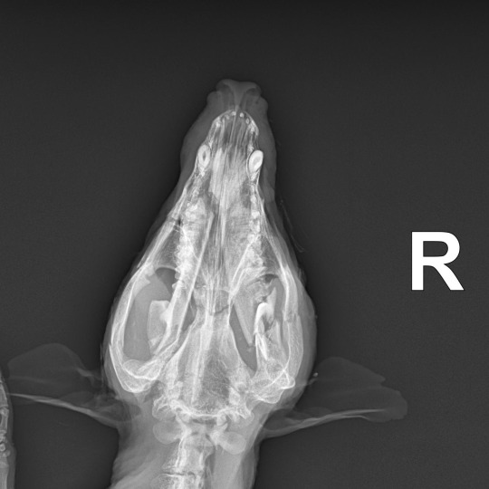
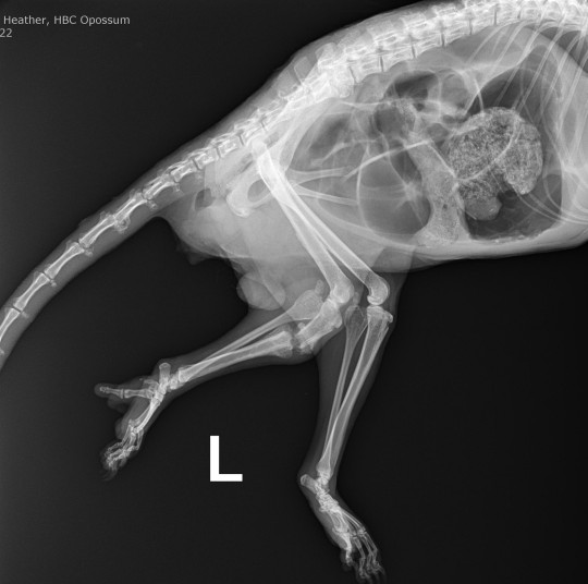
While driving I found a possum that had been hit by a car and suffered severe head trauma, but was still very much alive, unfortunately. I brought him to the clinic and put him to sleep.
Afterwards I took some radiographs for learning purposes. In addition to the mandibular fractures I was aware of, he had several skull fractures and broken teeth. He also had a healing tibia fracture, which may have contributed to getting hit by the car, some arthritic changes to his tail, and a large amount of air in his abdomen. I'm not sure what the latter is from, since his diaphragm was intact; he may have swallowed a large amount of air while panting after being hit. Anyone see any other pathology or other interesting things of note in the radiographs?


#veterinary#vet tech#trauma#animal death#wildlife#virginia opossum#marsupial#radiographs#euthanasia#blood#bones#skull#sad#vehicular trauma#veterinary education
11 notes
·
View notes
Text
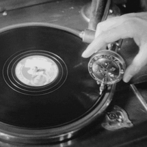
our radiology instructor brought in cardboard boxes for us to x-ray and see what the surprise inside of each was. simultaneously demonstrating the need for multiple angles when imaging.
anyway, so we x-ray the final box and it’s a glove. the glove is filled with… a liquid. she has us guess what the liquid is.
“lactated ringers” no
“water” no
“milk..” …no
“pee..” no!
it was a contrasting fluid used in certain types of radiography. she was introducing it to us in a fun way and we had the audacity to guess that she filled a glove with milk or PISS???
#vet tech#radiology#radiographs#veterinary technology#vet tech stuff#vet med#vet school#fun class today#college
5 notes
·
View notes
Text

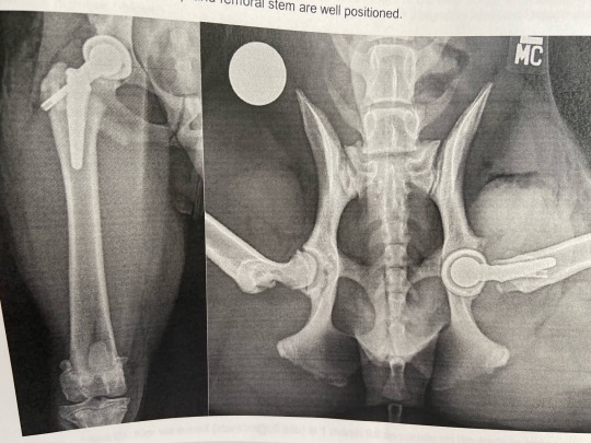
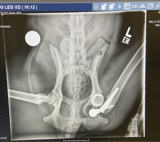

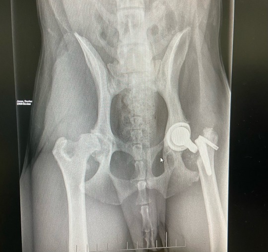
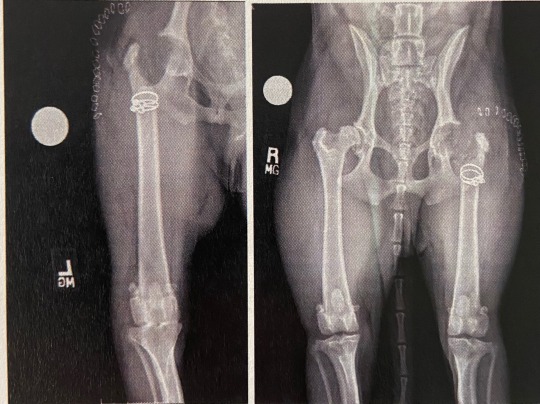
my dog’s fucked up hips: a progression
#booker#dogblr#radiographs#hip dysplasia#total hip replacement#femoral head ostectomy#this has all happened in the span of a year btw
2 notes
·
View notes
Text
Illuminating the Art and Science of Radiography: Honoring Dr. George P. Genereux on World Radiography Day

View On WordPress
#FriendsAreas#- pathologic#@FriendsAreas#computed tomographic scans#foundation#Friends of the Saskatoon Afforestation Areas#George Genereux Afforestation Area#George Genereux Urban REgional Park#illustrator#professor#radiographs#radiologic#Radiology#Saskatoon#University of Saskatcheawn
0 notes
Text
#DentalHealth#Radiographs#PeriodontalDisease#StayInformed#KnowledgeIsPower#HealthIsWealth#OralHealth#XrayRevealed#Prevention#HealthySmiles#KnowYourTeeth#newblogflo#secretstime#dentalhealth#oralhygiene#periodontaldisease#dentistry#dentalcare#dentalxray#teethcare#oralcare#periodontitis#healthy
0 notes
Text
How Vet Xray Systems Have Advanced
Just like all technologies that advance over time, radiography in both human medicine and veterinary practices has developed too. Radiography and Xray systems are used to understand the insides of patients, and to look for any internal abnormalities such as broken bones, tumours, and even diseases. However, over the past decades, imaging technologies have improved more and more, with CT, MRI, and MR scans available for increased functionality. Particularly in the veterinary industry, there are three main types of vet Xray systems available: conventional film-screen radiography, computed radiography (CR), and direct digital radiography (DR).

So, how are these new technologies different to the older ones?
Film Screen or Digital
In this digital age, the majority of things that undergo digital advancements will offer more benefits than those that are more traditional. There are many companies that offer a wide range of modern imaging technologies, click here.
Traditional Film Screen Radiography
Uses an Xray cassette
Uses intensifying screens to convert Xrays into visible light
The radiographic film undergoes film processing, to convert the ‘latent’ image into a permanent one
Digital Radiography (including CR and DR)
Digital radiography uses the same generator as conventional film; however, the image is printed on a plate, which is converted to digital to be displayed on a monitor
CR – uses a portable imaging plate to record the raw image. The plate is then placed into a reader, where the interior screen is scanned and produced into a digital image
DR – doesn’t need a reader to convert the image, the digital image can be sent directly to the computer utilising sensors
Digital radiographs are saved as DICOM (digital imaging and communications in medicine), stored within a PACS (picture archiving and communication system) network for all staff members to see
The Benefits of Modern Vet Xray Systems
Faster imaging – digital radiographs can produce images instantly, offering a more productive and efficient working environment. Traditional film radiographs, need to undergo a long process of converting the image, which can slow down diagnosis time.
Less radiation – compared to traditional methods, digital radiographs don’t need as much radiation, which can offer a range of health benefits to the patient.
Cost-effective – for veterinary practices, digital radiographs are relatively low cost. Conventional Xray films can be rather expensive and cannot be reused, whereas sensor plates can be. Other than the initial upfront cost, CR and DR are cost-effective.
Computerisation benefits – as the images are digitalised, they can be edited (contrast and brightness) and altered (cropped, zoomed in, and notations), which allows veterinarians to interpret the images a lot easier. Plus, as they are stored in a PACS network, all members of staff have access to the images.
Although conventional radiographs are still used, modern technologies are taking over, as they offer a range of advantages that makes jobs easier!
https://xray4vets.com
0 notes
Text
the guys <33
@williamy3w lil welcome (back) to tumblr gift for you guys :]]
82 notes
·
View notes
Text

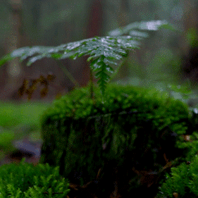
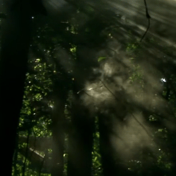


Radiograph (William Yew x Henry Bicknell) stimboard
✭ with radio/ forest stims
✫ RQ'd by @southernambrosebassford!
+ | + | +
+ | - | +
+ | + | +
#stimboard#stim gifs#stimblr#stimboard account#visual stim#sensory#william yew#henry bicknell#radiograph#en abime#radio stim#forest stim#nature stim#southernambrosebassford
61 notes
·
View notes
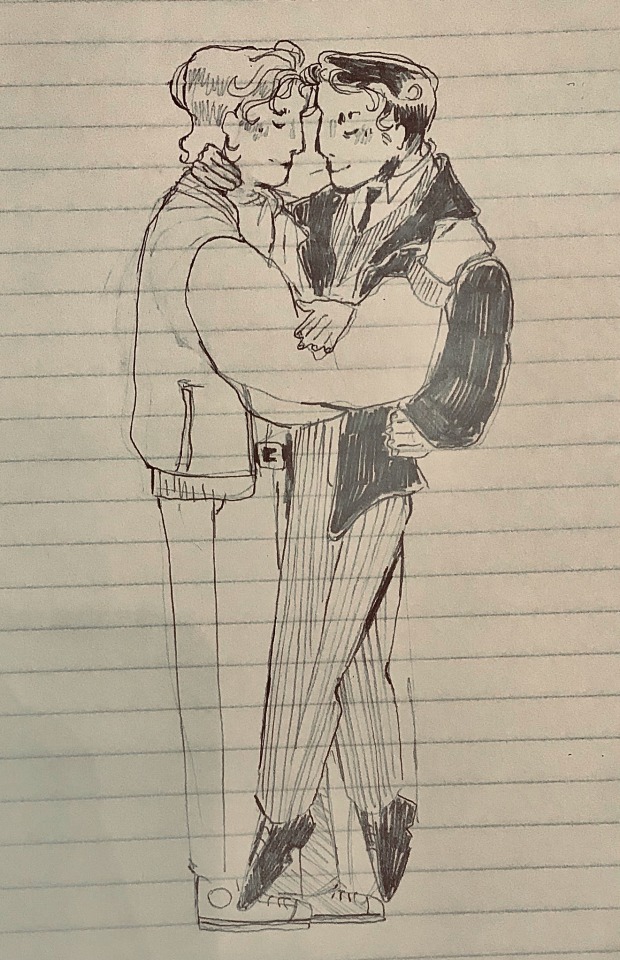
Text

“Till the end of forever do us part” 💙💚
#my art#en abime#william yew#henry bicknell#drawing#artists on tumblr#fanart#digital art#procreate#illustration#art#radiograph
28 notes
·
View notes
Text

sorry bout the spam reblog yesterday (it was for the greater good/j) Anyways quick Radiograph drawing<3
#jk posts#jks art tag#en abime#En Abime Radiograph#Radiograph#If there are no more radio graph fans then i'm dead<3#Anyways sorry about the shadowban as well William#☺️✌️
34 notes
·
View notes
Text

this is where the new url is from btw
1 note
·
View note
Note
Good luck for Duncan's appointment tomorrow! I hope everything goes as smoothly as possible. Our thoughts are with you and the little man.
Thank you!! I am so very excited and if I told him what’s happening tmrw he would be so very NOT excited! His appointment is at 10:00am and then we’ve got an hour and a half drive back home.
I will make sure to update you guys as soon as I can with how the consult went! I’m also going to make sure I specifically request his scans when he has them done tmrw, not just their finding/notes
#idk how the neurology department at Madison is but a lot of times when other clinics send us records they skip things like radiographs#and A) I want to see them B) Doc wants to see them#and C) I want to show you guys#my post#Duncan#ask
103 notes
·
View notes
Text
#en abime#operation country roads#radiograph#a bit late with this one but. it was literally like that
12 notes
·
View notes
Text
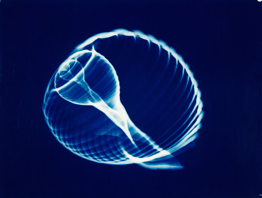
Hilja Raviniemi ~ Shell design, 1970. Suomen valokuvataiteen museon kokoelma. | src Fall 2023 exhibition
view & read more on wordPress
After her more traditional early work, Raviniemi explored the infinite creative possibilities offered by the darkroom, especially in the 1960s. Her recognizable blue era, which differed from the stark black-and-white art photography of the time, began in the late 1960s. Chemist by profession, Raviniemi was an ingenious artist in the darkroom.
In addition to blue-tinted prints, she also created completely abstract photographic artworks using different techniques. Raviniemi’s workplace at the University of Helsinki photography department laboratory also allowed her to make the first artistic radiographic images in Finland. Hundreds of Raviniemi’s radiographic works have been preserved and make up an exceptional ensemble of works in the history of Finnish art photography. read more on wordPress
#Hilja Raviniemi#x-ray#x-ray photography#radiogram#1960s#nature photography#seashell#shelldesign#shell structure#women artists#women photographers#finnish photographer#valokuva#blue tinted#radiographic image#radiograph#Finnish museum of photography
45 notes
·
View notes
Text
Ssbine had a really bad wheezing fit last night but I'm trying to remain optimistic 🤧
#it always sounds she's trying to pass a hairball but more breathy#the fact she CLEARLY has something but radiographs and sample tests cant detect it is driving me insane dude#I have one last idea (getting an autovac to keep my home totally dust free) before I do yet another vet visit#if its not internal its GOTTA be external and thats the only thing I haven't tried yet
14 notes
·
View notes
Text
This arg is so gay dear god—
11 notes
·
View notes
Last Seen Blogs

throatadmirer
guys with hot necks

mastaramguru
Hindi Sex Story
naoxslingo
the bloody adventures of a hman learnin' languages

yourfriendlyneighborhoodkiller
— i hate everyone

discoveringtechnoita
DiscoveringTechnoITA