#Glenoid labrum repairs
Text
Best Bankart Repair Doctors in Noida
The recurring partial dislocations of the shoulder joint lead to a medical condition called a bankart lesion. It occurs when the labrum and the capsule get torn off the glenoid. The condition was named after British surgeon Arthur Blundell Bankart, who first defined it in the British Medical Journal in 1923.

As noted above, repeated partial dislocations of the shoulder joint cause it to occur. The shoulder is the body's most flexible joint, which makes it more likely to dislocate than the other joints.
Symptoms of Bankart Lesions
Symptoms of Bankart Lesions include pain, swelling, numbness, and shakiness in the concerned area. Additional symptoms include difficulty moving the joint, visible deformity in the shoulder, etc.
Bankart Repair in Noida
There exist several bankart repair doctors in Noida. Treating bankart lesions may involve non-surgical methods such as rest, restriction of movement, and physical therapy. However, attaching a torn labrum to the shoulder socket is necessary.
Bankart lesion procedure involves fixing repeated dislocations of the shoulder joint to restore it by joining the torn ligaments to the joint of the shoulders. The goal of the process is to reconnect and constrict the torn labrum and ligaments of the shoulder. An unhealed Bankart Lesion may cause shoulder instability. Multiple dislocations lower the chances of healing without surgery.
Bankart repair surgery may be arthroscopic (with the use of a camera) or an open bankart repair operation. Physiotherapy services are essential after a bankart lesion surgery and must be availed.
The department of orthopaedics at Global Orthopaedic and Sports injury Centre specialises in the surgical and medical management of the disorders of the Foot and Ankle, General Orthopaedics, Hand and Upper extremities, Paediatric Orthopaedics, Podiatry, Shoulder disorders, Spine-Back and neck and paediatric spine, Hip and Knee Joint Replacement, Hip Scope Surgery, Knee disorders, Sports Medicine (Non-Surgical), Sports Concussions, Sports Medicine (Surgical), and Trauma and Fractures.
Here, you are under the care of the best sports medicine physicians and physiotherapists as we have advanced training certifications. We also house one of the best bankart repair doctors in Noida, Dr A K Sharma, an extremely devoted orthopaedic surgeon.
0 notes
Text
Best Orthopedic doctor in Aurangaba
What is Rotator Cuff Surgery?
Surgery to repair a torn rotator cuff is known as Rotator Cuff Surgery. It most often involves re-attaching the tendon to the head of the humerus known as the Upper arm bone.The tendon is stitched back to its original location to heal a full tear. The four little muscles that make up the rotator cuff in the shoulder allow the upper arm to spin. The tendon portion of the rotator cuff muscle separates from the upper arm’s bone when it is ripped. A rotator cuff tear can be caused by an accident, such as a fall or heavy lifting, or by repeated motions over a long time.
According to Dr. Gajanan Kathar, Best orthopaedic doctor in Aurangabad, Surgery might not be necessary to cure certain small tears. However, for many to recuperate correctly, surgery is necessary.
What procedure is used to treat a torn rotator cuff? An orthopedic surgeon uses an absorbable suture anchor to reconnect the torn rotator cuff tendon to the humerus (upper arm bone). The outcome of the procedure depends on how effectively the tendon-bone junction recovers.
How is a rotator cuff operation carried out? Arthroscopy is used to conduct rotator cuff surgery. Small incisions are made during a shoulder arthroscopy to guide an arthroscope—a short tube attached with a camera—to the area of the tear. Then, with the use of specially created tools, we may restore the rotator cuff while causing the least amount of damage possible to the nearby muscles and tissues. Instead of using a general anesthetic, patients at Hospital for Special Surgery have this surgery done while under a nerve block (regional anesthetic) and partial sedation. This reduces the quantity of anesthetic and pain medication used, which in turn lessens nausea and vomiting. The majority of our patients may now return home within a few hours following their operation thanks to advancements in anesthetic and less invasive procedures.
To diagnose and treat any related issues, such as a biceps tendon injury, loose bodies, or a torn labrum, an arthroscopy of the glenohumeral joint (the ball-and-socket component of the shoulder joint) is first done. The glenoid, the cup-shaped part of the shoulder joint, is surrounded by the shoulder labrum, which is a rim of cartilage. At this stage, a shaver can be used to trim a partial rotator cuff tear (a tear through less than 50% of the thickness of the rotator cuff tendon). The subacromial space, under which the rotator cuff tendon runs on the upper surface of the scapula or shoulder blade, is the next location for the arthroscope. The subacromial space is then made accessible by doing a subacromial decompression. A bone spur and abnormalities on the bottom of the acromion, which can lead to rotator cuff discomfort and tears, are removed during this treatment, known as an acromioplasty. Suture anchors and sutures are then used to restore the damaged tendon to the bone.
When should you get rotator cuff surgery? For individuals with a full thickness tear or those whose partial thickness tear has not responded to conservative treatment, surgery is advised (two months of physical therapy). After trying conservative therapy for two or three months and still experiencing discomfort and difficulties with activities, a patient may think about having surgery to repair the tear.
Exists a rotator cuff surgery substitute? Two months of physical therapy may be used to address minor or partial rotator cuff injuries.
Do partial rotator cuff injuries require surgery? Surgery is the sole choice if physical therapy has not successfully improved strength and function. Before initiating physical therapy, a patient must feel that more than 70% of their shoulder strength and function have been recovered by nonsurgical means.
What percentage of rotator cuff surgeries succeed?
For minor injuries, rotator cuff surgery has a success rate of greater than 95%. The success rate is still more than 70% for two tendon rips. It is recommended to fix tears when they are little since untreated tears tend to enlarge. Surgery, however, may still be beneficial for individuals with very significant tendon rips. We occasionally have the option to employ a method known as margin conversions. To keep the tendon in the joint, grafted tissue is used to cover the humeral head. A patch of donor tissue is applied when the tendon tissue is inadequate for immediate healing.
If you have any questions and are seekingorthopaedic doctor in Aurangabad, Contact Dr. Gajanan Kathar
0 notes
Text
Shoulder Specialists in Pune
Shoulder is the most common joint in the body to dislocate. Due to its ball and socket design we have a great deal of mobility within the shoulder, which is directly at the cost of stability.
Acute shoulder dislocation needs to be reduced by orthopaedic surgeon under suitable anaesthesia.
Rest in sling, ice , medicines and physical therapy are important
In recurrent shoulder instability surgical intervention is needed depending on age, dislocations and bone loss.
Arthroscopic Bankart repair – it is a keyhole surgery where anterior labrum can be repaired to glenoid with the help of suture anchors.
Latarjet procedure – this procedure is required if significant bone loss is present. Coracoids process (part of bone scapula) along with its muscle attached is fixed to glenoid rim.
Dr. Mayur Purandare is Orthopaedic & Joint Replacement consultant who has extensive knowledge and training in the field. He is also trained arthroscopy & shoulder Surgeon. He has more than 14 years’ experience of in the field. He has been outstanding throughout his academic career. He has done his fellowship in the joint replacement from DMH Pune & fellowship in shoulder surgery from the esteemed Institute – Wrightington Hospital, England. He is owner and chief orthopaedic surgeon at Orion Hospital, Wakad, Pune.
#orthopedics #orthopedicsurgeon #surgery #surgeon #physicaltherapy #doctor #ortho #orthopaedics #medical #orthopaedic #ortopedia #health #hospital #sportsmedicine #physiotherapy #injury #orthopedicdoctor #spine #wellness #surgical #orthop #knee #sports #kneereplacement #backpain #bowlegs #ortopedicsurgeons #orthopedic #orthopedicsurgery #backpain #pain #relief #neckpain #neck #back #pune #wakad #pimpri #maharashtra #painrelief #diagnosis #treatment #joint #jointreplacement #replacement #joint #kneepain
#orthopaedician#hospital#joint#pimpri#surgeon#surgery#orthopedic#muscle#maharashtra#pune#doctor#orthopedicsurgeon#orthopaedicsurgery#orthopaedist
0 notes
Text
Commonly Asked Questions by People Related to Shoulder Arthroscopy
What is arthroscopic surgery?
Arthroscopic surgery is a method that orthopedic surgeons use to detect and repair structural damage within a joint. The shoulder surgeon makes 3 or 4 small incisions around the joint, about ½ inch each. Here, a fiber-optic camera is used to see inside the joint and miniature surgical tools are used to perform the repair. Arthroscopic surgery of the shoulder is an outpatient surgery, which means that you will not have to be admitted to the hospital. You will return home shortly after the surgery is over. Arthroscopy of the shoulder joint has been a major advancement in surgical technique. This method allows less cutting of intact tissue to perform repairs, allowing for quicker recovery time than with open surgery.
What types of shoulder surgeries can be performed using arthroscopic technique?
Surgeons perform many types of shoulder arthroscopy. Among those are:
Rotator cuff repair
Glenoid labrum repairs
Repair of shoulder instability (dislocation)
Biceps tendon repair
Subacromial decompression (removal of bone spurs)
SLAP tears
Bursitis
Frozen shoulder release
How long will my surgery take?
Most surgeries will take 45 minutes to 1 hour. You will then be required to stay in the recovery room for about another hour & then discharged home.
How much pain will I have following my shoulder surgery?
This varies greatly from patient to patient. You will be prescribed few pain medications following surgery, along with instructions for icing the shoulder, which will help control too much swelling. During physical therapy sessions, you will be advised to move the shoulder joint in order to restore full arm motion. This may cause an increase in your pain level, and for this reason it is suggested that you take your pain medication 50 minutes prior to the start of therapy sessions. In time, you will need to take less pain medication.
How long is the rehabilitation process after shoulder arthroscopy?
This depends on the type of procedure you had. It is important to understand that physical therapy is an important part of recovery. You must be committed to post-operative rehab if you wish to achieve the best outcome.
#Shoulder arthroscopy#shoulder arthroscopy recovery#arthroscopy#shoulder injury treatment#shoulder pain#shoulder surgery#rotator cuff injury#rotator cuff treatment#rotator cuff surgery#Arthroscopic shoulder surgery#treatment of rotator cuff tears#Glenoid labrum repairs#Repair of shoulder instability (dislocation)#Biceps tendon repair#SLAP tears#Subacromial decompression#Frozen shoulder release
0 notes
Text
Arthroscopic Bankart Repair Treatment in Surat | By Dr. Nandan Rao
0261-2607040 | 0261-2607000 The shoulder joint (glenohumeral joint) is a ball joint, where the head of the arm bone (humerus) joins the shoulder socket (glenoid socket). The shoulder socket is extremely shallow and therefore needs additional support to prevent the shoulder bones from dislocating. The labrum, a cartilage sleeve that surrounds the shoulder socket, helps accomplish this purpose by forming a cup for the humeral head to move inward. Provides stability to the joint, allowing a wide range of movements.

Sometimes the labrum can rupture during a shoulder injury. A specific type of labrum tear that occurs when the shoulder is dislocated is called a Bankart tear. This is a tear in a part of the labrum called the inferior glenohumeral ligament and is common in younger patients who have a shoulder dislocation. A Bankart tear makes the shoulder prone to repeat dislocation in patients under 30 years of age.
Diagnosis
Your doctor will ask about your medical history and perform a complete physical exam of your shoulder. Your doctor may recommend additional tests such as X-rays or MRIs.
Treatment
Conservative treatment measures for a Bankart tear include rest and immobilization with a sling followed by physical therapy.
Bankart repair surgery is indicated when conservative treatment measures do not improve the condition and repeated dislocations of the shoulder joint occur.
Procedure description
Bankart surgery can be performed using a minimally invasive surgical technique called arthroscopy.
During an arthroscopic Bankart procedure, your surgeon makes a few small incisions over the shoulder joint. An arthroscope, a thin tubular device connected with a light and a small video camera on the end, is inserted through one of the incisions in the shoulder joint. The video camera transmits the image of the inside of the shoulder joint to a television monitor for the surgeon to see. Your surgeon then uses small surgical instruments through the other small incisions to trim the edges of your glenoid socket. Suture anchors are then inserted to reattach the detached labrum to the glenoid. The small incisions are then closed and covered with a bandage.
Arthroscopy causes minimal disruption to the other shoulder structures and does not require your surgeon to separate and reattach the overlying (subscapular) shoulder muscle as with the open technique.
Postoperative care
After your surgery, you will spend about an hour in the recovery room.
Your physical therapist will start you on shoulder exercises the day after surgery to strengthen and improve the range of motion of the shoulder joint.
You will be allowed to do your daily activities as tolerated, but without lifting heavier than a plate or glass while you heal.
Your arm may be put in a sling for three weeks to restrict use of the operated shoulder.
You may resume mild, low-risk activities, such as jogging and swimming, 8 to 10 weeks after surgery, and you may be advised to avoid contact sports for some time.
Risks
Arthroscopic Bankart repair is a relatively safe procedure. Being minimally invasive, it is associated with fewer risks and faster recovery. Some of the associated potential risks include:
Infection
Injury to adjacent nerves or blood vessels
Joint stiffness
Pain
To book your appointment call us on- 0261-2607040 | 0261-2607000
0 notes
Text
Here’s an idea: don’t invalidate the healing plan set down by another person’s physician.
I had surgery today. I had multiple tears in my labrum. As of today my healing plan from both the physician and the PT on site, I’m incredibly restricted. No driving is a part of that plan.
So in order to let people know what happened and not have to make multiple calls, I posted a blanket update on Facebook. Mostly imbuing the text with humour and thanking people for the kindness they’ve shown me. The lion share of messages were lovely, kind, and funny.

Then...
Here’s the deal:
The shoulder may be where both the rotator cuff and labrum reside, but the labrum is between the Glenoid and the Humeral Head. The rotator cuff resides ABOVE the humeral head. Different locations.
Per what my specialist and surgeon told my mom, I had MULTIPLE tears to my labrum.
These are the recommendations set down by him and the PT staff on site.
At this point, I’ve been on light duty for 6 months. I need to heed what the doctor and PT tell me in order to fully recover. And even two months after, I cannot lift the amount required for my job.
What your doctor recommended versus what mine will is depending on the intensity of the repair and what they see as the best route to my convalescence.
Do not compare your procedure to mine. Do not compare your recovery plan to mine. Keep your less than professional opinion to yourself. I’m not here for it.
/Rant.
#shut up Karen#no one asked you patrice#Facebook keyboard doctor offering unneeded commentary#piss off
0 notes
Text
300+ TOP ORTHOPAEDICS Objective Questions and Answers
ORTHOPAEDICS Multiple Choice Questions :-
1.Which is not a principle of compound fracture treatment?
a) No tendon repair
b) Aggressive Antibiotic cover
c) Wound debridement
d) Immediate Wound closure
Ans:d
2.Medial meniscus is more vulnerable to injury because of?
a) Its fixity to tibial collateral ligament
b) its semicircular shape
c) action of adductor magnus
d) its attachment to fibrous capsule
Ans:a
3.Injury to the popliteal artery in fracture lower end of femur is often due to?
a) Distal fragment pressing the artery
b) Proximal fragment pressing the artery
c) Tight plaster
d) Hematoma
Ans:a
4.In transverse fracture of the patella, the treatment is
a) Excision of small fragment
b) Wire fixation
c) Plaster cylinder
d) Patellectomy
Ans:b
5.monster type of dislocation of the hip is
a) Anterior
b) Posterior
c) Central
d) Dislocation with fracture of the shaft
Ans:b
6. March fracture affects
a) Neck of 2nd metatarsal c) Neck of 1 st metatarsal
b) Body of 2nd Metatarsal
c) Neck of 1 st metatarsal
d) Fracture of lower end of tibia
Ans:a
7.Commonest complication of extracapsular fracture of neck of femur is
a) Non Union
b) iscnemic necrosis
c) Maiunion
d) aTPuTmonary complications
Ans:c
9.Blood coagulation profile in pregnancy is
a) Increase in fibrinogen level of 10-25 %
b) Decrease in factor X, XI, XII
c) Decrease in plasminogen activity
d) Increase in platelet count
Ans:a
10.In classical caesarean section more chances of rupture of uterus is in
a) upper uterine segment
b) lower uterine segment
c) utero cervical junction
d) posterior uterine segment.
Ans:a

ORTHOPAEDICS MCQs
11.Only indication for Internal version nowadays is
a) Brow presentation
b) Face presentation
c) Second fetus of twins
d) Breech
Ans:c
12.Internal fixation is done in all fracture Except
a) Compound
b) Multiple
c) Elderly person
Ans:a
13.Myositis ossificans is commonly seen at tlie joint
d) Hip
a) Knee
b) Elbow
c) Shoulder
d) Hip
Ans:b
14.The most important factor in fracture healing is
a) Good alignment
b) Organization of blood clot
c) Accurate reduction and 100% apposition of fractured fragments
d) Immobilization
e) Adequate calcium intake
Ans:d
15.The most preferred treatment of fracture of neck of femur in a young person is
a) Hemiarthroplasty
b) Total hip treatment
c) conservative treatment
d) closed reduction & internal fixation
Ans:d
16.Lisfranc dislocation is
a) Tarsometatarsal dislocation
b) Lunate dislocation
c) Scaphoid dislocation
d) Posterior dislocation of elbow
Ans:a
17.Position of immobilization in fracture both bones of forearm in an adult male will be
a) Prone
b) Mid prone
c) Supine
d) 10° Supine
Ans:c
18.Carpal bone which fractures commonly
a) Scaphoid
b) Lunate
C) Hamate
d) Pisiform
Ans: a
19.True about clavicular fracture is
a) Most common at medial 1/3 & 2/3
b) comminuted fracture common
c) malunion occurs
d) usually due to fall on elbow
Ans:c
20.Commonest site of fracture scaphoid
a) Waist
b) Proximal third
c) Distal third
d) Tuberculosis
Ans:a
21.Excision of fractured fragment is practised in all fractures except
a) Patella
b) Olecranon
c) head of radius
d) lateral condyle humerus
Ans:b
22.The complication not common in colle's fracture is
a) malunion
b) non union
c) sudeck's atrophy
d) stiffness of wrist
Ans:b
23.In 65 year old male with history of fracture neck of femur 6 weeks old, treatment of choice
a) SP nailing
b) Mc Murray's osteotomy
c) hemiarthroplasty
d) none.
Ans:c
24.In Colles fracture not seen in
a) Proximal impaction
b) lateral rotation
c) dorsal angulation
d) medial rotation
Ans:d
25.In fracture medial epicondyle of humerus,which of the following can be affected
a) Flexion of fingers
b) Adduction of fingers
c) Abduction of fingers
d) Flexion of thumb
Ans:a
26.Most common cause of pathological fracture in a child is
a) malignancy
b) bone cyst
c) fibrous dysplasia
d) paget's disease
Ans:b
27.A lady presents with a history of fracture radius, which was put on plaster of paris casts for 4 weeks. After that she developed swelling of hands with shiny skin. What is the most likely diagnosis.
a) Rupture of extensor pollicis longus tendon
b) Myositis ossificans
c) Reflex sympathetic dystrophy
d) Malunion.
Ans:c
28.AH are components of Rotator cuff except
a) Supraspinatus
b) Infraspinatus
c) Subscapularis
d) Teres major
Ans:d
29.Complication of fracture scaphoid is
a) Injury to radial artery
b) avascular necrosis of proximal part
c) avascular necrosis of distal part
d) injury to radial nerve
Ans:b
30.Which fracture neck of femur has a poor prognosis
a) Intra capsular
b) Extracapsular
c) Both
d) None.
Ans:a
31.Fracture blisters commonly appear on how many days ?
a) 1- 3 days
b) 3- 5 days
c) 5-7 days
d) 5-9 days
Ans:b
32.If the greater tuberosity of the humerus is lost, which of the following movements will be affected
a) Adduction and flexion
b) Abduction and lateral rotation
c) Medial rotation and adduction
d) Flexion and medial rotation
Ans:b
33. The most common cause of a sprained ankle is injury of
a) Deltoid ligament
b) lateral ligament
c) Inferior tibiofibular ligament
d) Anterior Talofibular ligament.
Ans:d
34.In the case of 65 year old person with fracture neck offemur the treatment of choice is
a) close reduction
b) close reduction with internal fixation
c) open reduction
d) replacement of head and neck of the femur with a prosthesis
Ans:d
35. Tardy ulnar nerve palsy is seen in :
a) Cubitus valgus c) Fracture scaphoid
b) Dislocation of elbow
c) Fracture scaphoid
d) Supracondylar fracture of humerus
Ans:a
36.Common injury to baby is
a) Fracture humerus
b) Fracture clavicle
c) Fracture radius-ulna
d) Fracture femur
Ans:b
37.Young man with # tibia of left side 2 months ago, is having popliteal cast, Now needs mobilization with single crutch. Which will be the preferred site ?
a) Left sided crutch
b) Right sided
c) Any side
d) Both sides
Ans:a
38.Bryant's triangle is useful in diagnosis of following except
a) Supratrochanteric shortening
b) Infratrochanteric shortening
c) anterior dislocation hip
d) posterior dislocation hip
Ans:b
39.Line joining Ant Sup. iliac spine to tip of gluteal tuberosity, should normally touch the Greater trochanter, this is
a) Shoemakers line
b) Nelaton's line
c) Von-rossen
d) Perkins
Ans:b
40.Which of the following is least common in supracondylar fracture
a) Non union
b) Median nerVe injury
c) volkmanns ischemic contracture
d) cubitus varus
Ans:a
41.Earliest symptom of Volkmann's ischemia is
a) Pain in flexor muscles
b) Absence of pulse
c) Pain on passive extension
d) Cyanosis of limb
Ans:a
42.A patient develops compartment syndrome (swelling, pain and numbness) following manipulation and plaster for fracture of both bones of leg. What is the best treatment?
a)Split the plaster
b)Infusion of law molecular weight dextran
c)Elevate the leg after splitting the plaster
d)Do operative decompression of fascial compartment
Ans:d
43.Anterior dislocation of shoulder causes all except
a) Circumflex artery injury
b) Avascular necrosis head of humerus
c) Brachial plexus injury
d) Chip fracture scapula
Ans:d
44.In Colles# following is most common complication
a) Non union
b) Malunion
c) Sudeck's dystrophy
d) Volkmann's ischemic contracture
Ans:b
45. Patient with supracondylar fracture following reduction presented with claw hand. The likely diagnosis is
a) Volkmann's ischaemic contracture
b) median nerve injury
c) Ulnar nerve injury
d) Dupuytrens contracture
Ans:a
46.Pivot test for
a) anterior cruciate ligament
b) posterior cruciate ligament
c) medial meniscus injury
d) lateral meniscus injury
Ans:a
47.Triangular relation of Elbow is maintained in
a) Fracture ulna
b) Anterior dislocation of Elbow
c) Posterior dislocation of Elbow
d) Supracondylar fracture
Ans:d
48.Fracture which most often requires open reduction & internal fixation
a) Lateral condyle of humerus
b) Femoral condyle
c) Distal tibial epiphyseal separation
d) Fracture both bones forearm
Ans:a
49. A patient had injury to the upper limb 3 yrs earlier, now he presents with paresthesia over the medial border of the hand and anaesthesia over medial two finger. The injuryis likely to have been
a) supracondylar fracture
b) lateral condyle fracture humerus
c) medial condyle fracture humerus
b) lateral condyle fracture humerus
Ans:b
50.Commonest type of lesion causing recurrent shoulder dislocation is
a) Shallow glenoid labrum
b) Bankarts lesion
c) Weakness of subscapularis muscie
d) Injury to humeral head
Ans:b
ORTHOPAEDICS Objective Questions with Answers
51.Menisci calcification is a feature of
a) Gout
b) Hyperparathyroidism
c) pseudogout
d) ankylosing spondylosis
Ans:c
52.A young adult presenting with oblique, displaced fracture olecranon treatment of choice
a) Plaster cast
b) Percutaneous wiring
c) Tension band wiring
d) Removal of displaced piece with triceps repair
Ans:c
53.Volkmann's ischemic contracture mostly involves
a) Flexor digitorum superficialis
b) Pronator teres
c) Flexor digitorum profundus .
d) Flexor carpi radialis longus
Ans:c
54.Avascular necrosis is commonest in one of the following fractures
a) Gorden 1 & 2 fracture of femoral neck
b) Gorden 3 & 4 fracture of femoral neck
c) Sub-trochanteric fracture of femoral neck
d) Baso-trochanteric fracture
Ans:b
55.On measurement, the base of Bryant's triangle on the left side is found to be short by 2 cms as compared to the right side. This indicates
a) Fracture of the neck of the femur
b) Fracture of the shaft of the femur
c) Osteoarthritis of hip joint
d) Rheumatoid arthritis of the hip joint
Ans:a
56.All the following requires open reduction & internal fixation almost always except
a) Lateral condyle of humerus
b) Olecranon
c) Patella
d) Volar Barton's fracture
Ans:d
57.Pathognomic sign of traumatic fracture
a) Swelling
b) Tenderness
c) Redness
d) Crepitus
Ans:d
58. A football player, while playing, twists his knees over the ankle. He still continues to play.
a) Medial meniscus tear
b) Anterior cruciate ligament tear
c) Medial collateral ligament injury
d) Posterior cruciate ligament injury.
Ans:a
59.K-wire is used in
a) Circlage
b) fixing forearm bones
c) prior to plating
d) All of the above.
Ans:d
60.Treatment of Acute myositis Ossificans is
a) Active mobilization
b) Passive mobilization
c) Infra Red therapy
d) Immobilization
Ans:d
61. The treatment of choice for non-union of extracapsular fracture neck femur
a) Hip spica
b) Intramedullary nailing
c) Internal fixation
d) Compression plating
Ans:c
62.Duga's test is helpful in
a) Dislocation of hip
b) Scaphoid fracture
c) Fracture neck of femur
d) Anterior dislocation of shoulder
Ans:d
63.Open reduction in children is done for
a) Supracondylar fracture
b) forearm both bone fracture
c) femoral condyle fracture
d) lateral condyle of humerus fracture
Ans:d
64.Avascular necrosis of the head of femur is not seen in
a) Subcapital Fracture
b) Intertrochanteric fracture
c) Transcervical fracture
d) Central dislocation of hip
Ans:b
65.Stiffness in knee is maximum when traction is at
a) Skin
b) lower end femur
c) upper end tibia
d) calcaneum
Ans:b
66. Intramedullary fixation is ideal in a case of fracture of shaft of femur when there is
a) A transverse fracture
b) A compound fracture
c) Soft tissue interposition between the fractured ends
d) Such a fracture in a child
Ans:a
67.Meyer's operation is done for
a) Recurrent dislocation of patella
b) Dislocation of shoulder joint
c) Dislocation of hip joint
d) Scaphoid
Ans:b
68.Treatment of choice for old non-united fracture of shaft of femur
a) compression plating
b) bone grafting
c) nailing
d) compression plating with bone grafting.
Ans:d
69.The last step in the healing of a fracture is
a) Hematoma formation
b) Consolidation
c) Remodelling
d) Callus formation
e) Demineralization of bones.
Ans:c
70.A Bennet's fracture is difficult to maintain in reduced position because of the pull of
a) Extensor pollicis longus
b) Extensor pollicis brevis
c) Abductor pollicis longus
d) Abductor pollicis brevis
Ans:d
71.Inter trochanteric fracture has trendelenberg sign negative because of the action of
a) Gluteus medius
b) Gluteus minimus
c) G.maximus
d) Tensor fascia lata
Ans:a
72.A segmental compound fracture tibia with 1cm skin wound is classified as
a) Type I
b) Type II
c) Type ILIA
d) TypeIIIB
Ans:a
73.Steinman pin is used for all except
a) fracture of upper end of tibia
b) fracture through lower end of tibia
c) fracture through lower end of femur
d) skull traction
Ans:d
74.Treatment after removal of plaster for supracondylar fracture of humerus is
a) active mobilization at elbow joint
b) massage
c) no treatment
d) passive movements at elbow
Ans:a
75. Lateral condyle can cause
a) Genu valgum
b) Genu varus
c) Genu recarvatum
d) Dislocation of ankle
Ans:a
76. Muscles involved in Volkmann's ischemic contracture
a) Flexor pollicis longus
b) Flexor profundus
c) Flexor sublimis
d) All
Ans:d
77. Medial meniscus tear is more common than lateral meniscus because of its decreased
a) Nerve supply
b) Vascularity
c) Mobility
d) Fibroelasticity
Ans:c
78.Volkmann's ischaemic contracture is due to
a) Arterial injury
b) Venous injury
c) Nerve injury
d) Increase of compartment pressure in the limb
Ans:d
79.Attitude of the limb in anterior dislocation of hip
a) Flexion, abduction, external rotation
b) Flexion, adduction, external rotation
c) Flexion, Abduction, internal rotation
d) Flexion, adduction, internal rotation
Ans:a
80.Treatment of fracture clavicle in an infant is best treated by
a) Cuff and sling
b) Figure of 8 bandage
c) Open reduction
d) Shoulder cast
Ans:b
81.Dislocation of hip joint palpable on per rectal examination
a) Cogenital dislocation of hip
b) Posterior dislocation of hip
c) Fracture neck of femur
d) Anterior dislocation of hip
Ans:a
82.Fractures common in elderly women are all except
a) Clavicular
b) Colles
c) Intertrochanteric
d) neck of femur
Ans:c
83.Late complication of Acetabular fracture
a) Avascular necrosis of head of femur
b) Avascular necrosis of iliac crest
c) Fixed deformity of the hip joint
d) secondary osteoarthritis of hip joint
Ans:a
84.Treatment of anterior dislocation of shoulder is by
a) Kocher's manoeuvre
b) Dennis browne splint
c) Barlows manoeuvre
d) Surgery
Ans:a
85.Patient comes with fracture of femur in an acute accident, the first thing to do is
a) Secure airway and treat the shock
b) Splinting
c) Physical examination
d) X-Rays
Ans:a
86.Multiple bone fractures in a new born is seen in
a) Scurvy
b) Syphilis
c) Osteogenesis imperfecta
d) Morquio's syndrome
Ans:c
87. Most common bone to fracture in body is
a) Radius
b) Clavicle
c) femur
d) vertebra
e) Pelvis
Ans:b
88.Cock up splint is used in management of
a) Ulnar nerve palsy
b) brachial plexus palsy
c) radial nerve palsy
d) combined ulnar & median nerve palsy
Ans:c
89.The type of displacement of fractured fragment in which bone is not remodelled
a) Anterior angulation
b) Posterior angulation
c) Lateral angulation
d) Rotation
Ans:b
90.Carrying angle is decreased in
a) Cubitus varus
b) Cubitus valgus
c) Genu valgum
d) Genu varum
Ans:a
91. Transverse fracture of medial malleolus is caused by
a) Abduction
b) Adduction
c) Rotation of foot
Ans:a
92.Bennet's fracture is fracture dislocation of base of matacarpal
a) 4th
b) 3rd
c) 2nd
d) 1st
Ans:d
93. Most common type of supracondylar fracture is
a) extension type
b) flexion type
c) abduction type
Ans:a
94.Intramedullary nailing is contraindicated in fracture shaft femur if
a) The fracture is compound
b) The fracture is near the knee joint
c) The epiphysis have not fused
d) Any of the above is present
e) None of the above is present
Ans:d
95.A man was diagnosed to have myositis ossificans progressiva at the age of 20 yrs.
He died 5 yrs later. What is the most probable cause of death ?
a) Starvation and chest infection
b) Myocarditis
c) Hypercalcemia
d) Hyperphosphatemia
Ans:a
96. The commonest elbow injury in children is
a) Extension type of supracondylar fracture of humerus
b) Di slocation of elbow
c) Fracture lateral condyle of humerus
d) Fracture medial epicondyle of humerus
Ans:a
97. Ideal treatment with fracture neck of humerus in a lady will be
a) Triangular sling
b) Hemiarthroplasty
c) Chest arm bandage
d) Internal fixation
Ans:a
98. In Intertrochanteric #has most common complication of
a) Non union
b) Malunion
c) Avascular necrosis
d) Nerve Injury
Ans:b
99. Luxatio erecta
a) tear of the glenoidal labrum
b) inferior dislocation of shoulder
c) anterior dislocation of shoulder
d) defect in the humeral head
Ans:b
100. McMurray's osteotomy operation is based on the following principle
a) Mechanical
b) Biological
c) Bio-mechanical
d) None
Ans:c
ORTHOPAEDICS Questions and Answers pdf Download
Read the full article
0 notes
Photo

#Repost @physicaltherapyresearch ・・・ 🔬📚 𝐒𝐋𝐀𝐏 𝐓𝐞𝐚𝐫𝐬 Collaboration with @mskrehab . 💭 SLAP tears are injuries of the superior portion of the glenoid labrum extending from anterior to posterior in a curved fashion. 💭 Tears are common in overhead throwing athletes and laborers involved in overhead activities. 💭 SLAP tears are caused by forceful eccentric traction exerted on the biceps tendon. 🗓🗓 𝐇𝐢𝐬𝐭𝐨𝐫𝐲: The history is often vague and symptoms include: Anterior shoulder pain. Episodic clicking, particularly in the cocking position of throwing. Decline in function/throwing velocity. 🗓 Acute trauma cases may involve FOOSH or sudden arm traction/jerking while lifting a heavy object. 🔍🔍 𝐄𝐱𝐚𝐦𝐢𝐧𝐚𝐭𝐢𝐨𝐧: Motion, strength, and basic neurovascular function. 🔍 The proximal biceps tendon should be palpated; focal tenderness suggests tendon injury. 🔍 Recommended tests include: Anterior glide Compression rotation Active compression (O’Brien’s) Crank Speeds 💢💢 𝐃𝐢𝐚𝐠𝐧𝐨𝐬𝐢𝐬: Definitive diagnosis of a SLAP tear requires arthroscopy or MRA 💢 Clinical diagnosis is often adequate if: Patient is not a good surgical candidate. History and clinical findings strongly suggest the diagnosis. Other differential diagnoses ruled out. 🚫🚫 𝐃𝐢𝐟𝐟𝐞𝐫𝐞𝐧𝐭𝐢𝐚𝐥 𝐃𝐢𝐚𝐠𝐧𝐨𝐬𝐢𝐬: Rotator cuff tear or tendinopathy. Shoulder impingement. Biceps tendinopathy. 👐🏼👐🏼 𝐌𝐚𝐧𝐚𝐠𝐞𝐦𝐞𝐧𝐭: Most cases require orthopedic referral, especially high-level throwing or overhead athletes. 👐🏼 Management depends upon patient age, activity, and tear type. 👐🏼 Nonoperative management is preferred given the 6-12 month recovery following surgical repair. 👐🏼 In overhead athletes & patients without pain relief or functional improvement, surgical treatment should be considered. 👐🏼 Biceps tenodesis is the preferred primary procedure in non-athletic individuals due to high failure rate of SLAP repair. 📚📚📚 𝐒𝐨𝐮𝐫𝐜𝐞: 1. Up To Date article. 2017. Superior labrum anterior posterior (SLAP) tears. 2. Huri et al. 2014. Treatment of slap lesions: a literature review. ActOrthTraum. 48(3), 290-297. 3. Edwards, et al. 2010. Non operative treatment of SLAP tears https://www.instagram.com/p/BstBZuDF4B4/?utm_source=ig_tumblr_share&igshid=a2it8764thgd
0 notes
Text
Report: McKinley to undergo shoulder <b>surgery</b> after combine
The UCLA defensive end said he will undergo shoulder surgery to repair a torn labrum and a broken glenoid, and will be out for 5-6 months, according ...
from Google Alert - surgery http://ift.tt/2lCCIT0
0 notes
Text
Arthroscopic Shoulder Surgery in Hyderabad
Two Common Shoulder Injuries That Can Be Treated with Arthroscopic Surgery
Shoulder injuries are common because the shoulder is the most mobile joint in the body. Nonsurgical treatment options like rest, anti-inflammatory medications, and physical therapy treat the majority of shoulder injuries. Serious shoulder injuries that do not respond to nonsurgical treatment may require surgical intervention.
Arthroscopic shoulder surgery is more advanced and effective than ever. Many surgeries that were once performed using a large open incision are now performed arthroscopically, which translates to less postoperative pain and a quicker recovery.
Board Certified and Fellowship Trained Orthopaedic and Sports Medicine Specialist have extensive experience and training in performing the latest technology for shoulder arthroscopic procedures such as;
Rotator Cuff Repair
The rotator cuff is four muscles and tendons that attach to the top of the shoulder. The rotator cuff is important because it raises, rotates and stabilizes the shoulder. A partial or complete rotator cuff tear is a serious injury that requires medical attention as soon as possible. Dr. Kirthi Paladugu specialist diagnoses a tear and prescribes a treatment plan. The majority of rotator cuff repairs are performed arthroscopically:
· 2-3 tiny shoulder incisions are made
· An arthroscopic camera is used to visualize the tear
· Tiny instruments and sutures anchors are used to repair the tear and attach the tendon to the bone
The incisions are closed and sterilely dressed, and the shoulder is placed in a comfortable sling. Total procedure time is approximately 1 hour. Physical therapy after surgery is an important part of recovery because it helps the patient regain shoulder strength and flexibility.
Labrum Repair
The labrum is a rim of soft tissue that lies on the outer edge of the glenoid cavity. The labrum is important because it holds the humeral head in the glenoid cavity to stabilize the shoulder. A torn labrum is a serious injury because it causes shoulder instability and possibly shoulder subluxations or dislocations. Many times, a torn labrum needs to be arthroscopically surgically repaired:
· 2-3 tiny shoulder incisions are made
· An arthroscopic camera is inserted into the shoulder joint and the torn labrum is visualized
· Tiny instruments and suture anchors are used to repair the tear and attach the labrum to the glenoid cavity
· Incisions are closed and sterilely dressed, the shoulder is placed in a comfortable sling
Total procedure time is approximately 1 hours depending on the seriousness of the tear. Physical therapy after surgery is important because it helps stretch the shoulder muscles, ligaments and tendons.
Rotator cuff tears and labrum tears are serious injuries that should be seen as soon as possible.
Injury symptoms include:
· Difficulty lifting or raising the arm
· Instability
· Pain
· Subluxation/Dislocation
· Stiffness
· Swelling
Sports, physical activities, and osteoarthritis are common injury causes.

MAKE AN APPOINTMENT
DR. KIRTHI PALADUGU ORTHO SURGEON
If you’ve recently suffered an Shoulder injury, or if you’ve been living with Shoulder pain for an extended period of time, make an appointment with us at Srikara Hospital
If you are having symptoms of a shoulder injury, we encourage you to contact free and to make an appointment with Dr. Kirthi Paladugu, give us a call at +91 9177 679 797.
If you find the post useful and worth sharing, don’t forget to share it on Twitter, Facebook, and Google Plus!
Follow us on :
LinkedIn http://bit.ly/2KDon4X
Facebook http://bit.ly/2NS3cy1
Twitter http://bit.ly/2KDon4X
Contact us
Dr. KIRTHI PALADUGU
Consultant Orthopaedic Surgeon,
Srikara Hospitals,#222 & 223,
Mythri Nagar,Phase-II,
Miyapur,Hyderabad-500 049.
Srikara Hospital, Hyderabad.
To Contact: + 91 9177 679 797
E-Mail ID: [email protected]
#Shoulder dislocations Repair Arthroscopy#Bankarts Repair#Rotator Cuff Repair#frozen shoulder surgery#orthopedic doctor#Shoulder Replacement Surgery#Best Shoulder Replacement Surgeon in Hyderabad#arthroscopic shoulder surgery in hyderabad#shoulder arthroscopy in hyderabad
0 notes
Text
What Are Prolotherapy Injections?
Prolotherapy injections are for anyone who suffers from chronic joint pain or degenerative joint disease.
If you are not aware of this effective regenerative medicine, then it’s time you learn about what prolotherapy injections are and how they can benefit you. Prolotherapy is an orthopedic procedure wherein a “natural irritant” is injected into the soft tissue of an injured joint. This stimulates the body’s healing response so it can repair and strengthen the damaged joints and tissue.
How Does Prolotherapy Work?
When the patient is injected, the body is “tricked” into repair mode. The mild inflammatory response caused by the injection stimulates the growth of new or normal ligament, which “tightens” the weakened structure.
Prolotherapy has the “unique ability” to address the cause of instability, repair the damaged sites and produce new collagen tissue. This, in turn, leads to the “permanent stabilization” of the affected joints.
A common formula for the prolotherapy injection is a natural solution of dextrose and saline. This is called dextrose prolotherapy. Another type of injection is Platelet Rich Plasma (PRP) therapy.
In this procedure, patients are injected with a PRP solution generated from the patient’s own blood. PRP is a purified and concentrated solution of platelets that can “supercharge the healing process” of soft tissue injuries.
Prolotherapy injections are not a one-off procedure. Intervals can vary depending on the area being treated and its severity.
According to The American Osteopathic Association of Prolotherapy Regenerative Medicine (AOAPRM), the term ‘prolotherapy’ came from the term ‘sclerotherapy’. Sclerotherapy was discovered in the 1930s. The latter term was used for both vein and joint injections. Today, sclerotherapy is used to refer to treatment that addresses “vascular abnormalities” like hemorrhoids and varicose veins.
What Is Prolotherapy Used For And Who Can Benefit From This Treatment?
youtube
Problem areas that can be treated with prolotherapy include:
Low or mid-back pain including degenerative disc disease;
Hip pain including arthritis;
Neck pain including whiplash and migraine;
Knee pain and knee tears;
Wrist or hand pain including carpal tunnel;
Osteoarthritis;
Tendonitis;
Shoulder pain including rotator cuff and glenoid labrum tears;
Elbow pain,
Foot and ankle pain including sprains;
Hypermobility,
Temporal mandibular joint syndrome (TMJ); and
Other musculoskeletal pain or sports-related injuries.
Is Prolotherapy Effective?
The success of this procedure will depend on factors such as the patient’s overall health. This would include the ability to heal and nutritional deficiencies that can affect the healing process.
In one randomized controlled trial, doctors gave a group of knee osteoarthritis patients a prolotherapy injection in the knee joint. This included up to 15 surrounding tender areas with pain and swelling. This was done thrice, four weeks apart.
A WOMAC score was used to compare levels of pain, stiffness, and physical function before and after treatment. The results showed that those who received the treatment improved 24% compared to the salt-water group (11%) and the exercise group (12%).
In another trial, albeit with a smaller sample size, patients with thumb or finger osteoarthritis who received a dextrose prolotherapy injection. These patients noted that they experienced “less pain” when moving their fingers compared to those who only received lidocaine.
Source of Health provides prolotherapy in Scottsdale. We offer a complimentary consultation to determine if you can be a candidate for this type of treatment. Contact us now to learn more and find out how prolotherapy and regenerative medicine can help you.
About the Author:
Dr. Steven Sorr is the founder and chief medical officer at Source of Health in Scottsdale, AZ and has been in clinical practice using regenerative medicine since 2013. He received his doctorate in naturopathic medicine from SCNM and is a licensed healthcare provider in Arizona.
Dr. Sorr brings a huge passion for life and a diverse educational background of food, yoga, and medicine to Source of Health. His goal is to revolutionize the standard of care mindset by making significant strides in evidence-based therapies that are drug and surgery-free to restore high-level health for all.
The post What Are Prolotherapy Injections? appeared first on Source of Health: Functional And Regenerative Medicine.
0 notes
Text
Bankart Shoulders Repair Procedure And Recovery
A Bankart Shoulder Repair is a surgical procedure for the treatment of repeated dislocations of the shoulder joint. In the surgery, worn-out ligaments are reattached to their appropriate position in the shoulder joint to restore normal function.
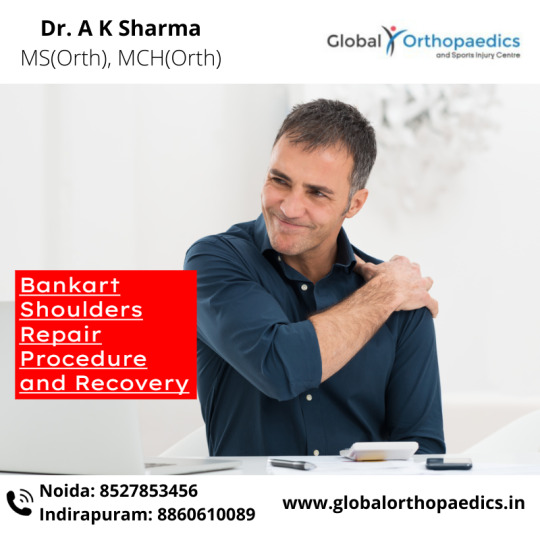
The shoulder is the primary joint most frequently dislocated following significant trauma, such as a car accident or a fall onto an extended arm. Approximately 96 percent of shoulder dislocations occur in the front (anterior), with 1-3 percent happening in the back (posterior) (posterior).
Falls and automobile accidents are major causes of first-time dislocations. Still, in many cases, recurring dislocations are caused by seemingly harmless actions such as extending the arm above the head or combing hair. Shoulder dislocations are more prevalent in males than females and adolescents. If you are looking for Bankart repair doctors in Noida, then you are in the right spot!
Bankart Shoulder Repair Procedure
Small incisions are made around the shoulder area for the Bankart Shoulder Repair operation. The orthopedic doctor can then implant a little camera to see the structures on a television screen.
Using tiny devices, the labrum is secured to the glenoid bone. The surgeon puts tiny anchors into the bone and sutures the labrum in place to retain the tissue in place.
The Bankart lesion is treated, and the shoulder's stability is restored with the open shoulder stabilization operation. This procedure necessitates a big incision across the front of the shoulder joint for the surgeon to realign the labrum and ligaments. This procedure is ideal for patients with a fully separated labrum due to a severe dislocation.
Both procedures are conducted under general anesthesia and might last between 1 12 and 2 12 hours. The arm nerves are muted with a local anesthetic to provide up to 24 hours of pain relief (called a regional block). Additionally, a thick dressing is put on the surgical sites after any of the two surgeries.
Recovery after the Bankart Shoulder Repair
The Bankart Shoulder Repair operation may necessitate an overnight hospital stay. The sutures will remain in place for 10 to 14 days. Anticipate that your arm will be placed in a cumbersome immobilizer sling. Following surgery, most surgeons provide painkillers and antibiotics to reduce pain and reduce infection.
A physical therapist will meet with you one week following surgery to teach you exercises and home maintenance. Expect to use just your non-surgical hand for the first four weeks following the Bankart Shoulder Repair in Noida.
You will be permitted to drive a car in around six weeks, and you should be utilizing the shoulder by week ten.
Typically, contact sports are not permitted until six months after surgery, although activities like swimming are permitted within three months. Dr. AK Sharma is the best doctor for bankart repair in Noida. So, go and contact him now for bankart shoulder repair!
0 notes
Text
ARTHROSCOPIC LABRAL REPAIR
Arthroscopic Labral Repair is performed for recurrent shoulder dislocations, SLAP tears and Bankart’s tears. These are injuries of the labrum, a ring of tough and flexible tissue on the rim of the glenoid (socket of shoulder). In labral repair surgery, via 2-3 puncture holes, suture anchors (small screws with stitches loaded) are placed in the glenoid bone socket. The labrum is then stitched to the glenoid. The puncture holes are then closed with 1 stitch each and the arm placed in a sling. You will be in a sling for a few weeks. Progressive exercises start the day after surgery. It takes at least 3-6 months to get the strength back in your shoulder. You are ready for contact sports only 9 months after surgery. The suture anchors used are generally made of bioabsorbable materials and do not need to be removed later.
visit www.jointclinic.in to know more
0 notes
Text
Minimally Invasive Repair for Rotator Cuff Injuries
Fast Facts
The rotator cuff is comprised of four musculotendinous units. The muscles help stabilize and move the shoulder joint.
Damage to any (or all) of the four muscles (alongside the ligaments that attach the muscles to the bones) can be attributed to chronic overuse, gradual aging, or acute weakness
The damage can result in -pain, decreased motion range (and use of the shoulder joint), weakness in raising the arm and in some instances, disability.
Anatomy
The shoulder is a ball and socket joint.
It makes moving the arm in several directions possible.
It is made up of the upper end of the upper arm’s bone (humeral head) fitting into the shoulder blade’s (scapula) glenoid fossa.
The labrum and the joint capsule keeps the humeral head in place.
The rotator cuff muscles are considered the movers and the dynamic stabilizers of the shoulder joint.
It also adjusts the scapula and humeral head’s position during shoulder movement.
The four rotator cuff muscles are:
Supraspinatus
Subscapularis
Infraspinatus
Teres minor
Other muscles that help stabilize and move the shoulder include:
Long head of the biceps tendon
Corachobrachialis
Teres major
Deltoid
Pectoralis major
Latissimus dorsi
Causes
Injuries to muscle and tendons are called strains.
Strains are classified based on the damage to the tendon or muscle fibers.
For instance:
Grade I – the fibers are stretched but there are no tears.
Grade II – injury resulted to partial tendon or muscle tearing.
Grade III – injury resulted to complete muscle or tendon tear.
The tendons and muscles in the rotator cuff can become damaged in several ways.
Damage can be a result of acute injury (i.e. from accidents or falls), gradual degeneration of the muscle and tendon secondary to aging, or chronic overuse (i.e. lifting or throwing a ball).
Acute Rotator Cuff Tear
Injury can be from an attempt to cushion a fall (for instance, a fall on the shoulder) or from drastic and powerful raising of the arm against resistance.
This type of injury will often require a significant amount of force.
This is especially the case for people that are below 30 years of age.
Chronic Tear
This injury is common among people engaged in sports or jobs that require an excessive amount of overhead activity (i.e. tennis and badminton players, baseball pitchers, and painters).
Chronic injuries can also be traced to previous acute injuries that has caused structural issues within the shoulder and has also affected the rotator cuff function or anatomy (i.e. bone spurs that impinge upon a tendon or muscle, causing inflammation).
Tendinitis
Tendinitis can result from wearing out of the tendons and muscles (degeneration) secondary to age.
When the rotator cuff is damaged, several issues may arise as a result:
Spasm and pain will limit the shoulder’s range of motion significantly.
The muscles will not make the small adjustments within the joint that will make the smooth movement of the humeral head possible.
Inflammation may cause fluid accumulation within the joint that can limit movement.
Calcium deposit and arthritis that can develop over time may also result to limited movement.
Injury severity can range from inflammation of the tendon or mild strain (no permanent damage) to partial or complete muscle tears that might require surgery to repair.
Symptoms
Acute Rotator Cuff Tear
Sudden tearing sensation.
Severe pain coming from the upper shoulder area (both in the back and front) down to the arm toward the elbow.
Decreased motion range secondary to muscle spasm and pain.
Acute pain from muscle spasm and bleeding (may be resolved within a few days).
Large tears may result to inability to elevate the arm or raise it away from the body’s side (abduction).
Chronic Rotator Cuff Tear
Pain that is often worse at night and may interfere with sleep.
Reduced shoulder motion and gradual weakness.
Reduced ability to abduct the arm.
Rotator Cuff Tendinitis
Deep ache in the shoulder and the outside upper arm (over the deltoid muscle).
Tenderness in the area injured.
Gradual pain that can become worse during abduction or internal rotation.
Treatment
In the absence of tendon tears, pain medications, physical therapy, and range of motion exercises are recommended.
In certain cases, steroid injections into the shoulder joint can help, especially the elderly with chronic tears not suitable for repair.
However, in the event of tears and severe pain, checking with a orthopedic surgeon to discuss surgical repairs might be necessary.
Surgical intervention is often an option in the following scenarios:
There is complete rotator cuff tear.
Patient is below 60 years old.
Injury does not respond to conservative treatments (rest, anti-inflammatory medications, physical therapy) after 6 to 8 weeks.
Arthroscopic Rotator Cuff Repair
Traditionally, surgery to repair the rotator cuff is carried out through a medium incision in the shoulder (about 5 to 7 cm long).
However, newer and more advanced surgical techniques may now be used to help minimize pain and reduce recovery time.
Arthroscopic rotator cuff repair is a minimally invasive procedure performed through tiny incisions (about 1 cm each) using an arthroscope.
The arthroscope is a thin fiber-optic viewing instrument comprised of a video camera, light source, and tiny lens.
Surgical instruments utilized are often only around 3 to 5 mm in diameter but will appear much larger viewed through the arthroscope.
The television camera that is attached to the arthroscope will display the image of the joint on a television screen so the surgeon can look throughout the shoulder—cartilage, ligaments, and the rotator cuff.
From there, the surgeon can also determine the type and severity of the damage and repair and correct the problem. It is more useful for acute tears in sporting individuals but may not be applicable to chronic tears in older patients.
Unlike before, arthroscopic rotator cuff repair is now the preferred option of many instead of the traditional open shoulder surgery because of benefits which includes:
Minimal trauma to the soft tissues
Less pain
Smaller incisions
Quicker healing time
Less scarring
Lower infection rate
Earlier mobilization in some cases
0 notes
Text
WHAT SHOULD BE YOUR RECOVERY EXPECTATIONS AFTER A BANKART REPAIR SURGERY?
A bankart repair is a surgical process for preventing recurring anterior shoulder dislocations. This happens due to the instability in the back of the shoulder. Moreover, bankart lesion is one of the most common forms of shoulder ligament injury - the ligaments are torn from the front of the socket. Bankart repair doctors in Noida repair the same by re-anchoring and suturing the cartilage that was torn. This is done to restore stability and security to the shoulder.

The glenoid labrum – a fibrocartilage rim – surrounds the edge of the shoulder socket or glenoid fossa. This is prone to damage or tear in different ways. In the case of shoulder dislocation of a person, the front or anterior portion of the cartilage is often torn. This is what we refer to as a Bankart tear or lesion.
Moreover, a bankart repair in Noida is a minimally invasive option that spares the tissues to treat the shoulder instability at the site of the injury. The procedure aims at re-attaching and tightening the torn ligament and cartilage of the shoulder. The surgeon does the procedure by inserting an arthroscope into a small incision. They use sutures and small bone anchors to secure the ligament.
What are the recovery expectations?
Complete rehabilitation is needed on the patient’s part – both in the clinic as well as at home. It serves as the key to making a full recovery concerning a labrum tear. However, the recovery time varies for every patient.
Many individuals might feel that they have regained full use of their arm and shoulder between 3 to 6 months after surgery. However, for many individuals, it might last longer than that – may be from 9 to 12 months.
Recovering from a bankart repair in Noida is not an easy thing for many individuals. But with the right rehabilitation, activity and will, one gets a very good chance to return to an earlier range of strength and motion levels soon. The first few days can be sore and difficult but you will feel good with every day passing.
For the best bankart repair treatment in Noida, Global Orthopaedics and Sports Injury Centre is the name you can rely on. With expert doctors having years of experience, you can be assured of the best bankart repair treatment from us. For more information, get in touch with us today.
0 notes
Text
ARTHROSCOPIC LABRAL REPAIR
Arthroscopic Labral Repair is performed for recurrent shoulder dislocations, SLAP tears and Bankart’s tears. These are injuries of the labrum, a ring of tough and flexible tissue on the rim of the glenoid (socket of shoulder). In labral repair surgery, via 2-3 puncture holes, suture anchors (small screws with stitches loaded) are placed in the glenoid bone socket. The labrum is then stitched to the glenoid. The puncture holes are then closed with 1 stitch each and the arm placed in a sling. You will be in a sling for a few weeks. Progressive exercises start the day after surgery. It takes at least 3-6 months to get the strength back in your shoulder. You are ready for contact sports only 9 months after surgery. The suture anchors used are generally made of bioabsorbable materials and do not need to be removed later.
0 notes
Last Seen Blogs
http-chood
castaway
dangoren
Dangoren

slimmepink
LIVING
adragoncat
no thought, only vibes

jekacatrina
My ranting