#cardiacMRI
Text
Here's a nice case. Cine shows "septal bounce" during diastole. Post-contrast images revealed diffuse pericardial enhancement (image in comments).
..
..
..
..
Findings are compatible with constrictive pericarditis. Case courtesy of Dr. Thomas Anderson.
#TeachingRounds#FOAMEd#FOAMRad#Radiology#Cardiology#CardioThoracicImaging#MRI#MRIRad#CardiacMRI#CardiacImaging
3 notes
·
View notes
Photo

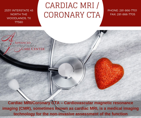
Cardiac MRI / Coronary CTA in Houston
Cardiac MRI/Coronary CTA – Cardiovascular magnetic resonance imaging (CMR), sometimes known as cardiac MRI, is a medical imaging technology for the non-invasive assessment of the function and structure of the cardiovascular system.

It is derived from and based on the same basic principles as magnetic resonance imaging (MRI) but with optimization for use in the cardiovascular system. These optimizations are principally in the use of ECG gating and rapid imaging techniques or sequences. By combining a variety of such techniques into protocols, key functional and morphological features of the cardiovascular system can be assessed.
Phone: 281-866-7701
Fax: 281-866-7705
Address: 25311 Interstate 45 North The woodlands, TX 77380
4 notes
·
View notes
Photo

"Cardiac MRI is the gold standard of all other imaging cardiac testing, so it provides the most accurate assessment of any given problem,” says Dr. Kamran Shaikh, Director of Cardiovascular MRI at Seton Hospitals. Watch the entire Ascension Seton Heart Report segment here to learn more about cardiac MRI with Dr. Shaikh. #SetonMedicalCenterHays #CardiacMRI #CardiacCare #MoreThanHome #CityofKyleEconomicDevelopment (at Kyle, Texas) https://www.instagram.com/p/Bvw4IlDg3YL/?utm_source=ig_tumblr_share&igshid=ai1p14r5lbsc
0 notes
Video
instagram
My valentine to Corinne - a lenticular card I had made using images from a Cardiac MRI I had done. My heart beats for you darling. Love you. ❤️ #valentines #noyouretheschmoopy #heartbeats #cardiacmri #gifpop
1 note
·
View note
Photo

S’ Wave: Tissue doppler imaging-Derived Tricuspid Lateral Annular Systolic Velocity S’ value less than 9.5 cm/sec indicating RV dysfunction. Mnemonic... If you stare at the number long enough you'll see 9.5 in it lol. . The first peak above baseline that is timed with the QRS represents isovolumetric contraction and not the tricuspid annular velocity. . -IkaN (Nakeya Dewaswala Bhopalwala) #medicine #medicalmnemonics #usmle #mbbs #medical #medicalstudent #medicalstudent #medicowesome #neet #pgcet #mrcp #internalmedicine #ABIM #internalmedicineresidency #internalmedicineresident #cardiology #vascularsurgery #echocardiography #sonographystudent #echo #JADEL #sonographer #cardiacMRI #cmr #whycmr #radiology #cardiowesome #echowesome https://www.instagram.com/p/CYF-RNJMmhH/?utm_medium=tumblr
#medicine#medicalmnemonics#usmle#mbbs#medical#medicalstudent#medicowesome#neet#pgcet#mrcp#internalmedicine#abim#internalmedicineresidency#internalmedicineresident#cardiology#vascularsurgery#echocardiography#sonographystudent#echo#jadel#sonographer#cardiacmri#cmr#whycmr#radiology#cardiowesome#echowesome
0 notes
Video
instagram
Granulomatous Cardiomyopathy This is a 48-years old with supraventricular tachycardia. The cardiac MRI shows mild to moderate hypokinesia of the basal anterior wall and antero-septal segment with mid myocardial enhancement suggestive of fibrosis with necrotic small mediastinal nodes. Granulomatous cardiomyopathy (GM) occurs due to sarcoidosis or tuberculosis. The distribution of nodes is more suggestive of TB in this situation, but it needs to be proven. #cardiacmri #mri #cardiomyopathy #tuberculosis #sarcoidosis (at Picture This by Jankharia)
0 notes
Text
Best Cardiac MRI/Coronary Treatments CTA in Houston
Cardiac MRI/Coronary CTA in Houston
Cardiac MRI/Coronary CTA – Cardiovascular magnetic resonance imaging (CMR), sometimes known as cardiac MRI, is a medical imaging technology for the non-invasive assessment of the function and structure of the cardiovascular system.
Contact Us On:
PHONE: 281-866-7701
FAX: 281-866-7705
ADDRESS: 25311 INTERSTATE 45 NORTH THE WOODLANDS, TX 77380
https://advancedcardiodr.com/Services/cardiac-mri-coronary-cta/
0 notes
Photo

Corinne also got chocolates, but these are from Rhea to her first Valentine - her Momma. #chocolate. #darkchocolate #gifpop #valentines #noyouretheschmoopy #cardiacmri
0 notes
Photo

Mitral valve anatomy and types of mitral regurgitation mnemonic #medicine #medicalmnemonics #usmle #mbbs #medical #medicalstudent #medicalstudent #medicowesome #neet #pgcet #mrcp #internalmedicine #ABIM #internalmedicineresidency #internalmedicineresident #cardiology #vascular#echocardiography #sonographystudent #echo #JADEL #sonographer #cardiacMRI #cmr #whycmr #radiology #medicalinspiration #studygram #studymotivation #studywithme #studynotes https://www.instagram.com/p/CXpHJafOfDE/?utm_medium=tumblr
#medicine#medicalmnemonics#usmle#mbbs#medical#medicalstudent#medicowesome#neet#pgcet#mrcp#internalmedicine#abim#internalmedicineresidency#internalmedicineresident#cardiology#vascular#echocardiography#sonographystudent#echo#jadel#sonographer#cardiacmri#cmr#whycmr#radiology#medicalinspiration#studygram#studymotivation#studywithme#studynotes
0 notes
Video
instagram
The Banana Heart This is 21 years old man with a restrictive physiology. The 4 chamber view shows a characteristic "banana" left ventricle with constriction at the level of or just distal to the AV grooves. Both LA and RA are dilated. The CT scan confirms the pericardial calcification and thickening. This is classic constrictive pericarditis as a sequelae of tuberculous pericarditis in the past with thickening and calcification. #cardiacmri #mri #constrictivepericarditis #heart #pericardium (at Picture This by Jankharia)
0 notes
Video
instagram
Cardiac MRI for Viability Imaging This is a 52 years old man with an LAD occlusion and a recent history of a myocardial infarct. Four weeks later, a decision needs to be taken on revascularization. The cardiac MRI shows a full-thickness, transmural infarct in the LAD territory involving the septum and anterior wall with areas of micro-vascular obstruction that carry a poorer prognosis. Revascularization is unlikely to be of help. #cardiacmri #viabilityscan #myocardialinfarction
0 notes
Video
instagram
The Cascading Effect of a Bicuspid Aortic Valve This is a 55 years old man with a diagnosis of polymyalgia rheumatica also known to have a dilated cardiomyopathy with an ejection fraction of approx. 30% for many years. He was finally referred for a cardiac MRI. _____________________________________ The MRI shows a bicuspid aortic valve missed on prior echoes with an associated dilated ascending aorta with a diameter of 46 mm, moderate AR and LV dilatation with an EF of 42% with mild mid-lateral wall fibrosis. _____________________________________This is no longer an idiopathic dilated cardiomyopathy #cardiacmri #bicuspidaorticvalve #dilatedcardiomyopathy (at Picture This by Jankharia)
0 notes
Video
instagram
Apical Hypertrophic Cardiomyopathy This lady presented with tachyarrhythmias and an echo showed apparent concentric LV hypertrophy. The MRI shows classic apical predominant hypertrophic cardiomyopathy (HCM) extending into the mid-cavity and partly into the basal segment. The 2C late Gd image shows diffuse enhancement, suggestive of fibrosis. #hcm #hypertrophiccardiomyopathy #mri #radiology #cardiacmri
0 notes
Last Seen Blogs

stardustneeko
It's Pronounced Ne-e-ko

salongrowthhacker
Salon Growth Hacker

daviddarmon
DAVID DARMON

alexiethymia
emotions without words

yamirami00
Sans titre