#medtechnews
Text
Atlas of human brain blood vessels highlights changes in Alzheimer’s disease

Atlas of human brain blood vessels highlights changes in Alzheimer’s disease
MIT researchers characterize gene expression patterns for 22,500 brain vascular cells across 428 donors, revealing insights for Alzheimer’s onset and potential treatments.
Your brain is powered by 400 miles of blood vessels that provide nutrients, clear out waste products, and form a tight protective barrier — the blood-brain barrier — that controls which molecules can enter or exit. However, it has remained unclear how these brain vascular cells change between brain regions, or in Alzheimer’s disease, at single-cell resolution.
To address this challenge, a team of scientists from MIT’s Computer Science and Artificial Intelligence Laboratory (CSAIL), The Picower Institute for Learning and Memory, and The Broad Institute of MIT and Harvard recently unveiled a systematic molecular atlas of human brain vasculature and its changes in Alzheimer’s disease (AD) across six brain regions, in a paper published June 1 in Nature Neuroscience.
Alzheimer's disease is a leading cause of death, affects one in nine Americans over 65, and leads to debilitating and devastating cognitive decline. Impaired blood-brain barrier (BBB) function has long been associated with Alzheimer’s and other neurodegenerative diseases, such as Parkinson's and multiple sclerosis. However, the molecular and cellular underpinnings of BBB dysregulation remain ill-defined, particularly at single-cell resolution across multiple brain regions and many donors.
Navigating vascular complexity
Embarking deep into the complexities of our gray matter, the researchers created a molecular atlas of human brain vasculature across 428 donors, including 220 diagnosed with Alzheimer's and 208 controls. They characterized over 22,514 vascular cells from six different brain regions, measuring the expression of thousands of genes for each cell. The resulting datasets unveiled intriguing changes in gene expression across different brain regions and stark contrasts between individuals afflicted with AD and those without.
“Alzheimer's therapy development faces a significant hurdle — brain alterations commence decades before cognitive signs make their debut, at which point it might already be too late to intervene effectively,” comments MIT CSAIL principal investigator and electrical engineering and computer science (EECS) Professor Manolis Kellis. “Our work charts the terrain of vascular changes, one of the earliest markers of Alzheimer's, across multiple brain regions, providing a map to guide biological and therapeutic investigations earlier in disease progression.”
Kellis is the study's co-senior author, along with MIT Professor Li-Huei Tsai, director of the Picower Institute and the Picower Professor in the Department of Brain and Cognitive Sciences.
The little cells that could
The threads of our human brain vasculature, and every part of our brain and body, are composed of millions of cells, all sharing the same DNA code, but each expressing a different subset of genes, which define its functional roles and distinct cell type. Using the distinct gene expression signatures of different cerebrovascular cells, the researchers distinguished 11 types of vascular cells.
These included endothelial cells that line the interior surface of blood vessels and control which substances pass through the BBB, pericytes that wrap around small vessels and provide structural support and blood flow control, smooth muscle cells that form the middle layer of large vessels and whose contraction and relaxation regulates blood flow and pressure, fibroblasts that surround blood vessels and hold them in place, and they distinguished arteriole, venule, and capillary veins responsible for the different stages of blood oxygen exchange.
The abundance of these vascular cell types differed between brain regions, with neocortical regions showing more capillary endothelial cells and fewer fibroblasts than subcortical regions, highlighting the regional heterogeneity of the BBB.
Clues and suspects
Armed with these annotations, the next phase was studying how each of these cell types change in AD, revealing 2,676 genes whose expression levels change significantly. They found that capillary endothelial cells, responsible for transport, waste removal, and immune surveillance, showed the most changes in AD, including genes involved in clearance of amyloid beta, one of the pathological hallmarks of AD, providing insights on the potential mechanistic implications of vascular dysregulation on AD pathology.
Other dysregulated processes included immune function, glucose homeostasis, and extracellular matrix organization, which were all shared among multiple vascular cell types, and also cell-type-specific changes, including growth factor receptors in pericytes, and transporter and energy in endothelial cells, and cellular response to amyloid beta in smooth muscle cells. Regulation of insulin sensing and glucose homeostasis in particular suggested important connections between lipid transport and Alzheimer’s regulated by the vasculature and blood-brain-barrier cells, which could hold promise for new therapeutic clues.
“Single-cell RNA sequencing provides an extraordinary microscope to peer into the intricate machinery of life, and ‘see’ millions of RNA molecules bustling with activity within each cell,” says Kellis, who is also a member of the Broad Institute. “This level of detail was inconceivable just a few years ago, and the resulting insights can be transformative to comprehend and combat complex psychiatric and neurodegenerative disease."
Maestros of dysregulation
Genes do not act on a whim, and they do not act alone. Cellular processes are governed by a complex cast of regulators, or transcription factors, that dictate which groups of genes should be turned on or off in different conditions, and in different cell types. These regulators are responsible for interpreting our genome, the ‘book of life,’ and turning it into the myriad of distinct cell types in our bodies and in our brains. These regulators might be responsible when something goes wrong, and they could also be critical in fixing things and restoring healthy cellular states.
With thousands of genes showing altered expression levels in Alzheimer’s disease, the researchers then sought to find the potential masterminds behind these changes. They asked if common regulatory control proteins target numerous altered genes, which may provide candidate therapeutic targets to restore the expression levels of large numbers of target genes. Indeed, they found several such ‘master controllers,’ involved in regulating endothelial differentiation, inflammatory response, and epigenetic state, providing potential intervention points for drug targets against AD.
Cellular murmurings
Cells do not function in isolation; rather, they rely on communication with each other to coordinate biological processes. This intercellular communication is particularly complex within the cellular diversity of the brain, given the many factors involved in sensing, memory formation, knowledge integration, and consciousness. In particular, vascular cells have intricate interactions with neurons, microglia, and other brain cells, which take on heightened significance during pathological events, such as in Alzheimer's disease, where dysregulation of this cellular communication can contribute to the progression of the disease.
The researchers found that interactions from capillary endothelial cells to neurons, microglia, and astrocytes were highly increased in AD, while interactions in the reverse direction, from neurons and astrocytes to capillary endothelial cells, were decreased in AD. This asymmetry could provide important cues for potential interventions targeting the vasculature and specifically capillary endothelial cells, with ultimate broad positive impacts on the brain.
“The dynamics of vascular cell interactions in AD provide an entry point for brain interventions and potential new therapies,” says Na Sun, an EECS graduate student and MIT CSAIL affiliate and first author on the study. “As the blood-brain barrier prevents many drugs from influencing the brain, perhaps we could instead manipulate the blood-brain barrier itself, and let it spread beneficiary signals to the rest of the brain. Our work provides a blueprint for cerebrovasculature interventions in Alzheimer's disease, by unraveling how cellular communication can mediate the impact of genetic variants in AD."
Going off script: genetic plot twists
Disease onset in our bodies (and in our brains) is shaped by a combination of genetic predispositions and environmental exposures. On the genetic level, most complex traits are shaped by hundreds of minuscule sequence alterations, known as single-nucleotide polymorphisms (or SNPs, pronounced snips), most of which act through subtle changes in gene expression levels.
No matter how subtle their effects might be, these genetic changes can reveal causal contributors to disease, which can greatly increase the chance of therapeutic success for genetically-supported target genes, compared to targets lacking genetic support.
To understand how genetic differences associated with Alzheimer’s might act in the vasculature, the researchers then sought to connect genes that showed altered expression in Alzheimer’s with genetic regions associated with increased Alzheimer’s risk through genetic studies of thousands of individuals. They linked the genetic variants (SNPs) to candidate target genes using three lines of evidence: physical proximity in the three-dimensional folded genome, genetic variants that affect gene expression, and correlated activity between distant regulatory regions and target genes that go on and off together between different conditions.
This resulted in not just one hit, but 125 genetic regions, where Alzheimer’s-associated genetic variants were linked to genes with disrupted expression patterns in Alzheimer’s disease, suggesting they might mediate these causal genetic effects, and thus may be good candidates for therapeutic targeting. Some of these predicted hits were direct, where the genetic variant acted directly on a nearby gene. Others were indirect when the genetic variant instead affected the expression of a regulator, which then affected the expression of its target genes. And yet others were predicted to be indirect through cell-cell communication networks.
ApoE4 and cognitive decline
While most genetic effects are subtle, both in Alzheimer’s and nearly all complex disorders, exceptions do exist. One such exception is FTO in obesity, which increases obesity risk by one standard deviation. Another one is apolipoprotein E (ApoE) in Alzheimer’s disease, where the E4 versus E3 allele increases risk more than 10-fold for carriers of two risk alleles — those who inherited one ‘unlucky’ copy from each parent.
With such a strong effect size, the researchers then asked if ApoE4 carriers showed specific changes in vascular cells that were not found in ApoE3 carriers. Indeed, they found abundance changes associated with the ApoE4 genotype, with capillary endothelial cells and pericytes showing extensive down-regulation of transport genes. This has important implications for potential preventive treatments targeting transport in ApoE4 carriers, especially given the cholesterol transporter roles of ApoE, and the increasingly recognized role of lipid metabolism in Alzheimer’s disease.
"Unearthing these AD-differential genes gives us a glimpse into how they may be implicated in the deterioration or dysfunction of the brain's protective barrier in Alzheimer's patients, shedding light on the molecular and cellular roots of the disease's development," says Kellis. "They also open several avenues for therapeutic development, hinting at a future where these entry points might be harnessed for new Alzheimer's treatments targeting the blood-brain barrier directly. The possibility of slowing or even halting the disease's progression is truly exciting.”
Translating these findings into viable therapeutics will be a journey of exploration, demanding rigorous preclinical and clinical trials. To bring these potential therapies to patients, scientists need to understand how to target the discovered dysregulated genes safely and effectively and determine whether modifying their activity can ameliorate or reverse AD symptoms, which requires extensive collaborations between medical doctors and engineers across both academia and industry.
“This is a tour de force impressive case series,” says Elizabeth Head, vice chair for pathology research and pathology professor at the University of California at Irvine, who was not involved in the research. “A novel aspect of this study was also the methodological approach, which left the vasculature intact, as compared to previous work where blood vessel enrichment protocol was applied. Manolis Kellis and his colleagues show clear evidence of neurovascular unit dysregulation in AD and it is exciting to see known and novel pathways being identified that will accelerate discoveries at the protein level. Many DEGs associated with AD are linked to lipid/cholesterol metabolism, to AD genetic risk factors (including ApoE) and inflammation. The potential for the ApoE genotype in mediating cerebrovascular function will also lead to possible new mouse models that will capture the human phenotype more closely with respect to the vascular contributions to dementia in humans. The regional differences in DEGs are fascinating and will guide future neuropathology studies in the human brain and drive novel hypotheses.”
"The predominant focus in AD research over the past 10 years has been on studying microglia, the resident macrophage-like cells of the brain,” adds Ryan Corces, an assistant professor of neurology at the University of California at San Francisco who was also not involved in the work. “While microglia certainly play a key role in disease pathogenesis, it has become increasingly clear through studies such as this one that vascular cells may also be critically involved in the disease. From blood-brain barrier leakage to an enhanced need for debris clearance, the vascular cells of the brain play an important part in this complex disease. This study, and others like it, have begun picking apart the underlying molecular changes that occur in vascular cells, showing which genes appear dysregulated and how those changes may interact to alter vascular cell functions. Together with the mounting evidence of vascular involvement in AD, this work provides an important foundation for guiding therapeutic interventions against blood-brain barrier dysfunction in AD, especially during the preclinical or prodromal stages of the disease, where the blood-brain barrier may be playing a central role.”
Sun, Kellis, and Tsai wrote the paper alongside Leyla Anne Akay, Mitchell H. Murdock, Yongjin Park, Fabiola Galiana-Melendez, Adele Bubnys, Kyriaki Galani, Hansruedi Mathys, Xueqiao Jiang, and Ayesha P. Ng of MIT and David A. Bennett of the Rush Alzheimer’s Disease Center in Chicago. This work was supported, in part, by National Institutes of Health grants, the Cure Alzheimer’s Foundation CIRCUITS consortium, the JPB Foundation, Robert A. and Renee Belfer, and a Takeda Fellowship from the Takeda Pharmaceutical Company.
Source: MIT
Read the full article
3 notes
·
View notes
Photo

We will be one of the 4,000+ designers, engineers, innovators and manufacturers from across the medical and healthcare sector exhibiting at Med-Tech Innovation Expo 2019 over 15th -16th May at the NEC in Birmingham @MedTechonline #medical #MedTechNews #testing
https://med-techexpo.com/medtechinnovationexpo2019/en/page/home
0 notes
Text
Magnetic guidewire steering at ultrahigh magnetic fields for medical imaging

Magnetic guidewire steering at ultrahigh magnetic fields for medical imaging.
Physicists and bioengineers can manipulate magnetically driven guidewires by using remote magnetic steering with scope for minimally invasive medical procedures. Magnetic steering strategies are presently limited by low magnetic fields, thereby preventing their integration in medical systems operating at ultrahigh fields, including magnetic resonance imaging (MRI) scanners. In a new study now published in Science Advances, Mehmet Tiryaki and a research team at the departments of physical intelligence, biomedical engineering, and medicine in Germany, Switzerland, and Turkey, developed a magnetic guidewire design alongside steering strategies at ultrahigh fields.
The work demonstrated an extensive research scope, alongside its potential for in situ re-magnetization. The outcomes illustrated steering principles of magnetic guidance made of neodymium magnets and a fiber optic rod in a preclinical magnetic resonance imaging scanner. The newly developed ultrahigh field magnetic actuation framework can facilitate next-generation magnetic automation to function in clinical MRI scanners.
Advancing the magnetic resonance imaging (MRI) system
Despite a decade-long development of methods for magnetic resonance imaging, the technology has shortcomings compared to X-ray fluoroscopy. The ionizing radiation-free nature of X-ray fluoroscopy alongside its superior soft tissue contrast, makes it a more advanced alternative. The MRI system is currently limited by the workspace area in the scanner and its lower resolution, leading to a range of new proposals to improve the method.
For instance, a completely remote MRI-powered actuation approach can integrate a ferromagnet permanent magnet for intuitive three-dimensional (3D) steering. However, the method requires real-time software access and added power to function inside an MRI scanner. In this work, Tiryaki and colleagues presented an ultrahigh field magnetic guidewire steering strategy in the MRI scanner and demonstrated its steering capacity in physiologically relevant 3D vascular phantoms with the arterial flow, as well as during MRI scanning in the kidney of an animal model.
Magnetizing permanent magnets at ultrahigh fields
Permanent magnets such as neodymium magnets are commonly used during magnetic actuation for high magnetic torque and force transmission at low magnetic fields. Permanent magnets are developed with a constant magnetization vector aligned to the easy axis of the magnet at low magnetic fields. While physicists have studied the magnetic theory of permanent magnets at ultrahigh fields, they remain to investigate effects of the concept during automated magnetic actuation.
For instance, at ultrahigh fields, permanent magnets assume the form of soft magnets. The team, therefore, examined the magnetization vector and calculated the magnetic force and torque acting on permanent magnets. They focused on bulk neodymium magnets and used a vibrating sample magnetometer to deduce magnetic material constants, and studied the effects of magnetic hysteresis, to verify the strength of magnetization.
Magnetic actuation method at UHF. (A) Magnetization at low fields, B < 0.1 T. The red and blue colors represent the direction of the easy axis (C). (B) Magnetization at high fields, B >> 0.1 T. The permanent magnet is magnetized substantially by the external magnetic field. (C) Magnetization curves of the cylindrical neodymium magnet measured inside a vibrating sample magnetometer at different θ. The magnet's magnetization in the x direction is measured, while the magnetic field is swept from 0 to 1.8 T in the x direction. (D) Magnetization vector alignment as a function of the easy axis alignment at different external field strengths. (E) Magnetic fields and gradients in the x direction of the MRI scanner. (F) Model-based magnetic torque acting on the cylindrical magnet as a function of the magnet orientation. The magnet is in its saturation regime for the solid lines and nonsaturated regime for the dashed lines. The direction of the torque is shown on the schematic. (G) Magnetic force as a function of the magnet position in the MRI scanner for different magnet orientations. The solid lines represent the force estimated with the magnetization model using the MRI magnetic field and gradient measurements. The dashed lines are the linear model estimation in the nonsaturated region. The dots are the force sensor measurements in the experiments. (H) The discretized Cosserat rod model, including the magnetic actuation. (I) Schematic of the magnetic actuation simulations. (J and K) The maximum rotation and rotation range in the simulations as a function of the guidewire thickness. Credit: Science Advances (2023). DOI: 10.1126/sciadv.adg6438
Magnetic actuation in the MRI scanner and guidewire design
Tiryaki and colleagues measured the magnetic field and magnetic gradient in the MRI scanner to model magnetic torque and force. They calculated the magnetization angle and torque acting on the permanent magnet at ultrahigh fields and investigated the design of the accompanying flexible construct that formed the elastic core of the guidewire, and optimized the stiffness of the flexible body to magnetically actuate guidewires and maneuver the magnetic actuation system.
The team used open-source software and developed a Cosserat rod model dynamic simulation to mimic the shape of the guidewire and included elastic and gravitational forces to understand their influence on MRI magnetic force and torque. They performed bending simulations to validate Young's moduli and other parameters underlying guidewire dynamics to proactively use the magnetic guidewires.
Magnetic steering modes and in situ magnetization
The scientists studied a variety of automated magnetic actuation systems with high degrees of freedom to achieve magnetic guidewire steering at low fields. In the absence of high degrees of freedom, interactions between the magnetic actuation system and ultrahigh fields led to blocked guidewire steering in the MRI scanner. The team, therefore, studied the cardinal configurations of the guidewire to understand this effect and placed the permanent magnets parallel, perpendicular, and antiparallel to the guidewire tip.
Tiryaki and colleagues explored a variety of steering modes with manual guidewire insertion in the MRI scanner to perform a variety of navigational tasks. The concept of in situ re-magnetization at ultrahigh fields led to an even more interesting magnetic guidewire design with dual stability, with two permanent magnets at the guidewire tip to conduct a variety of steering experiments in a two-dimensional plane.
Obstacle avoidance by magnetic steering at UHF. (A) Schematic illustration of the obstacle avoidance experimental setup. The obstacles are placed in the robotic platform with remotely controlled motion in the x direction and yaw angle. The guidewire is inserted into the platform through the insertion point, and an operator manually controls the insertion and twist of the guidewire by hand at the entrance of the MRI scanner bore. The same platform is used for demonstration with three different magnetic configurations. (B) Obstacle avoidance with guidewire with a parallel magnet. The guidewire is steered to three target points shown in (i) using the platform rotation and guidewire insertion at 7 T. The magnetic gradient is used as an assistive force in (iv). (C) Obstacle avoidance experiment with a guidewire with a perpendicular magnet. The guidewire is steered to three target points shown in (i) using the platform motion in the x direction and the base twist. The operator controlled the bending of the guidewire through the magnetic field strength. The platform angle is kept the same throughout the navigation. (D) Obstacle avoidance with guidewire with an antiparallel magnet. The guidewire is steered to three target points shown in (i) using the platform motion in the x direction, base twist, and in situ remagnetization. The first two target points are reached using the antiparallel configuration's larger bending. For the last target point, the guidewire is physically constrained using the obstacles (iii) and remagnetized into the parallel configuration. Credit: Science Advances (2023). DOI: 10.1126/sciadv.adg6438
Three-dimensional (3D) vascular navigation and guidewire steering during MRI
The team performed steering experiments in a realistic 3D vascular architecture of the renal arteries, aortic arch, common carotid artery, and middle cerebral arteries while emulating arterial flow with a cardiac flow simulation pump. The results emphasized the capacity to navigate 3D vessels across a variety of situations for clinical applications.
Additionally, they explored ultrahigh field magnetic actuation during MRI, by performing guidewire steering experiments in the renal cavity of a porcine kidney ex vivo, with guidewires in different magnetic configurations to target various regions of the organ. They performed a preclinical MRI to observe the boundaries of the renal cavity, followed by a series of visualization experiments across the renal cavity, lower calyx, and upper calyx to examine the steering capacity of the guidewire.
Outlook
In this way, Mehmet Tiryaki and colleagues introduced the concept of magnetic steering with magnetic guidewires at ultrahigh fields. They combined the theory of magnetism with mechanics to establish design principles for improved steering capabilities in the MRI scanner.
The physicists determined the feasibility of magnetic guidance steering during MRI imaging via a series of 3D navigation steps. They expect the new method of ultrahigh field actuation to impact clinical scenarios during MRI intervention to eventually facilitate the physical intelligence required for automated clinical intervention practices.
More information: Mehmet Efe Tiryaki et al, Magnetic guidewire steering at ultrahigh magnetic fields, Science Advances (2023). DOI: 10.1126/sciadv.adg6438
Martin Francis Phelan et al, Heat‐Mitigated Design and Lorentz Force‐Based Steering of an MRI‐Driven Microcatheter toward Minimally Invasive Surgery, Advanced Science (2022). DOI: 10.1002/advs.202105352
Journal information: Advanced Science, Science Advances
Source: Medical Xpress
Read the full article
2 notes
·
View notes
Text
New study on improving MRI image quality based on deep learning technology

New study on improving MRI image quality based on deep learning technology published in European Radiology.
SwiftMR™, an AI-powered MRI reconstruction solution from AIRS Medical proved its performance for enhancing the image quality of 3D high-resolution MRI.
SEOUL, South Korea, Nov. 25, 2022 - A recent study published in European Radiology demonstrated that SwiftMR, an AI-powered MRI reconstruction solution from AIRS Medical, successfully denoises 3D MR images and improves its image quality by using routine clinical scans only.
The study aimed to develop a deep neural network (DNN)–based noise reduction and image quality improvement by only using routine clinical scans and evaluating its performance in 3D high-resolution MRI. The study was conducted by AIRS Medical and Dr. Jinhee Jang, MD, Ph.D. of Seoul St. Mary's Hospital. The retrospective study included T1-weighted magnetization-prepared rapid gradient-echo (MP-RAGE) images from 185 clinical scans.
Qualitative evaluation between conventional MP-RAGE and DNN-based MP-RAGE was performed by two radiologists in image quality, fine structure delineation, and lesion conspicuity. Quantitative evaluation was performed with full sampled data as a reference by measuring quantitative error metrics and volumetry at seven different simulated noise levels. DNN application on VWI was evaluated by two radiologists in image quality.
The study shows that DNN-based MP-RAGE outperformed conventional MP-RAGE in all image quality parameters (average scores = 3.7 vs. 4.9, p < 0.001). In the quantitative evaluation, DNN showed better error metrics (p < 0.001) and comparable (p > 0.09) or better (p < 0.02) volumetry results than conventional MP-RAGE. DNN application to VWI also revealed improved image quality (3.5 vs. 4.6, p < 0.001).
"We are very pleased that we have proved SwiftMR™ contributes to not only reducing time but also helps make images good to great, eventually contribute radiologists could read images with confidence." remarked Hyeseong Lee, MD, CEO of AIRS Medical. "We are expecting more adaptions and collaborations with radiologists around the world thus we could grow together."
AIRS Medical recently announced its participation in the 108th Scientific Assembly and Annual Meetings of the RSNA 2022, held from November 27 to 30th in Chicago. During the event, AIRS Medical showcases its award-winning MRI reconstruction solution SwiftMR™and also delivering two oral presentations at scientific session.
"We are proud of ourselves since it is quite unusual for a startup to deliver oral presentations at RSNA scientific session. Being adopted as oral presentation means recognition of academically important achievements."
he added
Read the full article
4 notes
·
View notes
Text
MIT researchers develop advanced machine learning models to detect pancreatic cancer

MIT researchers develop advanced machine learning models to detect pancreatic cancer.
MIT CSAIL researchers develop advanced machine-learning models that outperform current methods in detecting pancreatic ductal adenocarcinoma.
Prismatic perspectives pancreatic cancer
The path forward
The first documented case of pancreatic cancer dates from the 18th century. Since then, researchers have embarked on a long and difficult journey to better understand this elusive and deadly disease. To date, early intervention is the most effective cancer treatment. Unfortunately, due to its location deep within the abdomen, the pancreas is particularly difficult to detect early on.
Scientists from the MIT Computer Science and Artificial Intelligence Laboratory (CSAIL), as well as Limor Appelbaum, a staff scientist in the Department of Radiation Oncology at Beth Israel Deaconess Medical Center (BIDMC), wanted to better identify potential high-risk patients. They set out to create two machine-learning models for the early detection of pancreatic ductal adenocarcinoma (PDAC), the most common type of cancer.
To gain access to a large and diverse database, the team collaborated with a federated network company and used electronic health record data from multiple institutions across the United States. This vast data set contributed to the models' reliability and generalizability, making them applicable to a wide range of populations, geographical locations, and demographic groups.
The two models—the “PRISM” neural network and the logistic regression model (a statistical technique for probability)—outperformed current methods. The team’s comparison showed that while standard screening criteria identify about 10 percent of PDAC cases using a five-times higher relative risk threshold, Prism can detect 35 percent of PDAC cases at this same threshold.
Using AI to detect cancer risk is not a new phenomenon; algorithms analyze mammograms, CT scans for lung cancer, and assist in the analysis of Pap smear tests and HPV testing, to name a few applications.
“The PRISM models stand out for their development and validation on an extensive database of over 5 million patients, surpassing the scale of most prior research in the field,” says Kai Jia, an MIT PhD student in electrical engineering and computer science (EECS), MIT CSAIL affiliate, and first author on an open-access paper in eBioMedicine outlining the new work. “The model uses routine clinical and lab data to make its predictions, and the diversity of the U.S. population is a significant advancement over other PDAC models, which are usually confined to specific geographic regions, like a few health-care centers in the U.S. Additionally, using a unique regularization technique in the training process enhanced the models' generalizability and interpretability.”
“This report outlines a powerful approach to use big data and artificial intelligence algorithms to refine our approach to identifying risk profiles for cancer,” says David Avigan, a Harvard Medical School professor and the cancer center director and chief of hematology and hematologic malignancies at BIDMC, who was not involved in the study. “This approach may lead to novel strategies to identify patients with high risk for malignancy that may benefit from focused screening with the potential for early intervention.”
Prismatic perspectives pancreatic cancer
The journey toward the development of PRISM began over six years ago, fueled by firsthand experiences with the limitations of current diagnostic practices.
“Approximately 80-85 percent of pancreatic cancer patients are diagnosed at advanced stages, where cure is no longer an option,” says senior author Appelbaum, who is also a Harvard Medical School instructor as well as radiation oncologist. “This clinical frustration sparked the idea to delve into the wealth of data available in electronic health records (EHRs).”
The CSAIL group’s close collaboration with Appelbaum made it possible to understand the combined medical and machine learning aspects of the problem better, eventually leading to a much more accurate and transparent model. “The hypothesis was that these records contained hidden clues — subtle signs and symptoms that could act as early warning signals of pancreatic cancer,” she adds. “This guided our use of federated EHR networks in developing these models, for a scalable approach for deploying risk prediction tools in health care.”
Both PrismNN and PrismLR models analyze EHR data, including patient demographics, diagnoses, medications, and lab results, to assess PDAC risk. PrismNN uses artificial neural networks to detect intricate patterns in data features like age, medical history, and lab results, yielding a risk score for PDAC likelihood. PrismLR uses logistic regression for a simpler analysis, generating a probability score of PDAC based on these features. Together, the models offer a thorough evaluation of different approaches in predicting PDAC risk from the same EHR data.
One paramount point for gaining the trust of physicians, the team notes, is better understanding how the models work, known in the field as interpretability. The scientists pointed out that while logistic regression models are inherently easier to interpret, recent advancements have made deep neural networks somewhat more transparent. This helped the team to refine the thousands of potentially predictive features derived from EHR of a single patient to approximately 85 critical indicators. These indicators, which include patient age, diabetes diagnosis, and an increased frequency of visits to physicians, are automatically discovered by the model but match physicians' understanding of risk factors associated with pancreatic cancer.
The path forward
Despite the promise of the PRISM models, as with all research, some parts are still a work in progress. U.S. data alone are the current diet for the models, necessitating testing and adaptation for global use. The path forward, the team notes, includes expanding the model's applicability to international datasets and integrating additional biomarkers for more refined risk assessment.
“A subsequent aim for us is to facilitate the models' implementation in routine health care settings. The vision is to have these models function seamlessly in the background of health care systems, automatically analyzing patient data and alerting physicians to high-risk cases without adding to their workload,” says Jia. “A machine-learning model integrated with the EHR system could empower physicians with early alerts for high-risk patients, potentially enabling interventions well before symptoms manifest. We are eager to deploy our techniques in the real world to help all individuals enjoy longer, healthier lives.”
Jia wrote the paper alongside Applebaum and MIT EECS Professor and CSAIL Principal Investigator Martin Rinard, who are both senior authors of the paper. Researchers on the paper were supported during their time at MIT CSAIL, in part, by the Defense Advanced Research Projects Agency, Boeing, the National Science Foundation, and Aarno Labs. TriNetX provided resources for the project, and the Prevent Cancer Foundation also supported the team.
Source: MIT
Read the full article
0 notes
Text
Inhalable sensors could enable early lung cancer detection

Inhalable sensors could enable early lung cancer detection.
The diagnostic, which requires only a simple urine test to read the results, could make lung cancer screening more accessible worldwide.
Inhalable particles
Accurate diagnosis
MIT, January 5, 2024 - Using a new MIT technology, diagnosing lung cancer could be as simple as inhaling nanoparticle sensors and then taking a urine test to see if a tumor is present.
The new diagnostic uses nanosensors that can be delivered via inhaler or nebulizer. When the sensors come into contact with cancer-linked proteins in the lungs, they generate a signal that accumulates in the urine and can be detected with a simple paper test strip.
This method has the potential to replace or supplement the current gold standard for lung cancer diagnosis, low-dose computed tomography (CT). According to the researchers, it could have a particularly large impact in low- and middle-income countries where CT scanners are not widely available.
“Around the world, cancer is going to become more and more prevalent in low- and middle-income countries. The epidemiology of lung cancer globally is that it’s driven by pollution and smoking, so we know that those are settings where accessibility to this kind of technology could have a big impact,”
says Sangeeta Bhatia, the John and Dorothy Wilson Professor of Health Sciences and Technology and of Electrical Engineering and Computer Science at MIT, and a member of MIT’s Koch Institute for Integrative Cancer Research and the Institute for Medical Engineering and Science.
Bhatia is the senior author of the paper, which appears today in Science Advances. Qian Zhong, an MIT research scientist, and Edward Tan, a former MIT postdoc, are the lead authors of the study.
Inhalable particles
To help diagnose lung cancer as early as possible, the U.S. Preventive Services Task Force recommends that heavy smokers over the age of 50 undergo annual CT scans. However, not everyone in this target group receives these scans, and the high false-positive rate of the scans can lead to unnecessary, invasive tests.
Bhatia has spent the last decade developing nanosensors for use in diagnosing cancer and other diseases, and in this study, she and her colleagues explored the possibility of using them as a more accessible alternative to CT screening for lung cancer.
These sensors consist of polymer nanoparticles coated with a reporter, such as a DNA barcode, that is cleaved from the particle when the sensor encounters enzymes called proteases, which are often overactive in tumors. Those reporters eventually accumulate in the urine and are excreted from the body.
Previous versions of the sensors, which targeted other cancer sites such as the liver and ovaries, were designed to be given intravenously. For lung cancer diagnosis, the researchers wanted to create a version that could be inhaled, which could make it easier to deploy in lower resource settings.
“When we developed this technology, our goal was to provide a method that can detect cancer with high specificity and sensitivity, and also lower the threshold for accessibility, so that hopefully we can improve the resource disparity and inequity in early detection of lung cancer,”
Zhong says
To achieve that, the researchers created two formulations of their particles: a solution that can be aerosolized and delivered with a nebulizer, and a dry powder that can be delivered using an inhaler.
Once the particles reach the lungs, they are absorbed into the tissue, where they encounter any proteases that may be present. Human cells can express hundreds of different proteases, and some of them are overactive in tumors, where they help cancer cells to escape their original locations by cutting through proteins of the extracellular matrix. These cancerous proteases cleave DNA barcodes from the sensors, allowing the barcodes to circulate in the bloodstream until they are excreted in the urine.
In the earlier versions of this technology, the researchers used mass spectrometry to analyze the urine sample and detect DNA barcodes. However, mass spectrometry requires equipment that might not be available in low-resource areas, so for this version, the researchers created a lateral flow assay, which allows the barcodes to be detected using a paper test strip.
The researchers designed the strip to detect up to four different DNA barcodes, each of which indicates the presence of a different protease. No pre-treatment or processing of the urine sample is required, and the results can be read about 20 minutes after the sample is obtained.
“We were really pushing this assay to be point-of-care available in a low-resource setting, so the idea was to not do any sample processing, not do any amplification, just to be able to put the sample right on the paper and read it out in 20 minutes,”
Bhatia says.
Accurate diagnosis
The researchers put their diagnostic system to the test in mice that have been genetically modified to develop lung tumors similar to those seen in humans. The sensors were given to the mice 7.5 weeks after the tumors formed, which corresponds to stage 1 or 2 cancer in humans.
The researchers measured the levels of 20 different sensors designed to detect different proteases in their first set of experiments in mice. The researchers identified a combination of only four sensors that was predicted to provide accurate diagnostic results using a machine learning algorithm. They then tested that combination in a mouse model and discovered that it could detect early-stage lung tumors accurately.
For use in humans, it’s possible that more sensors might be needed to make an accurate diagnosis, but that could be achieved by using multiple paper strips, each of which detects four different DNA barcodes, the researchers say.
The researchers now plan to analyze human biopsy samples to see if the sensor panels they are using would also work to detect human cancers. In the longer term, they hope to perform clinical trials in human patients. A company called Sunbird Bio has already run phase 1 trials on a similar sensor developed by Bhatia’s lab, for use in diagnosing liver cancer and a form of hepatitis known as nonalcoholic steatohepatitis (NASH).
In parts of the world where there is limited access to CT scanning, this technology could offer a dramatic improvement in lung cancer screening, especially since the results can be obtained during a single visit.
“The idea would be you come in and then you get an answer about whether you need a follow-up test or not, and we could get patients who have early lesions into the system so that they could get curative surgery or lifesaving medicines,”
Bhatia says
The research was funded by the Johnson & Johnson Lung Cancer Initiative, the Howard Hughes Medical Institute, the Koch Institute Support (core) Grant from the National Cancer Institute, and the National Institute of Environmental Health Sciences.
Source: MIT
Read the full article
0 notes
Text
MIT Engineers develop a vibrating, ingestible capsule that might help treat obesity

MIT Engineers develop a vibrating, ingestible capsule that might help treat obesity
Swallowing the device before a meal could create a sense of fullness, tricking the brain into thinking it’s time to stop eating.
When you eat a large meal, your stomach sends signals to your brain that cause you to feel full, allowing you to realize it's time to stop eating. These messages can also be sent by a stomach full of liquid, which is why dieters are often advised to drink a glass of water before eating.
MIT engineers have developed a new method for capitalizing on this phenomenon, employing an ingestible capsule that vibrates within the stomach. These vibrations activate the same stretch receptors that detect when the stomach is distended, giving the illusion of fullness.
The researchers discovered that giving this pill to animals 20 minutes before eating not only stimulated the release of hormones that signal satiety, but also reduced the animals' food intake by about 40%. Scientists still have a lot to learn about the mechanisms that influence human body weight, but if further research shows that this technology can be used safely in humans, such a pill could offer a minimally invasive way to treat obesity, according to the researchers.
“For somebody who wants to lose weight or control their appetite, it could be taken before each meal,” says Shriya Srinivasan PhD ’20, a former MIT graduate student and postdoc who is now an assistant professor of bioengineering at Harvard University. “This could be really interesting in that it would provide an option that could minimize the side effects that we see with the other pharmacological treatments out there.”
Srinivasan is the lead author of the new study, which appears today in Science Advances. Giovanni Traverso, an associate professor of mechanical engineering at MIT and a gastroenterologist at Brigham and Women’s Hospital, is the senior author of the paper.
A sense of fullness
When the stomach stretches, specialized cells known as mechanoreceptors detect it and send signals to the brain via the vagus nerve. As a result, the brain increases the production of insulin as well as other hormones like C-peptide, Pyy, and GLP-1. All of these hormones work together to aid digestion, feeling full, and stopping eating. At the same time, ghrelin, a hunger-promoting hormone, decreases.
Srinivasan became interested in controlling this process as a graduate student at MIT by artificially stretching the mechanoreceptors that line the stomach with vibration. Previous research had shown that applying vibration to a muscle can create the illusion that the muscle has stretched further than it actually has.
“I wondered if we could activate stretch receptors in the stomach by vibrating them and having them perceive that the entire stomach has been expanded, to create an illusory sense of distension that could modulate hormones and eating patterns,”
Srinivasan says.
As a postdoc at MIT's Koch Institute for Integrative Cancer Research, Srinivasan collaborated closely with Traverso's lab, which has pioneered many novel approaches to drug and electronic device delivery. Srinivasan, Traverso, and their colleagues created a capsule the size of a multivitamin that contains a vibrating element for this study. When the pill, which is powered by a small silver oxide battery, enters the stomach, acidic gastric fluids dissolve the capsule's gelatinous membrane, completing the electronic circuit that activates the vibrating motor.
In an animal study, the researchers discovered that when the pill begins to vibrate, it activates mechanoreceptors, which send signals to the brain via vagus nerve stimulation. The researchers monitored hormone levels while the device was vibrating and discovered that they mirrored hormone release patterns seen after a meal, even when the animals had fasted.
The researchers then examined how this stimulation affected the animals' appetites. They discovered that when the pill was activated for about 20 minutes before the animals were given food, they consumed 40% less on average than when it was not activated. The animals also gained weight at a slower rate when they were given the vibrating pill.
“The behavioral change is profound, and that’s using the endogenous system rather than any exogenous therapeutic. We have the potential to overcome some of the challenges and costs associated with delivery of biologic drugs by modulating the enteric nervous system,”
Traverso says.
The current version of the pill is designed to vibrate for about 30 minutes after arriving in the stomach, but the researchers plan to explore the possibility of adapting it to remain in the stomach for longer periods of time, where it could be turned on and off wirelessly as needed. In the animal studies, the pills passed through the digestive tract within four or five days.
The study also found that the animals did not show any signs of obstruction, perforation, or other negative impacts while the pill was in their digestive tract.
An alternative approach
According to the researchers, this type of pill could provide an alternative to current approaches to treating obesity. Nonmedical interventions such as diet and exercise are not always effective, and many existing medical interventions are quite invasive. Gastric bypass surgery and gastric balloons, which are no longer widely used in the United States due to safety concerns, are examples.
Drugs such as GLP-1 agonists can also help with weight loss, but the majority of them must be injected and are therefore out of reach for many people. According to Srinivasan, the MIT capsules could be produced at a low enough cost that they would be accessible to people who do not have access to more expensive treatment options.
“For a lot of populations, some of the more effective therapies for obesity are very costly. At scale, our device could be manufactured at a pretty cost-effective price point,” she says. “I’d love to see how this would transform care and therapy for people in global health settings who may not have access to some of the more sophisticated or expensive options that are available today.”
The researchers now plan to explore ways to scale up the manufacturing of the capsules, which could enable clinical trials in humans. Such studies would be important to learn more about the devices’ safety, as well as determine the best time to swallow the capsule before to a meal and how often it would need to be administered.
Other authors of the paper include Amro Alshareef, Alexandria Hwang, Ceara Byrne, Johannes Kuosmann, Keiko Ishida, Joshua Jenkins, Sabrina Liu, Wiam Abdalla Mohammed Madani, Alison Hayward, and Niora Fabian.
The research was funded by the National Institutes of Health, Novo Nordisk, the Department of Mechanical Engineering at MIT, a Schmidt Science Fellowship, and the National Science Foundation.
Read the full article
0 notes
Text
Samsung Announces New Medications Tracking Feature for Samsung Health App

Samsung Announces New Medications Tracking Feature for Samsung Health App.
Users can now utilize the Samsung Health app to conveniently track medication regimens and receive useful tips about intake
Samsung Electronics today announced a new Medications tracking feature that will be added to the Samsung Health app to help users manage their health more comprehensively. The new feature will help users easily keep track of both their prescription and over-the-counter medications and provide important, relevant information and tips about these medications. In particular, it can assist those who take medications regularly and those taking supplements for general well-being.
“Samsung Health aims to help people better understand and manage their health through a holistic platform by connecting devices, services and people,” said Hon Pak, Vice President and Head of Digital Health Team, MX Business at Samsung Electronics. “With the addition of the new Medications tracking feature, we believe users will be able to more conveniently manage their medications, improve adherence, and ultimately maintain better health overall.”
Upon entering the name of a select medication into Samsung Health, the Medications feature will provide users with detailed information that includes general descriptions as well as possible side effects. Adverse reactions that could occur from drug-to-drug interactions, or if taken alongside certain food and substances such as caffeine and alcohol, are also provided.
One example of this is that if a user is taking the prescription drug Simvastatin, Samsung Health will warn the user that the drug has been linked to serious side effects when combined with grapefruit juice. Users can even log the shape and color of their medications, allowing them to easily differentiate between the pills they are taking. Dosage, time of consumption and other details can also be added to avoid any potential confusion.
Users can set up alerts that remind them both when to take their medications and when they should consider refilling them. These alerts are fine-tuned to the individual user so the Medications feature is able to prioritize medications depending on their importance, with Samsung Health sending reminders ranging from “gentle” to “strong” depending on how important or urgent a given prescription is.
For crucial medications, users can set a “strong” reminder that will display a full-screen alert on their smartphone accompanied by a long tone. For supplements like vitamins, a simple pop-up reminder will appear that will not disturb the user. Galaxy Watch users will also receive reminders right on their wrist so they can stay on top of their medication schedules, even when away from their phones.
The Samsung Health app provides a range of advanced health offerings spanning sleep management, mindfulness programs and irregular heart rhythm detection capabilities. With the addition of the Medications tracking feature, Samsung Health delivers a truly holistic wellness experience that helps users maintain healthier lifestyles while keeping on top of their medication regimens.
The Medications tracking feature will first be available on Samsung Health app in the U.S. via the app updates rolling out later this week.
Read the full article
0 notes
Text
MIT engineers design a robotic replica of the heart’s right chamber

MIT engineers design a robotic replica of the heart’s right chamber.
The realistic model could aid the development of better heart implants and shed light on understudied heart disorders.
Robotic Replica - A ballet of beats
A heart’s shelf-life
December 08, 2023 - MIT engineers have created a robotic replica of the right ventricle of the heart that mimics the beating and blood-pumping action of a living heart.
The robo-ventricle is made up of real heart tissue and synthetic, balloon-like artificial muscles that allow scientists to control the ventricle's contractions while also observing how its natural valves and other intricate structures work.
The artificial ventricle can be programmed to simulate both healthy and diseased states. The researchers manipulated the model to simulate right ventricular dysfunction conditions such as pulmonary hypertension and myocardial infarction. The model was also used to test cardiac devices. For example, the researchers implanted a mechanical valve to repair a naturally malfunctioning valve, then observed how the ventricle's pumping changed as a result.
They claim that the new robotic right ventricle, or RRV, can be used as a realistic platform for studying right ventricle disorders and testing devices and therapies to treat them.
“The right ventricle is particularly susceptible to dysfunction in intensive care unit settings, especially in patients on mechanical ventilation,” says Manisha Singh, a postdoc at MIT’s Institute for Medical Engineering and Science (IMES). “The RRV simulator can be used in the future to study the effects of mechanical ventilation on the right ventricle and to develop strategies to prevent right heart failure in these vulnerable patients.”
Singh and her colleagues report details of the new design in an open-access paper appearing today in Nature Cardiovascular Research. Her co-authors include Associate Professor Ellen Roche, who is a core member of IMES and the associate head for research in the Department of Mechanical Engineering at MIT; along with Jean Bonnemain, Caglar Ozturk, Clara Park, Diego Quevedo-Moreno, Meagan Rowlett, and Yiling Fan of MIT; Brian Ayers of Massachusetts General Hospital; Christopher Nguyen of Cleveland Clinic; and Mossab Saeed of Boston Children’s Hospital.
Robotic Replica - A ballet of beats
The right ventricle is one of the heart’s four chambers, along with the left ventricle and the left and right atria. Of the four chambers, the left ventricle is the heavy lifter, as its thick, cone-shaped musculature is built for pumping blood through the entire body. The right ventricle, Roche says, is a “ballerina” in comparison, as it handles a lighter though no-less-crucial load.
“The right ventricle pumps deoxygenated blood to the lungs, so it doesn’t have to pump as hard,” Roche notes. “It’s a thinner muscle, with more complex architecture and motion.”
This anatomical complexity has made it difficult for clinicians to accurately observe and assess right ventricle function in patients with heart disease.
“Conventional tools often fail to capture the intricate mechanics and dynamics of the right ventricle, leading to potential misdiagnoses and inadequate treatment strategies,”
Singh says
To improve understanding of the lesser-known chamber and speed the development of cardiac devices to treat its dysfunction, the team designed a realistic, functional model of the right ventricle that both captures its anatomical intricacies and reproduces its pumping function.
The model includes real heart tissue, which the team chose to incorporate because it retains natural structures that are too complex to reproduce synthetically.
“There are thin, tiny chordae and valve leaflets with different material properties that are all moving in concert with the ventricle’s muscle. Trying to cast or print these very delicate structures is quite challenging,”
Roche explains
A heart’s shelf-life
In the new study, the team reports explanting a pig’s right ventricle, which they treated to carefully preserve its internal structures. They then fit a silicone wrapping around it, which acted as a soft, synthetic myocardium, or muscular lining. Within this lining, the team embedded several long, balloon-like tubes, which encircled the real heart tissue, in positions that the team determined through computational modeling to be optimal for reproducing the ventricle’s contractions. The researchers connected each tube to a control system, which they then set to inflate and deflate each tube at rates that mimicked the heart’s real rhythm and motion.
To test its pumping ability, the team infused the model with a liquid similar in viscosity to blood. This particular liquid was also transparent, allowing the engineers to observe with an internal camera how internal valves and structures responded as the ventricle pumped liquid through.
They found that the artificial ventricle’s pumping power and the function of its internal structures were similar to what they previously observed in live, healthy animals, demonstrating that the model can realistically simulate the right ventricle’s action and anatomy. The researchers could also tune the frequency and power of the pumping tubes to mimic various cardiac conditions, such as irregular heartbeats, muscle weakening, and hypertension.
“We’re reanimating the heart, in some sense, and in a way that we can study and potentially treat its dysfunction,”
Roche says
To show that the artificial ventricle can be used to test cardiac devices, the team surgically implanted ring-like medical devices of various sizes to repair the chamber’s tricuspid valve — a leafy, one-way valve that lets blood into the right ventricle. When this valve is leaky, or physically compromised, it can cause right heart failure or atrial fibrillation, and leads to symptoms such as reduced exercise capacity, swelling of the legs and abdomen, and liver enlargement.
The researchers surgically manipulated the robo-ventricle’s valve to simulate this condition, then either replaced it by implanting a mechanical valve or repaired it using ring-like devices of different sizes. They observed which device improved the ventricle’s fluid flow as it continued to pump.
“With its ability to accurately replicate tricuspid valve dysfunction, the RRV serves as an ideal training ground for surgeons and interventional cardiologists,” Singh says. “They can practice new surgical techniques for repairing or replacing the tricuspid valve on our model before performing them on actual patients.”
Currently, the RRV can simulate realistic function over a few months. The team is working to extend that performance and enable the model to run continuously for longer stretches. They are also working with designers of implantable devices to test their prototypes on the artificial ventricle and possibly speed their path to patients. And looking far in the future, Roche plans to pair the RRV with a similar artificial, functional model of the left ventricle, which the group is currently fine-tuning.
“We envision pairing this with the left ventricle to make a fully tunable, artificial heart, that could potentially function in people,” Roche says. “We’re quite a while off, but that’s the overarching vision.”
This research was supported, in part, by the National Science Foundation.
Source: MIT
Read the full article
0 notes
Text
NVIDIA BioNeMo Enables Generative AI for Drug Discovery on AWS

NVIDIA BioNeMo Enables Generative AI for Drug Discovery on AWS.
Pharma and techbio companies can access the NVIDIA Clara healthcare suite, including BioNeMo, now via Amazon SageMaker and AWS ParallelCluster, and the NVIDIA DGX Cloud on AWS.
New to AWS: NVIDIA BioNeMo Advances Generative AI for Drug Discovery
Also Available on AWS: NVIDIA Clara for Medical Imaging and Genomics
November 28, 2023 - Leading pharmaceutical and biotech companies' researchers and developers can now easily deploy NVIDIA Clara software and services for accelerated healthcare via Amazon Web Services.
The initiative, announced today at AWS re:Invent, allows healthcare and life sciences developers who use AWS cloud resources to integrate NVIDIA-accelerated offerings such as NVIDIA BioNeMo—a generative AI platform for drug discovery—which is coming to NVIDIA DGX Cloud on AWS and is currently available via the AWS ParallelCluster cluster management tool for high-performance computing and the Amazon SageMaker machine learning service.
AWS is used by thousands of healthcare and life sciences companies worldwide. They can now use BioNeMo to build or customize digital biology foundation models with proprietary data, scaling up model training and deployment on AWS using NVIDIA GPU-accelerated cloud servers.
Alchemab Therapeutics, Basecamp Research, Character Biosciences, Evozyne, Etcembly, and LabGenius are among the AWS users who have already started using BioNeMo for generative AI-accelerated drug discovery and development. This collaboration provides them with additional options for rapidly scaling up cloud computing resources for developing generative AI models trained on biomolecular data.
This announcement extends NVIDIA’s existing healthcare-focused offerings available on AWS — NVIDIA MONAI for medical imaging workflows and NVIDIA Parabricks for accelerated genomics.
New to AWS: NVIDIA BioNeMo Advances Generative AI for Drug Discovery
BioNeMo is a domain-specific framework for digital biology generative AI, including pretrained large language models (LLMs), data loaders, and optimized training recipes that can help advance computer-aided drug discovery by speeding target identification, protein structure prediction, and drug candidate screening.
Drug discovery teams can use their proprietary data to build or optimize models with BioNeMo and run them on cloud-based high-performance computing clusters.
One of these models, ESM-2, a powerful LLM that supports protein structure prediction, achieves almost linear scaling on 256 NVIDIA H100 Tensor Core GPUs. Researchers can scale to 512 H100 GPUs to complete training in a few days instead of a month, the training time published in the original paper.
Developers can train ESM-2 at scale using checkpoints of 650 million or 3 billion parameters. Additional AI models supported in the BioNeMo training framework include small-molecule generative model MegaMolBART and protein sequence generation model ProtT5.
BioNeMo’s pretrained models and optimized training recipes — which are available using self-managed services like AWS ParallelCluster and Amazon ECS as well as integrated, managed services through NVIDIA DGX Cloud and Amazon SageMaker — can help R&D teams build foundation models that can explore more drug candidates, optimize wet lab experimentation and find promising clinical candidates faster
Also Available on AWS: NVIDIA Clara for Medical Imaging and Genomics
Project MONAI, cofounded and enterprise-supported by NVIDIA to support medical imaging workflows, has been downloaded more than 1.8 million times and is available for deployment on AWS. Developers can harness their proprietary healthcare datasets already stored on AWS cloud resources to rapidly annotate and build AI models for medical imaging.
These models, trained on NVIDIA GPU-powered Amazon EC2 instances, can be used for interactive annotation and fine-tuning for segmentation, classification, registration, and detection tasks in medical imaging. Developers can also harness the MRI image synthesis models available in MONAI to augment training datasets.
To accelerate genomics pipelines, Parabricks enables variant calling on a whole human genome in around 15 minutes, compared to a day on a CPU-only system. On AWS, developers can quickly scale up to process large amounts of genomic data across multiple GPU nodes.
More than a dozen Parabricks workflows are available on AWS HealthOmics as Ready2Run workflows, which enable customers to easily run pre-built pipelines.
Read the full article
0 notes
Text
A new ultrasound wearable device can measure how full your bladder is

A new ultrasound wearable device can measure how full your bladder is
The wearable device, designed to monitor bladder and kidney health, could be adapted for earlier diagnosis of cancers deep within the body.
Ultrasound wearable device - Wearable monitoring
Bladder volume
MIT researchers have created a patch-like wearable ultrasound monitor that can image organs within the body without the use of an ultrasound operator or the application of gel.
The researchers demonstrated that their patch can accurately image the bladder and determine how full it is in a new study. According to the researchers, this could make it easier for patients with bladder or kidney disorders to determine whether these organs are functioning properly.
This method could also be used to monitor other organs within the body by relocating the ultrasound array and adjusting the frequency of the signal. Such devices may be able to detect cancers that form deep within the body, such as ovarian cancer, earlier.
“This technology is versatile and can be used not only on the bladder but any deep tissue of the body. It’s a novel platform that can do identification and characterization of many of the diseases that we carry in our body,”
says Canan Dagdeviren, an associate professor in MIT’s Media Lab and the senior author of the study.
Lin Zhang, an MIT research scientist; Colin Marcus, an MIT graduate student in electrical engineering and computer science; and Dabin Lin, a professor at Xi’an Technological University, are the lead authors of a paper describing the work, which appears today in Nature Electronics.
Ultrasound wearable device - Wearable monitoring
Dagdeviren's lab, which specializes in the development of flexible, wearable electronic devices, recently created an ultrasound monitor that can be incorporated into a bra and used to screen for breast cancer. The team used a similar approach in the new study to develop a wearable patch that can adhere to the skin and take ultrasound images of organs located within the body.
The researchers decided to focus on the bladder for their first demonstration, partly inspired by Dagdeviren's younger brother, who was diagnosed with kidney cancer a few years ago. He had difficulty completely emptying his bladder after having one of his kidneys surgically removed. Dagdeviren wondered if an ultrasound monitor that shows how full the bladder is could help patients like her brother or others with bladder or kidney problems.
“Millions of people are suffering from bladder dysfunction and related diseases, and not surprisingly, bladder volume monitoring is an effective way to assess your kidney health and wellness,”
she says
Currently, the only way to measure bladder volume is to visit a medical facility and use a traditional, bulky ultrasound probe. Dagdeviren and her colleagues wanted to create a wearable option for patients to use at home.
To accomplish this, the researchers created a flexible patch of silicone rubber embedded with five ultrasound arrays made from a new piezoelectric material developed specifically for this device. The arrays are arranged in the shape of a cross, allowing the patch to image the entire bladder, which measures approximately 12 by 8 centimeters when full.
The patch's polymer is naturally sticky and adheres gently to the skin, making it simple to attach and detach. When applied to the skin, underwear or leggings can help keep it in place.
Bladder volume
The researchers demonstrated that the new patch could capture images comparable to those taken with a traditional ultrasound probe in a study conducted with collaborators from the Center for Ultrasound Research and Translation and the Department of Radiology at Massachusetts General Hospital and that these images could be used to track changes in bladder volume.
The researchers recruited 20 patients with varying BMIs for the study. The subjects were photographed with a full bladder, then a partially empty bladder, and finally a completely empty bladder. The new patch produced images of comparable quality to traditional ultrasound, and the ultrasound arrays worked on all subjects regardless of body mass index.
Because the field of view is large enough to encompass the entire bladder, no ultrasound gel or pressure is required when using this patch, as with a regular ultrasound probe.
The researchers connected their ultrasound arrays to the same type of ultrasound machine used in medical imaging centers to view the images. The MIT team is now developing a portable device the size of a smartphone that could be used to view the images.
“In this work, we have further developed a path toward clinical translation of conformable ultrasonic biosensors that yield valuable information about vital physiologic parameters. Our group hopes to build on this and develop a suite of devices that will ultimately bridge the information gap between clinicians and patients,”
says Anthony E. Samir, director of the MGH Center for Ultrasound Research and Translation and Associate Chair of Imaging Sciences at MGH Radiology, who is also an author of the study
The MIT team also hopes to create ultrasound devices that can image other organs in the body, such as the pancreas, liver, or ovaries. The frequency of the ultrasound signal must be adjusted based on the location and depth of each organ, which necessitates the development of new piezoelectric materials. For some of these deep-seated organs, the device may be more effective as an implant rather than a patch.
“For whatever organ that we need to visualize, we go back to the first step, select the right materials, come up with the right device design and then fabricate everything accordingly,”
before testing the device and performing clinical trials, Dagdeviren says
“This work could develop into a central area of focus in ultrasound research, motivate a new approach to future medical device designs, and lay the groundwork for many more fruitful collaborations between materials scientists, electrical engineers, and biomedical researchers,”
says Anantha Chandrakasan, dean of MIT’s School of Engineering, the Vannevar Bush Professor of Electrical Engineering and Computer Science, and an author of the paper.
The research was funded by a National Science Foundation CAREER award, a 3M Non-Tenured Faculty Award, the Sagol Weizmann-MIT Bridge Program, Texas Instruments Inc., the MIT Media Lab Consortium, a National Science Foundation Graduate Research Fellowship, and an ARRS Scholar Award.
Source: MIT
Read the full article
0 notes
Text
Mobile healthcare dominates as most commonly used component in decentralized clinical trials, reveals GlobalData

Mobile healthcare dominates as most commonly used component in decentralized clinical trials, according to GlobalData.
The COVID-19 pandemic catalyzed the adoption of decentralized clinical trials (DCTs) even though they have been in use for decades. DCTs offer the advantage of enabling patients to take part in clinical studies from the convenience of their homes, eliminating the need for them to travel to clinical sites. This not only reduces the burden on patients but also leads to higher participation rates. Against this backdrop, out of the virtual components used in clinical trials, mobile healthcare is the most commonly used component, with 47% of DCTs using this element, reveals GlobalData, a leading data and analytics company.
GlobalData’s latest report “Thematic Intelligence: Digital Transformation and Emerging Technologies in the Healthcare Industry,” reveals that web-based technology is the second most commonly used virtual component, with 23% of DCTs using it for activities such as electronic data collection (eCOA, eConsent, eDiary, ePRO, and questionnaire). Mobile healthcare consists of activities such as remote patient monitoring, remote drug delivery, telemedicine, and home nursing.
“As technologies continue to improve, it has become easier to collect, transfer, and store data electronically. After the COVID-19 pandemic, there has been a drastic increase in patients becoming more comfortable with using gadgets. As people were forced to adapt to social distancing measures and lockdowns, many became more comfortable with using technologies for various purposes, including healthcare.”
Shiva Narayana, Associate Project Manager, Pharma at GlobalData, comments:
Progress in network technologies, connected gadgets, medical wearables, sensors, data analytics algorithms, and software is reshaping the healthcare and clinical trial environments. These breakthroughs are facilitating the more effective gathering of data and the provision of advanced healthcare through a variety of means.
With more patients using technologies such as wearable devices, smartphone apps, or other remote monitoring tools to collect and transmit data such as vital signs, medication adherence, and symptoms, clinicians have an opportunity to access real-world data and gain timely insights.
“By introducing virtual components, the study sponsors or clinical research organizations (CROs) can drive more cost-effective and efficient clinical trials. With fewer geographical constraints and increased patient engagement, decentralized trials can potentially be completed more quickly. However, there are also challenges and considerations in implementing decentralization in clinical trials, such as ensuring data privacy and security, addressing the digital divide, and maintaining the integrity of the study.”
Shiva Narayana concludes
Read the full article
0 notes
Text
'Lab on a chip' genetic test device can identify viruses within three minutes
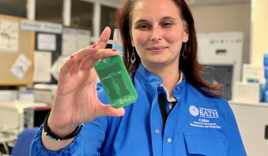
'Lab on a chip' genetic test device can identify viruses within three minutes with top-level accuracy.
Lab on a chip - How LoCKAmp works
Scope to track outbreaks via wastewater
A virus diagnosis device that gives lab-quality results within just three minutes has been invented by engineers at the University of Bath, who describe it as the "world's fastest COVID test."
The prototype LoCKAmp device uses innovative "lab on a chip" technology and has been proven to provide rapid and low-cost detection of COVID-19 from nasal swabs. The research team, based at the University of Bath, say the technology could easily be adapted to detect other pathogens, such as bacteria—or even conditions like cancer.
The device works by rapidly releasing and amplifying genetic material from a nasal swab sample by carrying out a chemical reaction to produce a result, which can be viewed on a smartphone app.
Unlike lateral flow assay tests, commonplace during the pandemic, the LoCKAmp employs the same 'gold standard' genetic-based testing techniques previously reserved for lab-based PCR (polymerase chain reaction) tests, thus enabling rapid testing at laboratory-scale standards for the first time.
As well as its accuracy, the speed of the LoCKAmp sets it apart. With results shown within three minutes, the research team say that to their knowledge, this makes LoCKAmp the fastest COVID-19 test reported to date.
Made with off-the-shelf components and factory-manufactured printed circuit boards, the prototype device could be made on a mass scale quickly and at low cost, presenting care providers and public health bodies around the world with an effective new tool in virus detection.
The research team says a commercial partner with the relevant design and manufacturing expertise could quickly redesigned the LoCKAmp into a small, portable device—with great potential for use in remote health care settings.
The research team is already engaging with academic and commercial partners and would welcome further approaches as it seeks to bring LoCKAmp into production.
The device and how it works is detailed in the research paper LoCKAmp: lab-on-PCB technology for
Read the full article
0 notes
Text
UK hospital AI-enhanced cancer treatment is better for patients
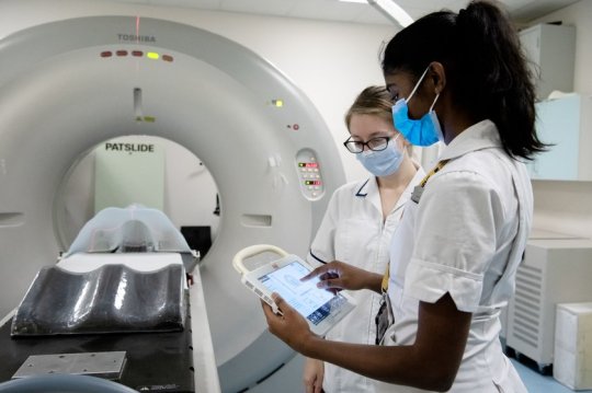
UK hospital AI-enhanced cancer treatment is better for patients.
AI-enhanced cancer treatment - Two-year pilot highlights benefits
Cambridge Cancer Research Hospital
Nov 1, 2023 - Addenbrooke's is the first NHS hospital to use an AI-enhanced superhot needle treatment to pinpoint and destroy life-threatening tumors in one go, including hard-to-reach cancers.
The treatment, known as thermal ablation, is highly-targeted and can treat multiple small tumors with less risk to surrounding healthy tissue. The target area is segmented, that is mapped in advance, using CT images. These images show the clinician where to guide the needles during the procedure.
But gases and blood in the treated area can make this mapping process less accurate meaning the more hard-to-find tumors potentially remain only partially treated. This can lead to local tumors recurrence, often not detected until post-treatment scans. When this happens, a patient may need to go through the process again.
Using AI to train computers to do the mapping makes the treatment quicker, less invasive for the patient, and more accurate. It also means the treatment is carried out in a CT-suite rather than in an operating theatre. Therefore, further scans can be taken during the actual treatment, providing near real-time monitoring which reduces the potential need for further treatment.
Combining the precision of AI and thermal ablation means hard-to-reach or very small tumors can be more easily and effectively treated without the need for repeat treatments. This means we can treat more patients and save and improve more lives. We are really grateful to ACT for making us the first NHS hospital to be able to offer this AI-enhanced treatment.
Dr Nadeem Shaida, Addenbrooke's Consultant Interventional Radiologist
https://youtu.be/cxcQLyV_cjU?si=9YlLC7ZeHNmIezow
AI-enhanced cancer treatment - Two-year pilot highlights benefits
The hospital's charity, Addenbrooke's Charitable Trust (ACT), bought the £250,000 thermal ablation machine following a two-year pilot involving 50 patients with liver cancer. Less than half of those treated during the pilot needed further treatment. The hospital is now looking at how this treatment can be expanded to kidney and other cancer patients.
Patients diagnosed with cancer deserve access to powerful, life-saving treatments. Generous donations from our supporters help us to make such treatments possible, making Addenbrooke's even better.
Shelly Thake, CEO, Addenbrooke's Charitable Trust
Cambridge Cancer Research Hospital
The development comes as Addenbrooke's forges ahead with plans to build the Cambridge Cancer Research Hospital, a world-class facility that will bring NHS staff and scientists together to deliver personalized precision treatments to patients.
Construction work on the new hospital’s site is due to begin in 2024. The hospital is set to be the first delivered in the East of England as part of the Government’s New Hospital Program.
Read the full article
0 notes
Text
Shanghai Unveils Advanced Synchrotron-Based Proton Therapy Facility for Enhanced Cancer Treatment

Shanghai Unveils an Advanced Synchrotron-Based Proton Therapy Facility for Enhanced Cancer Treatment
CHINA, October 29, 2023 - Proton therapy is an advanced form of radiation therapy that uses protons instead of X-rays to treat cancer. Due to its precise nature, it significantly reduces damage to surrounding normal tissue. The Bragg peak of proton beams ensures that there's almost no dose after the peak, protecting essential organs from unnecessary radiation. With over 103 proton therapy centers operational worldwide and more than 280,000 patients treated, this technology is revolutionizing the way cancer is treated.
This study is published in Nuclear Science and Techniques Volume 34, researchers from Shanghai Institute of Applied Physics, Chinese Academy of Sciences and Shanghai Advanced Research Institute, Chinese Academy of Sciences, has been successfully developed and certified by the National Medical Products Administration (NMPA). This cutting-edge proton therapy facility, based on synchrotron technology, is a significant leap forward in the realm of China made high-tech medical apparatus for cancer treatment.
The SAPT facility is located at the Shanghai Ruijin Hospital Proton Therapy Center. The construction of this sophisticated facility encompassed two phases. In the initial phase, four beam lines were developed in three treatment rooms. The second phase saw the introduction of a 360° rotating gantry room.
The facility's accelerator system includes a 7-MeV proton LINAC, a synchrotron, two rotating gantries, and corresponding transport beamlines. Each of the facility’s four treatment rooms is equipped with a beam delivery system, a positioning system, image-guided radiotherapy (IGRT), a treatment control system (TCS), a treatment plan system (TPS), an oncology information system (OIS), and a quality assurance (QA) system. With the pinpoint accuracy of the whole system, treatment is precise, ensuring optimal results with minimal side effects. Following rigorous third-party inspections and tests for medical devices, the SAPT facility underwent a clinical trial with 47 patients.
These patients, aged between 32 and 80, had tumors located in various regions, including the chest, abdomen, spine, and head and neck. Results were promising: the local control rate of the tumor was 100%, and no severe adverse reactions were noted. The SAPT facility demonstrated remarkable reliability, with an availability rate of over 98.5% during the clinical trial period.
With the SAPT facility now officially registered and certified, it will open its doors for patient treatment in July 2023.
This facility is expected to enhance the cancer treatment power in the region, offering a more precise method with less side effects to tackle various forms of tumors. As the need for such advanced treatment grows globally, the successful establishment and operation of the SAPT facility in Shanghai serve as a beacon of hope and an innovation model in the medical world.
Funding information: The work is supported by the major projects for strategic emerging industries of the Shanghai local government and National Key R&D Program of China.
More Information: DOI: 10.1007/s41365-023-01293-1
Read the full article
0 notes
Text
Advanced laser spectroscopy detects falsified vaccines

Advanced laser spectroscopy detects falsified vaccines.
Prevent, detect, respond
Tackling an important problem
New uses for advanced Raman Spectroscopy technology
Promising future applications
An international consortium of multidisciplinary researchers and specialists has developed a new method to counter the problem of COVID-19 vaccine falsification.
There have been numerous instances of vaccine supply chains being infiltrated by falsified products, both for vaccines before the pandemic and for COVID-19 around the globe.
In response to this, the Science and Technology Facilities Council (STFC) Central Laser Facility (CLF) and partners have demonstrated the use of a specialized laser spectroscopy technique to rapidly verify falsified vaccines.
Prevent, detect, respond
Apart from endangering the public, by not effectively protecting people from COVID-19, falsified vaccines also risk undermining trust in vaccines. As such, it is critically important that we maintain integrity of supply chains by detecting falsified products effectively.
To address this global health issue, the World Health Organization (WHO) member states adopted a prevent, detect, and respond strategy.
Although critical, the detection of falsified vaccines currently relies on analysis that can only be performed in specialized laboratories.
Tackling an important problem
In response to the need to develop new methods to counter falsified vaccines, a consortium of world-leading experts convened in 2020, consisting of representatives from:
- University of Oxford's Nuffield Department of Medicine, Department of Biochemistry, Department of Chemistry and Kavli Institute for Nanoscience Discovery
- STFC, part of UK Research and Innovation (UKRI)
- WHO, Geneva
- Agilent Technologies
- Serum Institute of India
- University of Huddersfield
- University of East London
In line with WHO strategy, this multidisciplinary and multi-institutional research consortium has developed a new method of effectively and efficiently detecting falsified vaccines.
Their study demonstrates the viability of the handheld spatially offset Raman spectroscopy (SORS) technique to rapidly authenticate COVID-19 vaccines through unopened vaccine vials. The full study can be read in the journal Vaccine.
New uses for advanced Raman Spectroscopy technology
SORS performs chemical analysis by shining a laser light into an intact vial of the vaccine and inspecting the light emanating from the vial to indicate the presence of different ingredients.
SORS was originally invented and developed into a spin-out company by CLF, which was acquired in 2017 by Agilent Technologies, where the technique continues to be developed for various applications.
SORS devices are currently used to screen for hazardous substances at airports and used widely by fire officers, the military, border protection, and law enforcement. Only minor modifications in sample compartment and software adaption are required for its deployment in the field to tackle falsified vaccines.
The fact that SORS can effectively screen for falsified vaccines without opening the vial is a major advantage in terms of speed of detection and the ability to use vaccines that pass the SORS testing.
"SORS is poised to become an important tool to protect against falsified vaccines. Since first being developed at the STFC Central Laser Facility, this innovative technology continues to find new areas of application which now include empowering governments and health care organizations to safeguard public health. It is a prime example of the enormous benefits of responding to global health challenges with strategic technological innovation."
Professor John Collier, director of STFC CLF, said
"The use of the handheld Resolve's SORS technology for swift and non-intrusive analysis within sealed containers is ground-breaking, attributed to both SORS technology, and the inherent sensitivity of the optical design. This approach not only advances vaccine authentication but also sets the stage for future high sensitivity analysis within sealed containers across diverse fields."
Dr. Rob Stokes, field detection marketing director at Agilent Technologies, said
Promising future applications
Although this study focuses only on COVID-19 vaccines, the method it describes may also be used for authenticating other vaccines, liquid and solid medicines. Further research is needed with more vaccines and to evaluate its effectiveness at various points in supply chains.
With the increasing importance of vaccines for many diseases with pandemic potential and their inequitable distribution, innovative tools to empower inspectors in detecting criminal falsification such as these are a vital asset.
More information: Sara Mosca et al, Innovative method for rapid detection of falsified COVID-19 vaccines through unopened vials using handheld Spatially Offset Raman Spectroscopy (SORS), Vaccine (2023). DOI: 10.1016/j.vaccine.2023.10.012
Journal information: Vaccine
Source: Medical Research Council
Read the full article
0 notes
Last Seen Blogs

bunnwing
Vibing

meilife328
MEILIFE

psychicduelistrbd
Drawing comics and fanart

syntonylife
Sintony.life

winter-swan
winter swan