#nodular swelling
Explore tagged Tumblr posts
Visit Tumblr Blog
Explore Tumblr blogs with no restrictions, modern design and the best experience.
Last Seen Tumblr Blogs


Fun Fact
Kazakhstan’s Minister of Communications and Informatics has blocked the Tumblr site because it contained 60 sites of terrorism, extremism, and pornography in 2015.
Text
Neoplasms of Soft Tissue
Alright, you can break these down by what's fucked up. We'll look at all kinds of them, but we'll get to that in a bit. First, we gotta talk about CELLS and TUMORS in general. Malignant soft tissue tumors are only about 1% of soft tissue tumors. The rest are benign. They are mostly sporadic, but some are linked with gene mutations, radiation, burns, toxins, etc. Basically, cells start dividing abnormally. Cells get fucked up all the time. It's only when our body doesn't kill these cells that they become a problem. Okay, that's enough of that, onto the fun stuff.
Adipocytic
You got lipomas and liposarcomas. Lipomas are benign fat tumors, and are the most common soft tissue tumor in adults. They are well-encapsulated, mature adipocytes that are typically found under the skin of the limbs. On rare occasions, they can be intramuscular and poorly-circumscribed. However, most a soft, mobile, and painless, and can just be cut out by a PCP. Lipomatosis is a condition in which there are a whole lot of lipomas on a limb.
A liposarcoma is malignant. They are the most common sarcomas in adults and are found in the deep soft tissue of the extremities, as well as the retroperitoneum. They most commonly occur in old people. Symptoms include pain, swelling, fatigue, and decreased range of motion. There are three subtypes based on karyotype: well-differentiated, myxoid, and pleomorphic. Pleomorphic ones are aggressive and frequently metastasize.
Fibroblastic
You got nodular fasciitis, fibromatosis (superficial and deep), and fibrosarcoma. Nodular fasciitis is a self-limited proliferation of fibroblasts and myofibroblasts that is seen in young adults. It's mostly going to be found on the forearms, chest, or back. These nodules feel firm, fixed, and may be tender. They usually regress by themselves.
Superficial fibromatoses is a benign growth that is found on the palms, bottom of the feet, or penis. It is a firm, painless thickening made of plump spindle cells and dense collagen. If it is on the penis, it can cause urethral blockage. Recurrence is common, even if you cut these off.
Deep fibromatoses (Desmoid tumors) are large (like about 10 cm in diameter) and infiltrative. They don't metastasize, but they are prone to recurring. They can be painful. They're going to be rubbery and fixed.
Fibrosarcoma are rare, malignant tumors of fibroblasts. They are typically found in the deep soft tissue of the legs and trunk, and are painful, fixed, and soft. A common first sign is unexplained weight loss. On histology, they have a characteristic herringbone pattern and variable collagen. The 5 year survival is ~41%.
Skeletal Muscle
We're talking about rhabdomyosarcoma. These are rare, malignant tumors of mesenchymal origin that differentiate to skeletal muscle. Typically seen in childhood, and present as a quickly growing, painful mass, as well as unexplained weight loss. A pleomorphic subtype is seen in adults. Rhabdomyosarcomas are aggressive, and require excision plus chemotherapy, and maybe radiation therapy. The fatality is based on the subtype, with alveolar types having a 4-year survival of 65% and pleomorphic being fatal.
Unknown
We're only gonna discuss synovial sarcoma and undifferentiated pleomorphic sarcoma. Synovial sarcoma is typically found in the deep soft tissues next to large joints (like the fucking knee). Sometimes, it throws us for a loop and says fuck it lets grow where there's no synovium. Fuck that. Be what you're told to be. Anyway, these mostly they appear in 20-40 year olds, and present with deep-seated pain, numbness, limited motion, and swelling. They are firm and fixed. The swelling and inflammation can be present for several years :D They can metastasize, typically to the lung and lymph nodes.
Undifferentiated pleomorphic sarcoma is a malignant, high-grade tumor of mesenchymal origin. The cells cannot be classified into a category. They typically appear in the deep soft tissues of the extremities of middle-aged adults. If they get big, they can cause numbness or pain. They can grow up to 20 cm, are gray-white fleshy masses, and commonly hemorrhage. Prognosis is poor.
3 notes
·
View notes
Text
Parietal Endometriosis in Abdominal Wall: Report of Eight Cases by Aymen laaliaoui in Journal of Clinical Case Reports Medical Images and Health Sciences
Abstract
Parietal endometriosis is a rare clinical entity, occurring after gynecological, obstetrical or abdominal surgery. Sometimes it is primitive. The etiopathogeny remains unclear.
Material and method: We report eight cases ofabdominal parietal endometriosis occurring in seven cases on laparotomy scars and in one case in a primitive way.
Discussion and Conclusion: Clinical features include swelling and pain that is rhythmic with the menstrual cycle, but this picture is rarely complete. Medical imaging is of little assistance. Only histological examination of thesurgical specimen will confirm the diagnosis. Surgical treatment is based on the complete removal of the lesions must be large enough to avoid any recurrence.
Introduction: External endometriosis is an ectopic localization oftissues whose morphological and functional characteristics are those of the endometrial mucosa. It is found in 10 to 20% of women in genital activity. It occurs in about 0.1% of scars from gynecological-obstetrical procedures.[1] On the other hand, in spontaneous skin localizations, it is 0.5%.[2] Diagnosisis relatively easy in women between 20 and 40 years of age with catamenial symptoms. [2] Abdominal parietalendometriosis has been described in various locations including the abdominal wall (rectus abdominis) and umbilicus,[3-4] caesarean section scars,[5-6] skin and adjacent tissue from abdominal or pelvic surgery scars.[7-9] at the site of amniocentesis needle passage.[4] and at laparoscopic trocar ports.[10] Here we report eight cases of parietal endometriosis observed during the last nine years (between 2001 and 2009) in the Gynecology
Obstetrics Department of the Ibn Rochd Hospital Center in Casablanca. These are young patients (between 34 and 39 years old) with parietal endometriosis on caesarean section scars, apart from one patient who had no previous surgery.
Patients And Methods: During nine years (between 2001 and 2009), we recorded eight cases of endometriosis of the abdominal wall,including seven cases of parietal endometriosis after laparotomy for gynecological or obstetrical pathology and one case of primary endometriosis, in the Gynecology-Obstetrics "C" department of the Ibn Rochd Hospital in Casablanca.
Results: The average age of the patients was 37 years (34 to 39 years). Seven patients were married and multiparous,four of them had delivered by cesarean section, three patients had delivered vaginally. One patient was single and had never given birth. The surgical history was marked by the presence of scarred uterus by Caesarean section in four patients, with a tricicatricial uterus in one patient whose last Caesarean section was performed ten years ago with tubal ligation and a bicicatricial uterus in another patient whose last Caesarean section was performed two years ago. In two other patients, the surgical history was marked by a cystectomy for an ovarian cyst of the nature of a sevencentimeter serous cystadenoma in a nulligest patient and by a myomectomy six years ago in two patients. The reason for consultation was paroxysmal pain with premenstrual recurrence at the scar level, without metrorrhagia, urinary or digestivedisorders in seven patients. The evolution was marked by the appearance of a swelling of the laparotomy scar, which was painful, catamenial with a bluish discoloration and gradually increasing in volume in seven patients (fig. 1). These pains appeared on average five years after laparotomy (one to nine years). In the patient with primary umbilical endometriosis, she consulted for painful and itchy umbilical swelling, a symptom that intensified during menstruation. This swelling had been evolving for nine months and was gradually increasing in volume. Clinical examination revealed a nodular mass in the middle of the Caesarean section scar, bluish in appearance and painful on palpation (Fig. 2). The size of the mass varied between20 and 100 mm, with an average of 40 mm. Clinical examination of the patient with a umbilical endometriosis found a tumour two centimetres in diameter which was located in the center of the umbilicus, mobile with respect to the deep, of firm consistency, slightly sensitive to palpation, without any surrounding skin lesion (Fig. 3). Gynecological examination and pelvic touching are without particularities in all cases.
A pelvic and endovaginal ultrasound scan did not reveal images in favour of pelvic endometriotic localizations. On the other hand, at the level of the laparotomy incision, Ultrasonography showed a hypoechoic, heterogeneous, parietal mass with a finely echogenic content averaging 30 mm (Figure 4). The pelvic CT scan was carried out in three patients, and in all cases it showed the presence, opposite the operative scar, at the expense of the skin tissue, of a hypodense formation, rising at the level of its periphery after injection of contrast agent, with discreet infiltration of the periwound fat. The size of the mass on the scanner was 40 mm. In one patient, the pelvic CT scan showed a tissue lesion process, with an anterior and medial parietal site, infiltrating subcutaneous fat and underlying muscle (Figure 5). The surgical procedure consisted of lumpectomy in all cases (Figures 6, 7 and 8). Surgical exploration did not detect any intra-abdominal processes. One patient had received medical treatment with LH-RH analogues for six months, with the aim of reducing the initial nodule size of 100 mm prior to surgery. This treatment resulted in the regression of half of the mass, measuring 50 by 40 mm. In this patient, surgical resection consisted of a subumbilical parietomy with extensive skin sacrifice, anterior fascial sacrifice and muscle sacrifice in the internal third of the rectus abdominis. The repair of the aponeurotic defect,extended over 10/10cm, was performed by sufficient external oblique flaps over 7 cm in the subumbilical area. This repair is reinforced by a plate on the omentum. The skin defect was closed by a T-shaped abdominal plasty, with externalization of the umbilicus. Follow-up at three and six months noted good abdominal tone (Fig. 9). In addition, surgical exploration did not find intra- abdominal processes in seven patients. An open laparoscopic examination was performed in the patient with umbilical endometriosis after removal of the tumour that had taken over the umbilicus (Fig. 10) through the same umbilical excision port. Exploration of the abdominal pelvic cavity allowed verification of the absence of any primary endometriotic lesion. The surgical procedure ended with suture of the sub umbilical fascia and umbilical plasty with creation of a new umbilicus (Fig. 11). Histological examination of the surgical specimen confirmed the diagnosis of endometriosis in all cases (Fig. 12). Followup of our patients showed no recurrence in seven patients. In one case, two local recurrences on the laparotomy scar had occurred two years apart. The last one was two years ago, when the patient was on progestins and had undergone surgical excision. The postoperative follow-up was simple. With a 14-month to 6-year follow-up, no recurrence was observed, apart from the above-mentioned case.
Discussion: Endometriosis is the ectopic implantation of endometrial tissue. Endometriosis of the abdominal wall is an uncommon pathology and the initial diagnosis is not always easy.[1] It is estimated to account for 0.03 to 2% of extra-genital endometriosis. Only 14.3% to 26% of cases are associated with pelvic endometriosis, in contrast to other atypical implantation sites such as the gastrointestinal tract or the urinary tract.[3] Abdominal parietal endometriosis has been described in various locations including the rectus abdominis.[1,11] the umbilicus. [12] scars from cesarean section, hysterectomy,[3-4] abdominal-pelvic surgery, amniocentesis needle site, laparoscopic trocar ports.[4] Other locations have also been reported, such as the cervix or vulva on episiotomy scars, but never described in the spleen.[4] The presence of isolated endometriosis sites in the umbilicus, rich in lymphatic and venous networks, and in the absence of previous surgery can only be explained by the venous or lymphatic metastatic theory.[1,2] Indeed, it was demonstrated the potential for remote implantation of endometrial glandular cells found in the blood and the migration of contrast material injected into the pelvic cavity towards the umbilicus via umbilical vestigial ducts. Recent work has shown the presence of endometrial tissue in the periumbilical lymphatic system.[4] This entity is rare after menopause.Indeed, at menopause the cytochorionic component regresses but the glandular component may persist. The reactivation of the glandular component under the effect of hormone replacement therapy or in the presence of a secreting adrenal or ovarian tumor (granulosa type) that produces a hyperestrogenic site is known. [1] In the multicenter study of the endometriosis study group,endometriosis of the abdominal wall mainly affects women with genital activity between 20 and 40 years of age.[4] The age of our patients ranges from 34 to 39 years with an average age of 37 years. Elabsi.[1] reports a case of scarring endometriosis in a postmenopausal woman.
Clinically: The clinical manifestation usually corresponds to anodular, inflammatory, persistent infiltration of an abdominal scar. This lesion is painful and catamenial.[4] This infiltrating, nodular, painful and cyclic nature of the lesion was found in all our patients. Frequently, the inflammatory area is associated with serous or serosanguinous discharge, which is exacerbated at the time of menstruation.[4,14] This notion was found in one of our patients. Parietal endometriosis is usually found on Caesarean section scars,[6] which is the case in four of our observations, or scars from gynecological surgery (laparoscopy or laparotomy),[4] as in two of our series. The frequency of endometriotic skin localizations after Caesarean section varies from 0.03 to 0.4%,[6] however, this frequency is much higher than that observed after conventional gynaecological surgery. [4,6] It can occur several weeks or years after surgery.[4] Koger et al.[17] report an interval of 1 to 20 years (mean 4.8 years) between surgery and the onset of symptoms. Zhao. [15] observed a correlation between the latency period and the age of patients at the onset of symptoms. It should be noted that the delay between the causal intervention and the onset of endometriosis is highly variable.[4] It is usually a few months but can sometimes be very delayed, as we observed in one of our observations. In our patients, the delay between the onset of symptoms and the date of obstetric surgery was 5.25 years on average (two to 10 years). The most common modes of disclosure are the discovery of a palpable mass or localized pain in patients with a history of Caesarean section.[4-6] Catameniality, i.e. the exacerbation of these non-specific signs during menstruation, is an important part of the diagnosis.[4] All of our patients were seen for assessment of a parietal mass. This mass was painful with catamenial exacerbation of the symptomatology in all cases, with pain being less predominant in two cases. It should be noted that signs of intra-abdominal pelvic endometriosis were found in only 26% of cases in the literature.[4,9] and none of our patients showed signs of intra-abdominal endometriosis. Thus, the preoperative diagnosis of scarring endometriosis of the abdominal wall can be suspected in the typical form that manifests itself by the presence of a nodule or swelling, purplish blue, painful with brown discharge or hemorrhagic in the period menstrual and catamenial evolution, occurring on a scar most often gynecological.
Ultrasonography plays an important role in the diagnostic orientation and preoperative assessment, even if it does not allow any formal diagnosis. It confirms the typically intramuscular parietal origin of the suspected mass on clinical examination. It also specifies the size, contours and relationships with adjacent structures.[18,21] The ultrasound aspect of parietal endometriosis is variable. Most often, it is a very limited, hypoechoic, tissue- typical mass. However, the lesion may be cystic, mixed or solid.[20-21] In our cases, a very limited hypoechoic parietal nodule was observed in all patients for whom ultrasonography was performed. Endometriomas can measure between 5 mm and 200 mm.[4,9] Most lesions measure less than 40 mm.[21] In our patients, they measured 30 to 40 mm in diameter in seven cases and 70 mm in one case. The color Doppler ultrasound shows an often highly vascularized mass with dilated afferent vessels. [21] Despite the absence of a specific ultrasound aspect of parietal endometriosis, ultrasound associated with the history of the disease should help to suspect the diagnosis of parietal endometriosis and exclude certain differential diagnoses. The differential diagnoses of a parietal mass on ultrasound are granulomas on scarring, post-operative events, benign tumours and exceptionally malignant tumours such as lymphomas and sarcomas. Granulomas may appear hypoechoic. They sit in contact with the scar. They cannot be differentiated from small wall endometriomas outside a catamenial context. In the case of postoperative echogenicity, echogenicity is variable depending on the contents of the sac, but the clinical examination is often evocative.[18,21] The differential diagnosis of spontaneous umbilical endometriosis is made with an irreducible umbilical hernia, pyogenic or foreign body granulomas, hemangioma, umbilical localization of Crohn's disease or melanoma. But it is especially with umbilical metastases of abdominopelvic tumors, or Sister Mary Joseph's nodule, that this diagnosis must be differentiated, especially in women.[2-5]
CT and MRI scans can be used to diagnose parietal endometriosis.[20-21] However, most authors report the absence of characteristic signs in imaging because the aspects observed depend on several parameters: distribution between stromal tissue and glandular elements, hemorrhagic character of the lesion and importance of the peripheral inflammatory reaction.[21] The diagnosis was made preoperatively by CT scan in four of our patients, showing an isodense tissue lesional process, anterior and medial parietal, infiltrating subcutaneous fat and underlying muscles. MRI, more than CT scan, is the examination of choice to confirm the diagnosis in case of doubt because it allows to highlight the iron content of the haemosiderin deposits in endometriomas. Observations of parietal endometrioma on MRI are exceptional. MRI is more sensitive than CT for the detection of small lesions.[20-21] The lesion signal is variable depending on whether the lesion is acute,hypersignal T1 and T2 or chronic, heterogeneous signal, and whether there is intra-lesional hemorrhage.[20] MRI was not performed in any of our patients. Thus, although it may be clinically suspected, parietal endometriosis can only be diagnosed by pathological examination of the lesion. Indeed, this is typical and highlights the existence of endometrial glands of varying sizes, often of cystic type, associated with a cytogenic chorion and lymphocyte inflammation. The ectopic situation of these endometrial glands thus corresponds to the diagnosis of external endometriosis. [4,9] Immunohistochemical techniques steroid receptor assays, using specific monoclonal antibodies, find estradiol and progesterone receptors in both the glandular and stroma, but the distribution is very heterogeneous.[4,22- 25] Recent progress in immunohistochemistry has shown that CD 10 is not expressed in glandular epithelial cells in endometriosis, but rather in the stroma, whereas it is expressed in other epithelial cells.[22] In contrast, COX2, a prostaglandin hydroperoxidase, is expressed in the endometrium with production of PGE2 and PGF2α.[22- 23] The combination of estrogen or progesterone receptor antibodies on the nuclei and CD10 or COX-2 antibodies on the cytoplasm may increase the certainty of diagnosis for ectopic endometriosis.[22-25] The treatment of these lesions is based on surgical excision. Surgical excision should be as wide as possible to remove the entire lesion, as the lesion may recur in the event of incomplete excision. It is the only way to confirm the diagnosis by pathological examination and to achieve healing.[1,4-6,29] Surgical treatment remains the most effective, especially since cancerization of parietal endometriosis has been described. [26-27] Indeed, although medical treatment (LHRH agonists or progestins) can improve the symptomatology by reducing the pain and inflammation of the lesions, it cannot bring about a cure and the lesions quickly recur when these therapies are stopped.[28-29] All our patients were treated by surgical excision. One patient had two recurrences for which she had undergone resections. Two patients were treated with LHRH agonists, one preoperatively in front of a 70 mm endometrioma and the second after surgical removal to prevent recurrence.
The surgical procedure can be disruptive, and parietal reconstruction often requires the use of devices such as nonabsorbable thread mesh to reinforce the aponeurotic scars.[6,29] In one of our patients, the repair of the fascial defect, which had spread over 10/10 cm, was performed with sufficient external oblique flaps over 7 cm subumbilically. This repair is reinforced by a plate on the omentum. Prevention may be proposed in patients with pelvic endometriosis lesions, without any evidence of efficacy. It consists of protecting the wall with surgical drapes during Caesarean section, irrigation or pressure saline cleansing of the wall at the end of the Caesarean section.[4,6,29] During the closure of a hysterotomy, it is necessary to ensure the quality of the closure and to put back in place any endometrial invagination, all the more so as the Caesarean section is performed early in the pregnancy.[4,6]
Conclusion: Parietal endometriosis is an infrequent and often unrecognized condition. Scarring endometriosis must be mentioned in front of any mass sitting on the scar from a gynecological-obstetrical operation. The diagnosis should be made in the presence of pain or a mass in the abdominal wall of a woman during genital activity, especially if this lesion presents catamenial changes and if the patient has a history of gynecological or obstetrical surgery. Color Doppler ultrasound is the morphological examination of choice to confirm the diagnosis and rule out other parietal pathologies by showing a hypervascularized hypoechoic mass. Due to the wide diffusion of CT scans, it is important for radiologists to be aware that these lesions appear as tissue nodules in the vicinity of a scar from obstetric or gynecological surgery. In case of diagnostic doubt before surgery, MRI has a definite place to detect the particular signal of hemorrhage in the endometrioma and confirm the diagnosis. However, the diagnosis is only confirmed by histological study. Healing is achieved by complete excision of the mass.
#Parietal Endometriosis#JCRMHS#Abdominal Wall#Report of Eight Cases#Journal of Clinical Case Reports Medical Images and Health Sciences#Journal of Clinical Case Reports Medical Images and Health Sciences (JCRMHS)| ISSN: 2832-1286#Clinical Images journal
2 notes
·
View notes
Text
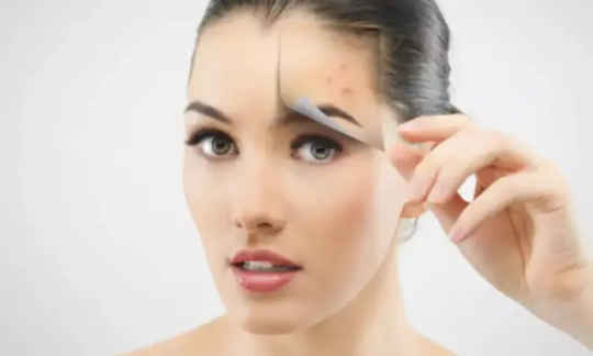
6 Cosmetic Issues a Facial Can Treat
Facials are often seen as a relaxing part of a spa day experience, but the cosmetic treatments actually do a lot of “heavy lifting” in terms of addressing specific cosmetic concerns. When it comes to facials, Albuquerque’s Spa @ Western Dermatology Consultants explains that the treatments can be tailored to the patient’s desires, with options ranging from “basic” to “extensive.”
While something like microneedling or a chemical peel might provide more dramatic results, these options do require more time for healing. Facials can deliver a beautiful effect with little to no down time.
A facial experience can include any of a number of steps, including steaming for opening pores and improving circulation, extraction of materials from out of the pores, masks for hydration and more, massages do enhance circulation, and even exfoliation to remove dead skin and debris.
Here are six cosmetic issues that facials are ideal for treating.
Dullness
The skin is constantly shedding dead cells in an ongoing turnover process, but these cells don’t always flake away on their own. If too many dead cells accumulate, they can give the face an overall dull appearance. This is because the dead cells are dry, lacking the natural hydration that makes living skin cells look vibrant and—yes—alive! Removing these cells via gentle exfoliation not only reveals the healthy and more radiant cells beneath, but also encourages circulation to provide a beautiful glow.
Clogged Pores
Pores are openings in the skin, each leading to a follicle that grows a hair. Usually these pores stay open, but they can become clogged by debris that remains on the surface. If dead cells mix with natural facial oils—made naturally by the skin, which can ���up” production in times of hormone shifts, such as puberty or menopause—the resulting substance can plug pores. A plugged pore can be more visible, as well as lead to more significant problems if certain bacteria are trapped within the follicle. Facials can help to remove the oils and dead skin to minimize future plugged pores, as well as open pores that are already gummed up.
Acne
If p. acnes bacteria become trapped in a plugged follicle, they can begin to feed on the natural material trapped with them, causing them to multiply. The body often sends specialized cells to fight the invaders, resulting in the creation of pus, which is made up of dead white blood cells, bacteria, and other material. The ongoing creation of this pus can eventually cause the follicle to swell, leading to a visible bump on the surface. This is known as a papule or pustule, but most people call it a pimple or zit. Keeping pores open and extracting the material inside minimizes ongoing acne breakouts and reduces the chances of future flares.
Acne Scars
Sometimes, follicles filled with acne-causing bacteria can become damaged or so full that they rupture. This results in the contents spilling into the surrounding tissue and forming a cyst or a nodule. These forms of acne extend deeper beneath the surface, causing harm that the skin can’t always fully repair. Cystic and nodular acne are known for leaving scars, which can be reminders of a skin problem best forgotten with time. Reducing surface dryness with an exfoliating facial can cause acne scars to be less noticeable, as they may appear shallower. More dramatic facials can also prompt skin regeneration that involves the creation of new collagen to promote a more even surface.
Cosmetic Sun Damage
Too much exposure to ultraviolet radiation can dry out the skin, as well as cause uneven texture, redness, and more. A soothing facial is ideal for calming irritation, as well as providing a smoother texture and more consistent tone. If cosmetic sun damage is of particular concern, talk to a facial provider about specific customizable options in order to address the various issues UV exposure can cause.
Wrinkles and Lines
Like many imperfections, wrinkles and fine lines both appear more pronounced in skin that is dry. Poorly hydrated skin or skin hidden under a layer of dead cells is prone to being thin, inelastic, and not as plump—all characteristics that magnify the look of facial wrinkles. Plumping the skin with a hydrating facial can make wrinkles a less-noticeable issue.
Ready to find out more about facials for improving the skin? Contact the team at Western Dermatology Consultants by messaging them online or calling the Spa @ WDC at (505) 855-9267.
0 notes
Text
Liver Cirrhosis: Symptoms, Causes, and Treatment Options

Introduction
The liver is one of the most vital organs in the human body, performing essential functions such as detoxification, protein synthesis, bile production, and nutrient storage. When the liver becomes severely scarred due to chronic damage, it leads to a condition called cirrhosis. This irreversible scarring disrupts liver function and can lead to life-threatening complications if not managed properly.
In Zambia, liver cirrhosis is a growing health concern, often linked to alcohol abuse, viral hepatitis, and fatty liver disease. Early detection and proper management can slow disease progression and improve quality of life. This article explores the symptoms, causes, diagnosis, and treatment options for liver cirrhosis, with a focus on prevention and care in Zambia.
What Is Liver Cirrhosis?
Cirrhosis occurs when healthy liver tissue is replaced by fibrous scar tissue, impairing the organ’s ability to function. The liver becomes hard and nodular, leading to portal hypertension (increased blood pressure in the liver’s blood vessels) and liver failure in advanced stages.
Stages of Cirrhosis
Compensated Cirrhosis – The liver is damaged but still functioning; symptoms may be mild or absent.
Decompensated Cirrhosis – Severe scarring leads to complications like jaundice, fluid retention, and internal bleeding.
Common Causes of Liver Cirrhosis in Zambia
1. Excessive Alcohol Consumption
Chronic alcohol abuse is a leading cause of cirrhosis. Alcohol metabolizes into toxic substances that inflame and destroy liver cells, leading to scarring over time. Even moderate drinking over many years can contribute to liver damage.
2. Viral Hepatitis (B and C)
Hepatitis B and Hepatitis C are major causes of cirrhosis in Zambia.
These viruses spread through contaminated blood, unsafe medical practices, unprotected sex, and mother-to-child transmission.
Without treatment, chronic hepatitis leads to liver inflammation, fibrosis, and cirrhosis.
3. Non-Alcoholic Fatty Liver Disease (NAFLD) & NASH
Linked to obesity, diabetes, and high cholesterol, NAFLD causes fat buildup in the liver.
In some cases, it progresses to NASH (Non-Alcoholic Steatohepatitis), leading to inflammation and cirrhosis.
4. Other Causes
Autoimmune liver diseases (Primary Biliary Cholangitis, Autoimmune Hepatitis)
Genetic disorders (Hemochromatosis, Wilson’s Disease)
Bile duct disorders (Primary Sclerosing Cholangitis)
Long-term medication use (e.g., excessive paracetamol)
Symptoms of Liver Cirrhosis
Early-stage cirrhosis often has no noticeable symptoms. However, as the disease progresses, the following signs may appear:
Early Symptoms
Fatigue and weakness
Loss of appetite
Nausea and weight loss
Mild abdominal discomfort
Advanced Symptoms
Jaundice (yellowing of skin and eyes)
Swelling in legs (edema) and abdomen (ascites)
Easy bruising and bleeding (due to reduced clotting factors)
Itchy skin (from bile buildup)
Confusion and memory problems (hepatic encephalopathy)
Spider-like blood vessels on the skin (spider angiomas)
Diagnosis of Liver Cirrhosis
Early detection is crucial to prevent further liver damage. Doctors use several diagnostic methods:
1. Blood Tests
Liver Function Tests (LFTs) – Check enzyme levels (ALT, AST), bilirubin, and albumin.
Complete Blood Count (CBC) – Low platelets may indicate cirrhosis.
Viral Hepatitis Screening – Detects Hepatitis B and C.
2. Imaging Tests
Ultrasound – Detects liver texture changes.
FibroScan (Elastography) – Measures liver stiffness (fibrosis).
CT Scan or MRI – Provides detailed liver images.
3. Liver Biopsy
A small liver tissue sample is examined to confirm cirrhosis and assess damage severity.
Treatment and Management of Cirrhosis
While cirrhosis is irreversible, treatment focuses on slowing progression and managing complications.
1. Lifestyle Changes
Stop alcohol completely – Even small amounts worsen liver damage.
Healthy diet – Low salt, high protein, and nutrient-rich foods.
Weight management – Crucial for fatty liver-related cirrhosis.
2. Medications
Antiviral drugs (for Hepatitis B & C)
Diuretics (reduce fluid retention)
Lactulose (prevents brain fog from toxins)
3. Managing Complications
Ascites (fluid buildup) – Low-sodium diet, diuretics, or drainage.
Variceal bleeding – Beta-blockers or endoscopic treatment.
Hepatic encephalopathy – Medications to reduce ammonia levels.
4. Liver Transplant
In end-stage cirrhosis, a liver transplant may be the only cure. However, availability in Zambia is limited, emphasizing the need for early intervention.
Prevention Tips for Liver Cirrhosis
Limit Alcohol – Avoid excessive drinking to prevent liver damage.
Get Vaccinated – Hepatitis B vaccine is highly effective.
Practice Safe Sex & Hygiene – Reduces Hepatitis B & C risk.
Control Diabetes & Obesity – Prevents fatty liver disease.
Avoid Unnecessary Medications – Some drugs harm the liver.
Regular Health Checkups – Early detection of liver issues.
Conclusion
Liver cirrhosis is a serious but preventable and manageable condition. In Zambia, raising awareness about alcohol abuse, viral hepatitis, and metabolic diseases is key to reducing cirrhosis cases. Early diagnosis, lifestyle changes, and proper medical care can slow disease progression and improve survival rates.
If you or a loved one experiences symptoms like fatigue, jaundice, or abdominal swelling, consult a doctor immediately. A healthy liver means a healthier life!
#Hepatic Cirrhosis#Cirrhosis of the liver#Liver cirrhosis#liver cirrhosis symptoms#Liver cirrhosis treatment#Fatty Liver Disease#Hepatitis C#liver disease
0 notes
Text
Hypothyroidism Pathophysiology of the disease development The thyroid, a gland located in the front of the neck, plays a critical role in the body's endocrine system, specifically in regards to cellular metabolism-i.e. how cells use energy (NLM, 2015). In a normal, healthy person the thyroid is in a state of homeostasis with the rest of the body; however, in a number of cases the body can produce too much (hyperthyroidism) or too little (hypothyroidism) of thyroid hormone needed to keep the body in a state of health. Hypothyroidism can have a number of causes, many of which are poorly understood. The immune system may attack the thyroid gland, mistaking it for a foreign invader. According to the American Thyroid Association (ATA): "autoimmune thyroiditis can begin suddenly or it can develop slowly over years. The most common forms are Hashimoto's thyroiditis and atrophic thyroiditis" (ATA, 2014). A variety of other conditions, including pregnancy and certain kinds of viruses can cause swelling and inflammation. Certain medications, radiation treatment, surgical removal of the thyroid gland, and even pregnancy (postpartum thyroiditis) can cause hypothyroidism (NLM, 2015). Surgical removal or radiation may be necessitated by cancer or due to hyperthyroidism. "Some people with graves' disease, nodular goiter, or thyroid cancer are treated with radioactive iodine (i-131) for the purpose of destroying their thyroid gland" (ATA, 2014). Unexplained weight gain is often the first and most common symptom which causes individuals with an underactive thyroid to go to the doctor. Constipation, a feeling of sluggishness, and, as symptoms advance, puffy hands and feet and brittle hair and nails may manifest themselves. Some patients may show no symptoms, however. Diagnosis is confirmed with a TSH (thyroid-stimulating hormone) test, which "measures how much of the thyroid hormone thyroxine (T4) the thyroid gland is being asked to make. An abnormally high TSH means hypothyroidism: the thyroid gland is being asked to make more T4 because there isn't enough T4 in the blood" ATA, 2014). Treatment The first and foremost objective of treatment is to restore thyroid levels to normal in a hypothyroid patient. This is almost always accomplished with medication. "T4 replacement can restore your body's thyroid hormone levels and your body's function. Synthetic thyroxine pills contain hormone exactly like the T4 that the thyroid gland itself makes" (ATA, 2014). Dosages must be carefully monitored -- any changes in dosing should be verified between 6 to 10 weeks. Too much synthetic hormone can case the hypothyroid patient to develop hyperthyroidism, which poses its own risks and symptoms (such as weight loss, sweating, high blood pressure). Patients should be careful of ingesting over-the-counter medications such as calcium supplements and iron pills which may interfere with the effects of the drug (Mayo clinic, 2015). Natural remedies are also available for patients that are "derived from the thyroid glands of pigs" (Mayo Clinic, 2015). These products are available via prescription and should not be confused with extracts claiming to be thyroid treatments available at natural health food stores, which are not regulated by the FDA. In general, attempting to self-medicate for thyroid conditions can be extremely dangerous: although iodine levels, for example, can affect thyroid levels, patients should not attempt to adjust the level of iodine in their diet without consulting a physician. Similarly, while eating a healthy diet, exercising, and adopting regular sleep habits is important, this is not a substitute for a physician's care regarding treatment of hypothyroidism. Reference Hypothyroidism. (2014). American Thyroid Association (ATA). Retrieved from: http://www.thyroid.org/wp-content/uploads/patients/brochures/ata-hypothyroidism-brochure.pdf Hypothyroidism. (2015). Mayo Clinic. Retrieved from: http://www.mayoclinic.org/diseases-conditions/hypothyroidism/basics/alternative-medicine/con-20021179 Hypothyroidism. (2015). National Library of Medicine (NLM). Retrieved from: https://www.nlm.nih.gov/medlineplus/ency/article/000353.htm Read the full article
0 notes
Text
Best Homeopathic Drops for Cysts
Cysts are fluid-filled sacs that can develop in various parts of the body, including the skin, ovaries, and internal organs. homeopathic drops for cysts, reduce inflammation, and prevent recurrence.
1. Tumorin Drop (Doctor Bhargava) (Best for Cysts & Tumors)
Helps reduce benign tumors and cystic formations.
Supports immune system function and detoxification.
Check Tumorin Drop Here.
2. Thuja Occidentalis (Best for Skin & Sebaceous Cysts)
Helps shrink skin cysts, warts, and nodular growths.
Useful for sebaceous cysts and lipomas.
3. Calcarea Fluorica (Best for Hard & Fibroid Cysts)
Effective for hard, glandular, and fibroid cysts.
Helps dissolve ovarian and breast cysts.
4. Apis Mellifica (Best for Fluid-Filled & Painful Cysts)
Useful for cysts with inflammation and burning pain.
Reduces swelling and fluid retention.
5. Silicea (Best for Draining & Abscess Cysts)
Helps in expelling pus from cysts and abscesses.
Supports healing of long-standing cystic infections.
6. Conium Maculatum (Best for Glandular & Breast Cysts)
Works well for breast lumps, fibroids, and nodules.
Helps in slow-growing glandular swellings.
How to Use Homeopathic Drops for Cysts?
Tumorin Drop: Take 10-15 drops in water twice daily or as prescribed.
Other remedies should be used under the guidance of a homeopathic practitioner.
Combine with a healthy diet and detoxification practices for better results.
Final Thoughts
Homeopathy provides a gentle yet effective approach to managing cysts without side effects. Doctor Bhargava’s Tumorin Drop and other homeopathic medicines can support natural cyst reduction and healing.
👉 Try Tumorin Drop for a homeopathic approach to cyst management.
Would you like recommendations for a specific type of cyst?
0 notes
Text
How Prolonged Achilles Tendinitis Causes Chronic Pain: Causes, Symptoms, and Treatment Options

Introduction Achilles tendinitis is a common condition affecting athletes, runners, and individuals with active lifestyles. While it often resolves with rest and treatment, prolonged Achilles tendinitis can lead to chronic pain, significantly affecting quality of life. In this article, we explore how prolonged Achilles tendinitis develops into chronic pain, the underlying mechanisms, symptoms, and effective treatment options to manage and prevent it.
What is Achilles Tendinitis? Achilles tendinitis refers to inflammation of the Achilles tendon, the largest tendon in the body, connecting the calf muscles to the heel bone. The condition is often caused by repetitive stress, overuse, or sudden increases in physical activity.
When untreated or poorly managed, acute tendinitis can progress to a chronic state, resulting in long-term pain and functional limitations.
How Prolonged Achilles Tendinitis Leads to Chronic Pain
Degenerative Tendon Changes (Tendinosis) Chronic tendinitis causes the tendon fibers to weaken due to repeated microtears and incomplete healing. Over time, this leads to:
Collagen disorganization
Reduced tendon elasticity and strength
Formation of scar tissue
These degenerative changes, known as tendinosis, result in stiffness and chronic pain.
Neovascularization and Nerve Ingrowth Prolonged inflammation can stimulate the formation of new, abnormal blood vessels in the tendon (neovascularization). These blood vessels often bring new pain-sensing nerve fibers, increasing sensitivity and contributing to chronic pain, even with minimal activity.
Persistent Inflammation and Swelling Continued stress on the Achilles tendon can cause low-grade, unresolved inflammation. This inflammatory state sensitizes surrounding tissues, perpetuating the pain cycle.
Calcification and Bone Spurs Prolonged Achilles tendinitis may result in calcification of the tendon or the development of bone spurs where the tendon attaches to the heel bone. These structural changes can irritate the tendon and nearby tissues, causing pain and limiting mobility.
Symptoms of Chronic Achilles Tendinitis Chronic Achilles tendinitis is characterized by:
Persistent pain along the back of the heel or lower calf
Morning stiffness in the tendon
Swelling and tenderness
Thickened or nodular tendon
Pain that worsens during activity and improves with rest
Difficulty walking or running
Risk Factors for Chronic Achilles Tendinitis Certain factors increase the likelihood of Achilles tendinitis progressing to chronic pain, including:
Overuse without adequate rest
Poor biomechanics (e.g., flat feet, tight calf muscles)
Wearing improper footwear
Lack of warm-up or stretching before exercise
Age-related tendon degeneration
Treatment Options for Chronic Achilles Tendinitis
Conservative Management
Rest and Activity Modification: Reduce high-impact activities to allow the tendon to heal.
Physical Therapy: Eccentric exercises can strengthen the tendon and improve flexibility.
Orthotics and Footwear: Supportive shoes or custom orthotics can correct biomechanical issues.
Anti-inflammatory Medications: NSAIDs may help reduce pain and inflammation.
Advanced Treatments
Extracorporeal Shockwave Therapy (ESWT): Stimulates healing by promoting blood flow and collagen production.
Platelet-Rich Plasma (PRP) Injections: Enhance tissue repair by delivering growth factors directly to the damaged area.
Corticosteroid Injections: Reduce inflammation, though their use must be limited to avoid further tendon damage.
Surgical Intervention In severe cases, surgery may be necessary to remove damaged tissue, repair tears, or address bone spurs. Post-surgical rehabilitation is crucial for optimal recovery.
Prevention of Chronic Achilles Tendinitis To reduce the risk of chronic Achilles tendinitis:
Gradually increase activity levels
Incorporate stretching and strengthening exercises into your routine
Wear proper footwear with adequate arch support
Address any biomechanical issues with a healthcare professional
Prioritize rest and recovery after intense physical activity
#health#medicine#pain management#mental health#back pain#apdss#chiropractic#neckpain#neurostar#depressionhelp
0 notes
Text
All About Acne by The Best Skin Doctor in Indore Dr. Atul Kathed
Introduction:
Millions of individuals worldwide suffer from acne, a common skin ailment that may be emotionally and physically distressing. Its causes, varieties, and therapies must be understood in order to manage it effectively. The Best Skin Doctor in Indore, Dr. Atul Kathed, offers insightful commentary and professional acne treatment guidance.
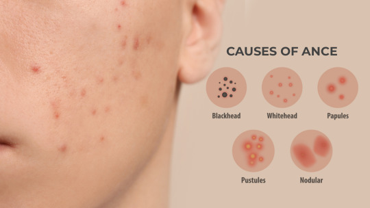
Understanding Acne by The Best Skin Doctor in Indore:
According to Dr Atul Kathed the Best Dermatology in Indore, a skin ailment known as acne arises from the accumulation of oil and dead skin cells in hair follicles. It usually appears on the face, forehead, chest, upper back, and shoulders and frequently takes the form of whiteheads, blackheads, pimples, and cysts. The main causes of acne are inflammation, germs, clogged hair follicles, and excessive oil production.
Types of Acne:
Dr. Atul Kathed, renowned as the best skin doctor in Indore, explains that acne can be classified into different types based on its severity and appearance:
Comedonal Acne: Comedone acne is characterized by blackheads and whiteheads and is brought on by clogged pores.
Inflammatory Acne: This kind contains red, swelling pustules and papules that are swelled as a result of inflammation.
Nodular Acne: Large, painful lumps under the skin’s surface called nodules frequently need to be treated by a professional.
Cystic Acne: The most severe kind, known as cystic acne, is characterized by painful, deep cysts that are packed with pus and may leave scars.

Causes and Triggers:
Acne can be brought on by or made worse by several things. Breakouts can result from hormonal fluctuations brought on by puberty, menstruation, pregnancy, and stress. Moreover, the use of comedogenic skincare products, a diet heavy in dairy and refined carbohydrates, and several medications can all exacerbate acne..
Treatment Options:
As the best skin doctor in Indore, Dr. Atul Kathed emphasizes a tailored approach to acne treatment, considering the severity and type of acne. Common treatments include:
Topical Treatments: Acne can be lessened with over-the-counter or prescription creams, gels, and lotions that contain retinoids, salicylic acid, or benzoyl peroxide.
Oral Medications: For moderate to severe cases, a doctor may prescribe antibiotics, hormonal therapies, or isotretinoin..
Procedures: To properly treat obstinate acne, professionals might use procedures including chemical peels, laser therapy, drainage, and extraction.
Skincare Tips for Acne-Prone Skin by The Best Skin Doctor in Indore:
Dr. Atul Kathed recommends the following skincare tips for managing acne:
Gentle Cleansing: Use a mild, non-comedogenic cleanser twice daily to remove excess oil and impurities.
Moisturizing: For hydrated skin that doesn’t clog pores, use moisturizers that are non-comedogenic and oil-free.
Sun Protection: Every day, use a broad-spectrum sunscreen to avoid hyperpigmentation and sun damage.
Avoid Picking: Refrain from picking or squeezing pimples to minimize the risk of scarring.
To Consult Best Dermatology in Indore Click Here
Conclusion:
Although acne can be difficult to treat, clear skin is possible with the correct advice and care. The Best Skin Doctor in Indore, Dr. Atul Kathed, provides knowledgeable counsel and individualized treatment regimens to assist people in successfully treating acne. Gaining knowledge about the origins, varieties, and remedies of acne will enable you to take proactive measures for improved, more radiant skin.
For more personalized advice and professional acne treatment, consult Dr. Atul Kathed, the best skin doctor in Indore, and embark on your journey to radiant skin
0 notes
Text
#Thyroid Disease
#Autoimmune Disease
There are several types of thyroid diseases, each impacting the production of vital hormones that lead to hypo- and hyperthyroidism. Here are some common thyroid-related conditions:
Hyperthyroidism: The thyroid produces hormones in excess, causing many body functions to speed up. It’s often associated with Graves’ disease or toxic nodular goiter.
Symptoms include restlessness, nervousness, rapid heart rate, weight loss, and bulging eyes (in Graves’ disease).
Diagnosis involves blood tests measuring thyroid hormone levels.
Treatment options include medications, radioactive iodine, or surgery.
Hypothyroidism: The thyroid does not produce enough hormones, leading to symptoms like fatigue, weight gain, and dry skin.
The most common cause is Hashimoto’s thyroiditis, an autoimmune disorder.
Treatment involves thyroid hormone replacement therapy.
Hashimoto’s Thyroiditis: An autoimmune disorder causing inflammation of the thyroid gland, leading to hypothyroidism and swelling (goiter).
Graves’ Disease: Another autoimmune disorder resulting in hyperthyroidism.
Thyroid Tumors: Noncancerous growths (nodules or adenomas) that can release excess thyroid hormones.
Thyroid Cancer: Malignant tumors affecting the thyroid.
Postpartum Thyroiditis: Thyroid inflammation following childbirth.
Remember that some thyroid diseases can be cured, while others require ongoing management. An estimated 20 million people in the United States have thyroid disease, and hypothyroidism is more common than hyperthyroidism.
For individuals with thyroid conditions, including hypothyroidism and Hashimoto’s thyroiditis, there are several reasons why certain foods are recommended to be limited or avoided. Let’s explore each of these:
Dairy:
Sensitivity to Dairy: People with Hashimoto’s thyroiditis tend to have a greater sensitivity to certain proteins found in dairy products. Additionally, a higher incidence of lactose intolerance is observed in those with Hashimoto’s thyroiditis.
Inflammatory Response: Consuming dairy may trigger an inflammatory response in the body, leading to the production of inflammatory chemicals. Chronic inflammation can negatively impact energy levels, mental health, and physical health.
Processed Dairy: Most forms of dairy available today are highly processed, containing added preservatives and hormones that can disrupt the digestive system and trigger inflammation.
Gluten:
Autoimmune Connection: Both hypothyroidism and Hashimoto’s thyroiditis have an autoimmune component. Gluten, found in wheat, barley, and rye, can exacerbate autoimmune responses.
Cross-Reactivity: Some proteins in gluten can cross-react with thyroid tissue, potentially worsening inflammation.
Leaky Gut: Gluten consumption may contribute to leaky gut syndrome, which can affect nutrient absorption and overall health.
Soy:
Goitrogens: Soy contains compounds called goitrogens, which can interfere with thyroid function by inhibiting iodine uptake. This can affect thyroid hormone production.
Estrogen-Like Compounds: Soy contains phytoestrogens that can mimic estrogen in the body. Excessive estrogen levels may impact thyroid health.
Genistein: Genistein, found in soy, can inhibit thyroid peroxidase, an enzyme involved in thyroid hormone synthesis.
Wheat:
Gluten and Autoimmunity: Wheat contains gluten, which can contribute to autoimmune responses. In some cases, gluten may worsen inflammation and thyroid dysfunction.
Gut Health: Wheat consumption may affect gut health, leading to leaky gut and impaired nutrient absorption.
Important Note:
Not all dairy products are problematic. Raw and fermented dairy can have redeeming nutritional qualities.
If you suspect sensitivity to any of these foods, consider lab tests or try eliminating them to observe how you feel.
Consult a healthcare provider or a dietitian for personalized advice based on your specific condition and need
0 notes
Text
How to Manage Acne with Acutret 10 MG ?
Acutret 10 MG Capsule is a medicine that contains isotretinoin. It is a naturally occurring derivative of vitamin A. This medicine is used for the treatment of a severe form of acne called nodular acne characterized by large, inflamed, painful acne lesions deep under the skin. It acts on skin glands to reduce the production of a substance called sebum that can cause acne or pimples. It also has an anti-inflammatory action which decreases the redness and swelling associated with acne.
Acutret 10 MG Capsule has some common side effects like cracked lips, dry eye, dry nose, dry skin and dry mouth. Consult your doctor if any of these side effects persist or bother you. Do not take this medicine if you are allergic to it. Acutret 10 MG Capsule should be taken with food.
Take this as prescribed by your doctor. Complete the whole treatment, even if you feel better after taking a few doses. Use skin protective methods like wearing sunscreen and having a balanced diet along with Acutret 10 MG Capsule for the Acne treatment. Acutret 10 MG Capsule is not recommended to be used in pregnancy and breastfeeding.
Inform your doctor if you have liver problems before taking this medicine. It is not recommended for use in children under 12 years of age. Acutret 10 MG Capsule may cause skin sensitivity to sunlight so it is advised that you avoid going out in harsh sunlight or take precautions like sunscreen.
You should avoid alcohol consumption while taking this medicine as it may cause severe side effects. Inform your doctor immediately if you experience and serious side effects like changes in mood or depression, dizziness, ringing in ears, headache, problems with vision, pain behind the eyes, increased thirst and urination while using this medicine.
When not to use?
Allergy
Avoid using Acutret 10 MG Capsule if you are allergic to isotretinoin or any other component present in this medicine. Seek medical help immediately if you notice any symptoms such as skin rashes, itching/swelling, Dizziness, breathing difficulty, etc.
Hypervitaminosis A
Hypervitaminosis A is a condition that occurs when you have too much vitamin A in your body. Symptoms of hypervitaminosis A include vision problems, changes in the skin, and bone pain. In such cases Acutret 10 MG Capsule is not recommended for use as it is also a derivative of vitamin A and it may worsen your condition.
Severe liver disease
Acutret 10 MG Capsule is not recommended for use if you have severe liver conditions as it may worsen your condition.
Dosage
Missed Dose
Do not skip a dose of Acutret 10 MG Capsule. If you forget to take a dose of this medicine, take it as soon as you remember. If it is time for your next dose, skip the missed one. Do not double the dose to compensate for the missed one.
Overdose
If you have taken more than the prescribed dose of Acutret 10 MG Capsule you may experience symptoms like vomiting, flushing, severely chapped lips, stomach pain, headache and dizziness. Consult with your doctor immediately in case of an overdose.
General Instructions
Take Acutret 10 MG Capsule as instructed by your doctor. Follow all the directions mentioned on the label. Do not take more or fewer doses than prescribed.
This medicine should be taken with food. The symptoms might get worse in the initial phase of treatment. It is advisable to avoid sunlight during the course of treatment. Take plenty of water and have a clean nutritious diet. Keep away from the reach of children and pets. Ensure that unused medicine is disposed of properly.
0 notes
Text
Skin abscesses, also referred to as cutaneous or epidermal abscesses, are localized accumulations of pus that form under the skin as a result of bacterial infections. An abscess initially starts as a reddened, tender swelling that progressively fills... #Mirari #MirariDoctor #MirariColdPlasma #ColdPlasma
0 notes
Text
Why Is Breast Self Examination So Important -Gibmed International Hospital

How should you check your breasts for signs of cancer?
It is widely recognised that when those who may be at risk of breast cancer take the time to examine their breasts, this can help them to identify any potentially concerning changes, which they can then report to a medical professional.
If you are reading this article, you are likely to appreciate how crucial breast self-examination can be for detecting any possibly worrying symptoms at the earliest possible stage.
It is important not to panic if you do think you feel a lump in your breast, as some women do have some lumps or lumpy areas in their breasts, and the majority of breast lumps turn out to be benign (non-cancerous). Examples of non-cancerous conditions in the breasts, which account for the majority of women visiting a breast clinic, include (but are not limited to) breast cysts, cyclical nodularity (fibrocystic disease), and benign nipple discharge.
Nonetheless, with breast cancer being the most common malignant disease among women, it is important to be alert to any potentially abnormal changes in the breasts. If such changes do turn out to be cancer, detecting them as early as possible will maximise the likelihood of the condition being treatable with good results.
So, with all the above in mind, here are some good tips to follow to ensure you carry out a thorough and effective breast self-examination.
Get accustomed to how your breasts normally look
Before we go any further, we should probably revisit the way we have phrased the title of this article. There is no “should” when it comes to checking one’s own breasts, as there is no right or wrong way to do it. However, if there is one thing that you might be especially well-advised to be mindful of, it is the importance of being aware of how your breasts normally look and feel.
One way in which you might ensure this, is by regularly looking at your breasts in the mirror, with your shoulders straight and your arms on your hips. Indeed, it can be a good idea to begin by looking at yourself in the mirror with your arms down at your sides, and then with your arms up in the air.
Doing this routinely will enable you to maximise your awareness of such things as the normal size, shape, and colour of your breasts, and ensuring they are evenly shaped, with no visible swelling or distortion.
Feel around your breasts for signs of anything unusual
As we referenced above, positioning your body in different ways can aid your efforts to spot anything potentially abnormal about your breasts, and you can further help this process by not only looking, but feeling.
For this reason, many women check their breasts for lumps or anything else unusual from a lying-down position, as well as while standing or sitting. Women have often found that they can feel their breasts more easily when their skin is wet and slippery, so you might try doing your standing-up checks in the shower.
As for how you might do those checks, you may begin by feeling around each breast in a circular motion, before feeling under your arm, and then around the nipple.
Educate yourself on the symptoms that might require medical attention
Of course, even knowing the above processes well might be of limited use if you have little sense of what would constitute a potentially concerning symptom (other than the aforementioned lumps).
However, there are various breast changes that, if you notice them, would make it a good idea to reach out to a medical professional. These include any unexpected changes in the size, outline, or shape of your breast, as well as any rash, redness, puckering or dimpling on the skin of the breast.
A discharge of fluid from either of the nipples, any change in nipple position, and/or a new lump, swelling, thickening, or bumpy area in one breast or armpit that you do not recall coming across previously, could give you further reason to contact a medical expert as soon as possible.
Seeking help in this way will enable you to rule out breast cancer as a possible cause of the concerning symptoms – and if cancer is detected, it will allow for quick and suitable treatment to be provided.
To learn more about the breast screening in Gibraltar that we can provide here at GibMedInternational in support of your breast health, please don’t hesitate to contact us.
0 notes
Text
About Resoten 20 Capsule 10's

Resoten 20 Capsule 10's belongs to a class of drugs called retinoids (a synthetic form of vitamin A) primarily used to treat a severe form of acne known as nodular acne that cannot be cleared by any other treatments, including antibiotics. Nodular acne is severe acne that occurs due to excessive production of oil in the skin cells, causing swollen, red and tender lumps in the skin. Acne nodules are larger in size than typical pimples.
Resoten 20 Capsule 10's contains 'isotretinoin', a form of vitamin A that works by reducing sebum (skin's natural oil) production on the skin's surface by decreasing the activity and size of sebaceous glands. Thereby unblocks pores and kills acne-causing bacteria.
Take Resoten 20 Capsule 10's as prescribed. Your doctor will advise you on how often you need to take Resoten 20 Capsule 10's based on your medical condition. Some people may experience chapped lips, dryness of skin, eyes, nose, or lips. Most of these side effects of Resoten 20 Capsule 10's do not require medical attention and gradually resolve over time. However, if the side effects persist or worsen, please consult your doctor.
If you are known to be allergic to Resoten 20 Capsule 10's or any other medicines, please tell your doctor. Do not take Resoten 20 Capsule 10's if you are pregnant or breastfeeding as it may cause adverse effects on the baby. If you have depression or any suicidal thoughts while taking Resoten 20 Capsule 10's or after stopping treatment with Resoten 20 Capsule 10's, please consult a doctor. Avoid sun exposure while using Resoten 20 Capsule 10's as it may make the skin more sensitive to sunlight and cause sunburn. Wear protective clothing and use sunscreen while going out to protect your skin from sunburn.
Uses of Resoten 20 Capsule 10's
Nodular acne
Medicinal Benefits
Resoten 20 Capsule 10's is a synthetic form of vitamin A used to treat a severe form of acne. Resoten 20 Capsule 10's reduces sebum (skin's natural oil) production on the skin's surface by decreasing the activity and size of sebaceous glands. Thereby unblocking pores and killing acne-causing bacteria. Also, Resoten 20 Capsule 10's reduces swelling in the skin.
Directions for Use
Take Resoten 20 Capsule 10's with food or as advised by the doctor. Swallow it as a whole with a glass of water. Do not break, crush or chew it.
Storage
Store in a cool and dry place away from sunlight
Side Effects of Resoten 20 Capsule 10's
Chapped lips
Dryness of skin, eyes, nose or lips.
0 notes
Text
Best Homeopathic Medicines for Tumors
Homeopathy provides a best homeopathic medicine for tumors to managing tumors by enhancing the body's self-healing ability. While homeopathic remedies do not replace conventional treatments, they help in reducing abnormal growths, alleviating symptoms, and boosting immunity.
1. Tumorin Drop (Doctor Bhargava) (Best for Benign Tumors & Cysts)
Helps reduce tumor size and abnormal tissue growth.
Supports immune system function and detoxification.
Try Tumorin Drop for a homeopathic approach.
2. Thuja Occidentalis (Best for Soft Tumors & Warts)
Effective for benign tumors, cysts, and fibroids.
Works well for skin growths, warts, and glandular swelling.
3. Conium Maculatum (Best for Hard Tumors & Glandular Swelling)
Helps in slow-growing, hard tumors in breasts, lymph nodes, and glands.
Useful for tumors associated with pain and tightness.
4. Calcarea Fluorica (Best for Bone Tumors & Fibroids)
Supports bone and connective tissue health.
Helps with bony growths, fibroids, and nodules.
5. Silicea (Best for Abscesses & Cystic Tumors)
Aids in draining pus-filled tumors and abscesses.
Works well for long-standing cysts and nodular swellings.
6. Carcinosin (Best for Genetic & Hereditary Tumors)
Helps in cases of family history of tumors.
Supports overall immune and cellular health.
7. Phytolacca Decandra (Best for Breast & Glandular Tumors)
Effective for breast lumps, mastitis, and nodular formations.
Helps in reducing inflammatory swelling of glands.
How to Use Homeopathic Remedies for Tumors?
Always consult a homeopathic practitioner for the correct dosage.
Combine with a healthy diet and lifestyle for better results.
Regular follow-ups are necessary for monitoring progress.
Final Thoughts
Homeopathy offers a gentle yet effective approach to managing tumors by addressing underlying causes and boosting immunity. Doctor Bhargava's Tumorin Drop and other homeopathic medicines can be beneficial for benign tumors and cystic growths.
👉 Check Tumorin Drop here for a natural approach to tumor management.
Would you like personalized recommendations based on specific tumor types?
0 notes
Text
A rare case of bilateral plantar fibromatosis (Ledderhose’s disease): A case report by Amrutha Viswanath in Journal of Clinical and Medical Images, Case Reports
Abstract
Ledderhose’s disease, also known as plantar fibromatosis is a rare, benign hyperproliferative disorder affecting plantar fascia with unknown etiology. Clinical presentation of the disease varies according to the stage of the disease and individual characteristics. Diagnosis of the disease is usually based on clinical findings. Histopathological examination, Ultrasound or MRI can be used to rule out other conditions and for confirmation of the disease. Plantar fibromatosis can mimic the features of plantar fasciitis especially in early stages of the disease, hence it should be considered as a differential diagnosis in patients with pain and nodules in plantar aspect of foot. In this case report, we present a case of 24-year-old male with bilateral plantar fibromatosis, which was managed by surgical excision of the nodules due to unresponsive conservative management.
Keywords: Plantar fibromatosis; ledderhose’s disease; heel pain; nodular swelling; plantar fasciitis.
Introduction
Plantar fibromatosis or Ledderhose disease, is a rare benign pathology of the plantar aponeurosis, first described by Dr. George Ledderhose. It is characterised by disordered fibrous tissue proliferation and the subsequent formation of lump or nodules over the plantar aspect of the foot. The Office of Rare Diseases of the National Institutes of Health listed it as a rare disease with frequency about 1–1.75/100,000 [1]. Although etiology of plantar fibromatosis is unknown, it is associated with Dupuytren’s disease (palmar fibromatosis), Peyronie’s disease (penile fibromatosis) [2]. Increased risk of its occurrence is associated with alcoholism, chronic liver disease, diabetes mellitus, long term anticonvulsive treatment for epilepsy and genetic factors [3]. Males are more commonly affected than females. 25% of cases with plantar fibromatosis present with bilateral disease [4]. Diagnosis of Ledderhose’s disease is usually established clinically. Initially the nodule is asymptomatic and it becomes symptomatic as it enlarges in size. Direct pressure on the nodule while walking barefoot, standing for long periods of time and use of restrictive shoes may exacerbate pain and walking disability. Over time, multiple nodules may develop and can cause exacerbation of symptoms, contractures and deformities [5]. Given the benign nature, initial phase of the disease can be managed conservatively and if symptoms persist, definitive management of surgical excision of nodule gives complete relief of symptoms. The nodular swellings affecting the plantar fascia is of greater significance in population with poor socioeconomic status as people prefer to walk barefoot in developing countries.
The similarities of plantar fibromatosis to Dupuytren's disease affecting palmar fascia support the theory that, two conditions are different expressions of the same disorder [6]. Even though much has been discussed about Dupuytren's contracture in the literature; only very few literatures are available regarding plantar fibromatosis. In this case report, we present a case of 24-year-old male with bilateral plantar fibromatosis and aims to discuss the clinical presentation and various management options in plantar fibromatosis.
Case Report
A 24-year-old male presented to our department with dull aching type of pain over the plantar aspect of both feet of 1-year duration. Pain prevented the patient from weight-bearing for long time and walking for small distances. There was no significant familial history of the disease or history of any associated trauma. No associated medical history in the patient. Patient gives history of treatment in another hospital as bilateral plantar fasciitis. Conservative management was given there in the form of analgesics, anti-inflammatory drugs, advice to use footwear with soft insole and gives a history of 3 steroid injections administered 4-6 weeks apart. With persistence of symptoms patient came to our department. On physical examination small, well circumscribed, palpable, firm, nodular, single swelling was present over the medial plantar aspect of his both feet. The swellings measured about 2 x 1.5 cm on the right foot and 1 x 1 cm on the left foot. The skin over the swellings appeared normal and there were no neurovascular deficits or deformities. Ankle joint and foot range of movements were within normal range. On further examination, we found a similar swelling of size 0.5 x 0.5 cm on the palmar aspect of right hand with no restriction of movements and clinical signs. FNAC report showed mild to moderately cellular oval to plumb spindle shaped fibroblastic cells with elongated nuclei arranged in clusters and dispersed pattern associated with myxoid matrix. Cytology findings were suggestive of benign fibroblastic lesion. A provisional diagnosis of bilateral plantar fibromatosis was made, based on clinical and cytological findings. Since conservative management was tried earlier and there was persistence of symptoms and limitation in function surgical excision of the nodules was planned. Surgery was performed under spinal anesthesia. Nodules on both sides were palpated and skin over it was marked for surgical incision. The dissection of skin and soft tissue exposed the nodules on both sides, which were greyish white in colour, firm in consistency and attached to plantar fascia (Figure 3).
Figure 1: Nodular swelling on right foot (dot circle).
Figure 2: Dot circle indicating the nodular swelling on right palm.
Figure 3: Exposed nodule ( Right foot ) intraoperative image.
Figure 4: Excised nodule from right foot (greyish white, measuring 1x0.8x0.2 cm).
Figure 5: Excised nodule from left foot (greyish white to greyish brown, measuring 1.7x1.5x0.4 cm).
Excision of the nodules were done in both feet and primary wound closure was done. The patient was advised for non-weight bearing for 2 weeks and use of soft insole footwear thereafter for 2 weeks. Postoperative period was uneventful and sutures were removed after 2 weeks of surgery.
Figure 6: Postoperative wound before suture removal.
The histopathological examination of the excised nodules revealed spindle-shaped cells with abundant collagen in a fibrous stroma background and features were consistent with the diagnosis of bilateral plantar fibromatosis. On follow up of 6 months, patient reported complete relief of symptoms and improvement in function.
Figure 7: Photomicrograph of HPE slide showing nodular lesion composed of spindle shaped cells in a fibrous stroma background. (H&E staining, x40).
Figure 8: Photomicrograph of HPE slide showing spindle shaped cells with abundant collagen in fibrous stroma(H & E staining , x100).
Discussion
Ledderhose’s disease (Plantar fibromatosis) is a fibrous hyperproliferative pathology affecting the plantar fascia characterised by formation of nodules [7, 8]. The diagnosis of Ledderhose’s disease is usually established clinically and rarely require further investigations for confirmation [9]. Histopathological analysis and diagnostic imaging helps to differentiate between other lesions that can present with similar symptoms such as plantar fasciitis ( The most common disorder of plantar fascia), lipoma , ganglion cyst, leiomyoma, epithelioid sarcoma, rhabdomyosarcoma and liposarcoma [10, 11]. According to the clinical and pathological studies, plantar fibromatosis can be classified into three stages. The first (proliferative) stage of the disease is characterised by cellular proliferation and increased fibroblastic activity. The second stage of the disease which is the active phase is characterised by formation of nodules. It is followed by the third (residual) stage where collagen maturation and tissue contractures occur [11, 12]. Therefore the normal plantar fascia is replaced progressively by abnormal collagen fibres and can present at any stage of the disease with pain, nodule, walking difficulty, contractures or deformities of toes and the treatment is planned accordingly.
Patients presenting in the early stage of the disease with no or mild pain can be conservatively managed with padded shoes with soft insoles or custom offloading to redistribute the weight from the nodules, analgesics, anti-inflammatory drugs and intralesional steroid injections[10,13]. If left untreated, nodules may gradually increase in size and number which in rare cases may result in deformities of the toes due to contractures in later stages. In cases with persistence of symptoms after conservative management, lesions which are progressive, severe limitation of function and in advanced stages of the disease surgical management is considered as the last resort of treatment [14, 15].
The nodular swellings affecting the plantar fascia is of greater significance in developing countries with poor socioeconomic status as people prefer to walk barefoot. For the same reason, early surgical management is indicated for symptomatic cases in the developing countries.
Conclusion
The diagnosis of Ledderhose’s disease can be done clinically alone. Diagnostic imaging such as Ultrasound or MRI and histopathological examination may be used, to exclude other conditions and to rule out malignancies [16]. The treatment of the disorder is planned accordingly. Even though plantar fasciitis is the commonest disorder affecting plantar fascia, plantar fibromatosis should be considered as a differential diagnosis in patients presenting with pain and nodules in plantar aspect of foot as it can mimic the features of plantar fasciitis. The recommended treatment approach is to start with conservative management in early stages of the disease and perform surgical excision in unresponsive cases and advanced stages. But the best treatment plan is to establish a personalised approach depending on the individual characteristics, type of symptoms, stage of the disease and recurrence.
Authors Disclosures:
Funding / Grants: Nil.
In this study, there was no competing interests or financial benefits to the authors.
Details of any previous presentation of the research, manuscript, or abstract in any form: Not presented anywhere.
Acknowledgement:
Authors declare no conflict of interest.
In this case report, there is no financial benefits to the authors.
For more details : https://jcmimagescasereports.org/author-guidelines/
#Plantar fibromatosis#ledderhose’s disease#heel pain#nodular swelling#plantar fasciitis#characterised#FNAC#Cytology#hyperproliferative#pathology#Amrutha Viswanath#jcmicr
0 notes
Text
5 TYPES OF ACNE YOU SHOULD KNOW ABOUT
Acne is one of the most common skin concerns, but there are many forms of it and hence the treatment is complex. You would have noticed that everyone’s acne is different. It is critical to recognize and diagnose the type of acne you're dealing with in order to treat it (you may have more than one).
Let’s try and understand the various types of acne, so you can manage it better.
PAPULES (REGULAR PIMPLES)
These are classic pimples that form when excess oil, dead skin cells, and bacteria become trapped in the deeper layers of the skin, causing inflammation which can later lead to swelling and redness.
Pustules (Large pimples)
Pustules are a type of acne that occur when papules (the mild type of acne) get worse. The initial pimple becomes larger, filled and enlarged with more oil and fluid because the skin attempts to deal with the problem on its own.
Most teens with acne have a mixture of papules and pustules.
NODULAR ACNE (DEEP PIMPLES)
Large and painful breakouts are called acne nodules which are caused by severe inflammation.
Acne nodules are larger than standard pimples, and they affect deeper layers of the pores and skin.
BLACKHEADS (OPEN COMEDONES)
One of the most common forms of comedonal acne is blackheads (a.k.a open comedones). Comedones form when the cells lining the sebaceous duct multiply (cornification) and sebum production increases. A blackhead appears when the debris blocks the sebaceous duct and hair follicle.
WHITEHEADS (CLOSED COMEDONES)
Tiny white bumps that appear on the skin are another type of clogged pore, called white heads. They’re occasionally referred to as closed comedones because, unlike blackheads, the head of the white bump is protected by a thin layer of skin. As the pore is not exposed to the air, it doesn’t oxidize or turn black.
Acne additionally is available in various severity and it affects everyone differently. The most widely used classification of acne is simple: mild, moderate, and severe. Your skin can move between these grades because acne tends to increase and decrease on its own.
Based on the severity, you can determine the best course of action.
Mild acne: Whiteheads and blackheads, with a few papules and pustules, are called mild acne. It can usually be treated with over-the-counter acne medications. However, if it is not treated immediately, it can develop into more serious forms.
Moderate acne: Both papules and pustules in multiples covering less than half of your face are called moderate acne.
Moderately severe acne: Numerous papules and pustules with occasionally inflamed nodules on more than half of your face are categorised as moderately severely acne.
Topical retinoids and topical antibiotics (for non-inflammatory acne) individually or a combination of both are the most commonly used topical acne treatments for moderate acne.
Severe Acne: Numerous painful, large and inflamed pustules, nodules, or cysts, usually affecting most of your face are categorised as severe acne. It's difficult to get severe acne under control, and hence, dermatologist consultation is required.
#causeacne#acnecauses#skincare#skincareproducts#suganda#darkspots#acnemarks#acnefree#acne#acnefacewash#acne face wash#facewash#moisturizer#vitaminc#whiteheads#PIMPLES#pimples#pimplescauses#acnescars#acneclear#clearacne#clear acne on face#acne free skin#acne fighter#acne solutions#acne removal#acne treatment#acne studios#skin#antiacne
3 notes
·
View notes