#x-ray diffraction
Photo
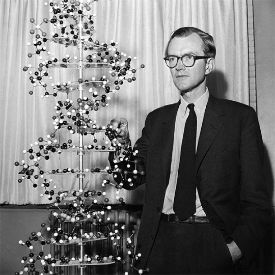


Maurice Wilkins – Scientist of the Day
Maurice Wilkins, a New-Zealand-born English physicist turned molecular biologist, died Oct. 5, 2004, at the age of 87.
read more...
#Maurice Wilkins#biophysics#DNA#X-ray diffraction#histsci#histSTM#20th century#history of science#Ashworth#Scientist of the Day
21 notes
·
View notes
Photo
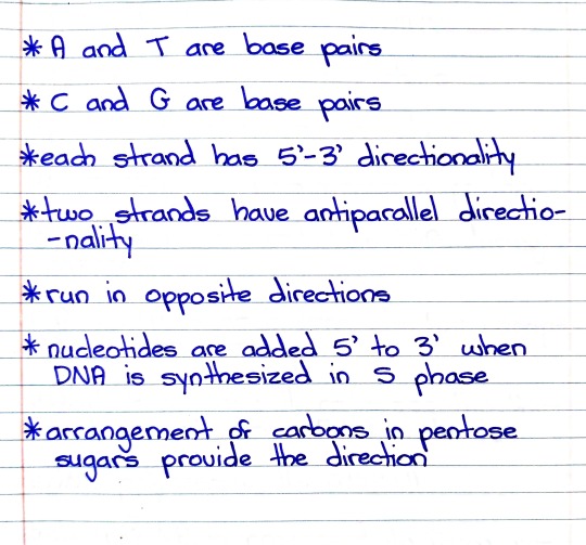
#studyblr#notes#genetics#genetics notes#dna#dna structure#structure of dna#nucleotides#dna strands#dna synthesis#pentose#x-rays#x-ray diffraction#helical structure#carbons#carbon#dna direction#dna strand direction
5 notes
·
View notes
Text
Conversation with Professor Albert Berghuis on Antiobiotics and the Only Synchrotronin Canada: Professor, Department of Biochemistry, McGill University
Publisher: In-Sight Publishing
Publisher Founding: March 1, 2014
Web Domain: http://www.in-sightpublishing.com
Location: Fort Langley, Township of Langley, British Columbia, Canada
Journal: In-Sight: Independent Interview-Based Journal
Journal Founding: August 2, 2012
Frequency: Three (3) Times Per Year
Review Status: Non-Peer-Reviewed
Access: Electronic/Digital & Open Access
Fees: None…
View On WordPress
#Albert Berghuis#antibiotic resistance#antibiotics#Canadian Light Source#McGill University#Synchrotron#University of Saskatchewan#X-ray diffraction
0 notes
Text

X-ray lasers: Why does brighter mean darker?
When we illuminate something, we usually expect that the brighter the source we use, the brighter the resulting image will be. This rule also works for ultra-short pulses of laser light—but only up to a certain intensity. The answer to the question why an X-ray diffraction image 'darkens' at very high X-ray intensities not only deepens fundamental understanding of the light-matter interaction, but also offers a unique perspective for the production of laser pulses that have significantly shorter pulse duration than those currently available.
The more light, the brighter? This observation might sound trivial, were it not for the fact that it is not always true. When silicon crystals are illuminated with ultrafast laser pulses of X-ray light, the resulting diffraction images are indeed initially brighter the more photons fall on the sample, i.e., the higher the beam intensity. Recently, however, a counterintuitive effect has been observed: when the intensity of the X-ray beam starts to exceed a certain critical value, the diffraction images unexpectedly weaken.
This puzzling phenomenon has just been explained, thanks to the efforts of the experimental and theoretical physicists from Japanese, Polish and German research institutions, including the RIKEN SPring-8 Centre in Hyogo, the Institute of Nuclear Physics of the Polish Academy of Sciences (IFJ PAN) in Cracow and the Center for Free-Electron Laser Science (CFEL) at the DESY laboratory in Hamburg.
Read more.
23 notes
·
View notes
Text
The Watson-Crick model (see figure 25.6) was based on molecular modelling and two lines of experimental observations: chemical analyses of DNA base compositions and mathematical analyses of X-ray diffraction patterns of crystals of DNA.

"Chemistry" 2e - Blackman, A., Bottle, S., Schmid, S., Mocerino, M., Wille, U.
#book quotes#chemistry#nonfiction#textbook#watson crick model#dna#deoxyribonucleic acid#james watson#francis crick#modeling#experimental#observation#chemical analysis#mathematical analysis#x ray#diffraction#patterns#crystals
2 notes
·
View notes
Text
The efforts, however, are well rewarded because calculations give very accurate locations for the atoms within the unit cell.
"Chemistry" 2e - Blackman, A., Bottle, S., Schmid, S., Mocerino, M., Wille, U.
0 notes
Text
Chapter 7: X-Ray Diffraction
X-ray diffraction is a powerful technique used to determine the atomic and molecular structure of crystalline materials. By analyzing the interference patterns produced by X-rays scattering off the crystal lattice, we can derive valuable information about the arrangement of atoms within the crystal. This chapter will introduce the basics of X-ray diffraction and its applications.
7.1…
View On WordPress
0 notes
Text
Powder x ray diffraction

#Powder x ray diffraction full#
Peak positions are determined by the crystal symmetry and dimensions, as well as the X-ray wavelength utilised. Indexing makes use only of the positions of the observed peaks. Generally, the unit-cell information is of greater interest than the Miller index labels. The size and shape of the unit cell is determined as part of the indexing procedure. Indexing gets its name from the assignment of Miller index labels to each of the peaks in a pattern. XRPD indexing is the process of determining the size, shape and symmetry of the crystallographic unit cell for a crystalline component responsible for a set of peaks in an XRPD pattern. XRPD indexing is one method that can be used to extract information and aid the interpretation of XRPD patterns. Extracting information from an XRPD pattern beyond visual interpretation adds significant value and greatly enhances understanding of crystal forms. Despite the wealth of information available, routine interpretation of XRPD patterns is often limited to a qualitative visual comparison that, at best, underutilises the available information and, at worst, leads to incorrect conclusions. Information encoded in the XRPD pattern includes whether a material is a single phase or a mixture of phases the size, shape and symmetry of the unit cell the position of the molecules in the unit cell and the crystallite strain, among other information. The XRPD pattern of a crystal form at a given thermodynamic state point serves as a fingerprint for the form under the given conditions. For these reasons, XRPD is used routinely for the characterisation of crystalline solids. X-ray powder diffraction (XRPD) is an ensemble analysis that is generally representative of a powder material, and utilises powder samples that are often easier to produce than single crystals. Furthermore, a single crystal is not always representative of the polycrystalline material from which it was obtained. The main drawback to single crystal diffraction is the need to grow a sufficiently large and defect-free crystal for analysis, which is not practical for all crystal forms.
#Powder x ray diffraction full#
A single crystal structure provides full structural characterisation of the form at the atomic scale. Crystal-structure determination using X-ray diffraction from single crystals is a well-developed technique (1).
0 notes
Photo

Max von Laue was born on October 9, 1879. A German physicist who received the Nobel Prize in Physics in 1914 for his discovery of the diffraction of X-rays by crystals. In addition to his scientific endeavors with contributions in optics, crystallography, quantum theory, superconductivity, and the theory of relativity, Laue had a number of administrative positions which advanced and guided German scientific research and development during four decades. A strong objector to Nazism, he was instrumental in re-establishing and organizing German science after World War II.
#max von laue#physics#x-rays#diffraction#optics#crystallography#quantum theory#superconductivity#relativity#nobel prize#nobel prize winners#science#science history#science birthdays#on this day#on this day in science history
0 notes
Text
Powder x ray diffraction

#POWDER X RAY DIFFRACTION SOFTWARE#
#POWDER X RAY DIFFRACTION SERIES#
When possible, parameters from real-life examples are used as the default. Bugs and problems should be reported to In particular, the so-called ‘Manipulate’ option of Mathematica is extensively used to visualize the impact of parameters in an interactive manner. Non-subscribers of Mathematica can run the scripts using the freely available Wolfram Player at. They are freely available at the TOPAS Wiki web site ( ). All scripts have been written in Wolfram Mathematica version 12.1.1.0 and are constantly updated.
#POWDER X RAY DIFFRACTION SERIES#
In this series of papers dealing with the visualization of mathematical functions used to describe a powder pattern, we present a collection of user-friendly, interactive and freely distributable Mathematica (Wolfram Research, ) teaching scripts.
#POWDER X RAY DIFFRACTION SOFTWARE#
Understanding the algorithms is necessary for correct use of the software and assessment of the reliability of results. In addition, many functions describing a physical effect show discontinuities, and the refined parameters are typically restricted by physical boundaries. In particular, among non-experts, a lot of confusion exists regarding the meaning and significance of fixed or refined parameters, their contribution to the Bragg peaks, the quality of the fit, the range of convergence, precision and accuracy, meaning and reliability of standard deviations etc. It is frequently the case that some parameters are little affected by the nonlinear least-squares procedure, while others change wildly. Many programs have in common the ability to work like a black box for the user. Bragg reflections are characterized by their position, intensity, breadth and shape, each containing a wealth of information (Dinnebier & Billinge, 2008 ▸ Dinnebier et al., 2018 ▸).Ĭomputer programs for fitting peak profiles in powder diffraction perform either single peak fits or whole powder profile fitting with or without a structural model. X-ray powder diffraction (XRPD), an established technique for the study of crystal structures, finds increasing application in qualitative and quantitative phase analysis and the study of microstructure for micro- and nanocrystalline materials.įor the study of crystalline materials, a powder pattern can be divided into Bragg reflections and background, where the latter among other contributions contains diffuse scattering from the sample.
0 notes
Text
Call me x-ray diffraction the way I am unintuitive and confusing and most people would prefer not to deal with me
724 notes
·
View notes
Text
A new technique that produces 3D models of individual crystals has opened a window for scientists to see the subtle deviations that emerge in their otherwise perfect patterns.
Researchers from New York University (NYU) went back to the drawing board on how to look deep inside solids made of repeating units, and determine how they grow.
With a short wavelength roughly the same size as many of the repeating units that make up crystals, X-rays have long allowed scientists to infer how a crystal's components fit together by measuring the angle at which the rays are diffracted.
Continue Reading.
46 notes
·
View notes
Text

While J. Watson and F. Crick are widely known for their discovery of the DNA double helix, Rosalind Franklin's crucial contributions, particularly her X-ray diffraction images of DNA, were essential in deciphering its structure. Her work was pivotal, yet often overshadowed in historical accounts.
15 notes
·
View notes
Text


Materials Characterization: X-ray Diffraction
X-ray diffraction (XRD) is a technique used for characterizing solid materials, either in powder form or bulk materials. The wavelengths of X-rays (on the order of 10^−8 m to 10^−12 m) are able to diffract directly from lattice planes in crystalline materials (images 2 and 3 above), and the resulting information provided by the scan can be used to collect crystal structure information. The technique can also be used on amorphous materials, but without any order to the atoms or molecules, the result will only be able to confirm the amorphous nature of the material.
However, XRD scans don't simply output a crystal structure. The most basic results are similar to what's shown in image 1 above, with the spots corresponding to lattice planes (an amorphous material would an indistinct ring; see Figure 4 of this link). Another form of the data output is the plot in image 5 above, wherein each peak corresponds to a lattice plane. In order to determine crystal structure from there, one must know the allowed and forbidden reflections for each crystal structure (too detailed to go into here, an example is that the 111 plane is a forbidden reflection in bcc, but allowed for fcc, so if that plane is present it helps narrow down your material). In addition, the height or intensity of the peaks reflects, qualitatively, the frequency of different orientations, thereby providing information about texture.
Though it can be quick and relatively easy to use, with minimal sample preparation, there are limitations of XRD. It works best on single phase materials; XRD can provide information on multiphase materials, and, indeed, provide an idea as to which phases are present if it is unknown, but it is not always reliable. Peak locations must typically be compared to known spectrum and different materials or crystal structures will have overlaps or peaks in the same location.
Sources/Further reading: ( 1 ) ( 2 - images 2 and 3 ) ( 3 ) ( 4 - image 4 ) ( 5 - image 5 ) ( 6 ) ( 7 )
Image 1.
#Materials Science#Science#X Rays#X Ray diffraction#Diffraction#Materials Characterization#MyMSEPost
11 notes
·
View notes
Text
In 1953, using X-ray diffraction photograph of DNA fibres (figure 7.50) obtained by Rosalind Franklin and New Zealand-born Maurice Wilkins, James Watson and Francis Crick came to the conclusion that the DNA structure consists of the now-famous double helix (see figure 7.51).
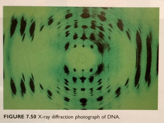
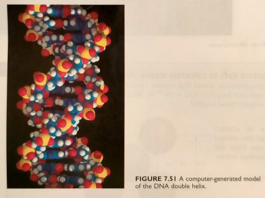
"Chemistry" 2e - Blackman, A., Bottle, S., Schmid, S., Mocerino, M., Wille, U.
#book quotes#chemistry#nonfiction#textbook#50s#1950s#20th century#x ray diffraction#dna#double helix#rosalind franklin#maurice wilkins#james watson#francis crick
6 notes
·
View notes
Text
In 1913, William Henry Bragg and his Australian-born son William Lawrence Bragg discovered that just a few variables control the appearance of an X-ray diffraction pattern.
"Chemistry" 2e - Blackman, A., Bottle, S., Schmid, S., Mocerino, M., Wille, U.
#book quote#chemistry#nonfiction#textbook#10s#1910s#20th century#william henry bragg#william lawrence bragg#william bragg#x ray#x ray diffraction#patterns
0 notes
Last Seen Blogs

theo-wyatt
Theo

solestixx
love of ur life

mirfanlb
Muhammad Irfan

lostthoughtsfindwords
Everyday Poetry

clavepuppy48
The Life of Klemmensen 183