
Statistics
We looked inside some of the posts by biomedicool and here's what we found interesting.
Average Info
Notes Per Post
15K
Likes Per Post
11K
Reblog Per Post
4K
Reply Per Post
26
Time Between Posts
23 days
Number of Posts By Type
Text
13
Link
2
Photo
2
Last Seen Tumblr Blogs


Fun Fact
Tumblr has 411 employees.
Text
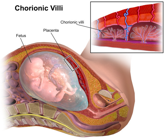
Molar Pregnancy (Hydatidiform Mole)
Molar pregnancy is an abnormal form of pregnancy in which a non-viable fertilized egg implants in the uterus and will fail to come to term (will not develop into a child). Instead, the cells divide and replicate into a growing mass (mole) of non-foetal tissue.

The mass formed is characterised swollen chorionic villi.
Molar pregnancies
Can develop when a fertilized egg does not contain an original maternal nucleus.
Usually contain no foetal tissue.
Characterized by the presence of a hydatidiform mole (or hydatid mole).
Approximately 20% of cases with a complete mole develop a trophoblastic malignancy (malignant disease // cancer)
Molar pregnancies make up 1 in 1,000 pregnancies in the US and up to 1 in 100 pregnancies in parts of Asia.
Symptoms
Vaginal bleeding - molar tissue separates from the decidua, causing bleeding.
Uterus may become distended by large amounts of blood, and dark fluid may leak into the vagina.
Hyperemesis - severe nausea and vomiting due to very high levels of human chorionic gonadotropin (hCG).
Hyperthyroidism - thyroid gland is stimulated by the high levels of circulating hCG or by a thyroid stimulating substance (ie, thyrotropin) produced by the trophoblasts.
Partial mole
Partial moles do not generate the same clinical features as a complete mole. Patients instead present with signs and symptoms consistent with an incomplete or missed abortion, including vaginal bleeding and absence of foetal heartbeat.
#pregnancy#maternity#ob gyn#biomedical science#medical science#science#biology#human biology#medblr#nursing#premed#studyblr#midwifery#midwife#reproduction#development#pathology#obstetrics#biomed#notes#2#molar pregnancy#educational#gynae
182 notes
·
View notes
Text
Stem cells for therapy - types
Bone Marrow Cells (BMCs)
Whole bone marrow cells (BMCs) and bone marrow mononuclear cells (BMMCs) are the most accessible and studied source of stem cells.
BMMCs are isolated from whole bone marrow, and contain a diverse cell population, including mesenchymal stem cells and hematopoietic progenitor cells.
Mesenchymal Stem Cells (MSCs)
MSCs can be isolated from a variety of tissues such as bone marrow, adipose, and umbilical cord; although it is not clear whether their properties are uniform (Selem, Hatzistergos and Hare, 2011).
MSCs are of particular note due to their immunopriveleged nature – a reduced expression of MHC class-I molecule, and lack of MHC class-II and co-stimulatory molecules, means they could potentially be used for allogeneic grafts (Zimmet et al., 2005). This means that they don’t produce an immune response and could be used in transplants - the body won’t reject them.
MSCs inhibit the activity of various immune cells, including T cells, B cells, natural killer cells, and dendritic cells via cell to cell contacts and soluble factors (Laflamme and Murry, 2005).
Foetal and Umbilical Cord Cells
Embryonic stem cells (ESCs), the prototypical stem cell, can develop into all cell types in the body. However, the practical application of human ESCs remains limited due to ethical problems, teratoma formation (cancer), and immune rejection. With rapidly expanding knowledge of molecular and genetic pathways for ESC differentiation, it may become possible to avoid contamination with undifferentiated ESCs, thereby inhibiting teratogenesis when transplanted into the body (Kucia et al., 2006).
Foetal-derived stem cells can also be isolated from the amniotic fluid, which include both pluripotent and committed stem cells.
Umbilical cord cells can be gathered at birth and stored, eg if for treatment later on if a defect is detected in utero.
Induced pluripotent stem cells (iPSCs)
Induced pluripotent stem cells are a more attractive alternative to ESCs, as they are autologous. This means cells can be taken from an individual, ‘reset’ back to their stem cell stage, and then administered to that same individual to avoid rejection. Pluripotency transcription factors are introduced to adult terminally differentiated somatic cells, such as dermal fibroblasts, in a novel strategy which ‘reprograms’ the cells back to an embryonic stem cell-like stage (Yu et al., 2007).
Despite slight epigenetic differences associated with reprogramming, iPSCs fully resemble ESCs in terms of differentiation capacity, morphology and gene expression profile; and have the ability to differentiate into other cells. Ethical and immune response dilemmas are bypassed by the autologous nature of iPSCs, however clinical application is not yet on the horizon due to their teratogenic potential and the oncogenes and virus vectors required for the current method of pluripotent induction (Yamanaka and Takahashi, 2006).
Skeletal myoblasts (SM)
Skeletal myoblasts (satellite cells) are derived from skeletal muscle and have the capacity to differentiate into muscle fibre, which makes them obvious candidates for treating conditions such as heart damage following infarction. However, clinical trials have been halted as SM have been observed to couple with resident cardiomyocytes, resulting in dysfunctional electrocardiology and arrhythmias, and have struggled to transdifferentiate into cardiomyocytes in vivo (Reinecke, Poppa and Murry, 2002).
#stem#stem cells#regeneration#therapy#biomedical science#biomedicine#biology#medicine#medblr#studyblr#premed#science#human biology#notes#2#3#biomed#sciblr#nursing#med
222 notes
·
View notes
Text
Stem cells
Stem cells are cells in the body that don’t yet have any role (undifferentiated or partially differentiated), and can change to become almost any cell type.
Can proliferate (divide to make more cells) indefinitely to make more of the same stem cells.
They are the earliest type of cell in a cell lineage (if a cell was an adult human, stem cells would be the foetus)
found in both embryonic and adult organisms, but they have slightly different properties in each
Can also be made in a lab by reprogramming other cells - resetting them back to stem cell stage.

In embryonic development (a baby forming in the womb), pluripotent stem cells develop at the blastocyst stage (3-4 days) and differentiate into all the cells of the human body as the foetus develops.
Stem cells do exist in the adult body, however they are not pluripotent - they are unipotent or multipotent - this means they can only differentiate into one or a few cell types respectively.
Adult stem cells
Adult stem cells are found in a few select locations in the body, known as niches, such as:
the brain
bone marrow
blood and blood vessels
skeletal muscles
liver
gonads
They exist to replenish rapidly lost cell types and include hematopoietic stem cells, which replenish blood and immune cells, basal cells, which maintain the skin epithelium, and mesenchymal stem cells, which maintain bone, cartilage, muscle and fat cells. Adult stem cells are a small minority of cells.
#stem cell#stem cells#Regeneration#cells#notes#2#biomedicine#biomed#biomedical science#medblr#sciblr#biology#human biology#medicine#premed#nursing#physiology
157 notes
·
View notes
Text

#happy holidays!#life's been busy so i've had a hiatus but i'm still around#science#memes#stats#science memes#meme#santa#christmas
2K notes
·
View notes
Text
IBS - Irritable Bowel Syndrome
Symptoms of irritable bowel syndrome include stomach cramps, bloating, diarrhoea and constipation. These may come and go over time.
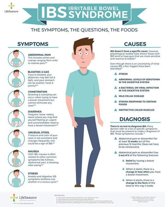
IBS is characterized by abdominal pain and altered bowel habit in the absence of a specific and unique organic pathology. This means that there are symptoms of colon irritation/damage, but no specific disease can be identified and there is vast variation between patients.
Pathology
Causes for IBS have not yet been identified, but current research suggests:
Altered GI motility
The myoelectric activity (electric potential of muscles - how muscles contract and relax, in this case to push food along) of the colon is composed of background slow waves with spike potentials.
Colonic dysmotility in irritable bowel syndrome manifests as variations in slow-wave frequency and a blunted, late-peaking, postprandial response of spike potentials.
This causes delayed meal transit (slow digestion) in patients prone to constipation, and in accelerated transit in patients prone to diarrhoea.
Current theories suggest generalized smooth muscle hyperresponsiveness - the muscle cells overreact to the electric currents.
Visceral hyperalgesia
IBS symptoms may be a result of an increased pain response to colon activity
Rectosigmoid and small bowel balloon inflation produces pain at lower volumes in patients than in controls.
Notably, hypersensitivity appears with rapid but not with gradual distention.
Patients who are affected describe widened dermatomal distributions of referred pain. Sensitization of the intestinal afferent nociceptive pathways that synapse in the dorsal horn of the spinal cord provides a unifying mechanism.
Psychopathology
IBS is both more common and more debilitating in patients with panic disorder, major depression, anxiety disorder, and hypochondriasis.
The psychopathology and stress caused by these disorders may contribute to IBS.
This is particularly concerning as IBS symptoms will worsen these conditions, which may in turn worsen the IBS.
Microscopic inflammation
Both colonic inflammation and small bowel inflammation have been discovered in a subset of patients. These studies are still in early stages.
Alterations in the intestinal biome
Small bowel bacterial overgrowth has been heralded as a unifying mechanism for the symptoms of bloating and distention.
The faecal microflora (bacteria in poo) also differs among patients with irritable bowel syndrome compared to those without.
#ibs#medicine#medblr#studyblr#biomedicine#Biomedical Science#biomed#premed#nursing#science#biology#colon#pathology#gi#colonoscopy
112 notes
·
View notes
Text
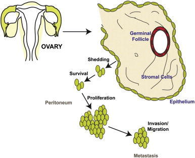
Ovarian Cancer
Malignant lesions of the ovaries include primary lesions arising from normal structures within the ovary and secondary lesions from cancers arising elsewhere in the body.
Signs and symptoms
Bloating; abdominal distention or mass
Pressure effects on the bladder and rectum
Constipation, indigestion, reflux
Vaginal bleeding
Shortness of breath, tiredness, weight loss
Diagnosis
Physical findings are uncommon in early stages. Advanced disease may present with ovarian or pelvic mass, ascites, pleural effusion, or abdominal mass or bowel obstruction.
Pathophysiology
Typically spreads to the peritoneal surfaces and omentum.
Occurs via local extension, lymphatic invasion, intraperitoneal implantation, hematogenous dissemination, or transdiaphragmatic passage.
Malignant cells can implant anywhere in the peritoneal cavity but are more likely to implant in sites of stasis along the peritoneal fluid circulation.
Epithelial tumours make up 90% of ovarian tumours. Other histologies include:
Sex-cord stromal tumors
Germ cell tumors
Primary peritoneal carcinoma
Metastatic tumors of the ovary
Epithelial ovarian cancer
Arises from epithelium covering the fimbria of the fallopian tubes, or the ovaries, both of which are derived from the coelomic epithelium.
Found primarily as cystic lesions with solid components.
Surface may be smooth or covered in papillary projections.
Cysts contain fluid from yellow to brown and haemorrhagic.
Four main histologic subtypes:
Serous (from fallopian tube)
Endometrioid (endometrium)
Mucinous (cervix)
Clear cell (mesonephros)
Tumours of low malignant potential
Tumours of low malignant potential (LMP) are a variety of much less aggressive epithelial ovarian cancer, with good prognosis.
LMP tumors can cause a range of symptoms similar to epithelial ovarian cancer, including increasing abdominal girth, an abdominal mass, abdominal pain, abnormal uterine bleeding, urinary symptoms, and gastrointestinal symptoms. They may be asymptomatic and found on routine physical examination or ultrasound scan.
#cancer#oncology#gynecology#ovarian cancer#ovaries#pathology#science#medblr#studyblr#sciblr#medschool#premed#nursing#medicine#biology#human biology#notes#2
162 notes
·
View notes
Text
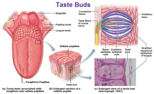
Taste
The tongue is covered with many little bumps called papillae. Taste buds are found in the walls of papillae and the grooves surrounding them. Each taste bud contains anywhere from 50 to 150 taste receptor cells.
Microvilli extend from taste receptor cells
and protrude through an opening (taste pore) into the mouth.
These microvilli come in contact with substances in the mouth that can be tasted, also known as tastants.
Tastants interact with taste receptor cells through a number of different mechanisms to depolarize the cells.
When taste cells are depolarized, they release neurotransmitters that stimulate sensory neurons that travel in cranial nerves VII, IX, and X.
These neurons terminate on neurons in the nucleus of the solitary tract in the medulla then continue on to the thalamus.
Taste information is sent to the gustatory cortex, ( ocated on the border between the anterior insula and the frontal operculum).
This information encodes for basic tastes, such as sweet, salty, sour, bitter, and savory or umami.
However, the actual flavour of a food---which is what we typically define as taste---is created by a combination of taste and olfactory (smell) information.
Sweetness
Produced by the presence of sugars, some proteins, and other substances.
Detected by G protein-coupled receptors T1R2+3 (heterodimer) and T1R3 (homodimer).
Saltiness
Saltiness is a taste produced best by the presence of cations (such as Na+, K+or Li+)
Directly detected by cation influx into glial like cells via leak channels causing depolarisation of the cell.
Sourness
Sourness is acidity and is also sensed using ion channels.
Undissociated acid diffuses across the plasma membrane of a presynaptic cell, where it dissociates in accordance with Le Chatelier's principle.
The protons that are released then block potassium channels, which depolarise the cell and cause calcium influx.
Bitterness
Current research suggests TAS2Rs (taste receptors, type 2, also known as T2Rs) such as TAS2R38 are responsible tasting bitter substances.
Savouriness
The amino acid glutamic acid is responsible for savouriness, but some nucleotides (inosinic acid and guanylic acid) can act as complements.
Glutamic acid binds to a variant of the G protein-coupled receptor, producing a savoury taste
#medicine#biomed#biomedicine#notes#medblr#studyblr#sciblr#premed#nursing#biology#human biology#med#biomedical science#science#taste#physiology#anatomgy#gustatory#gustatory system#gustatory cortex#mouth
265 notes
·
View notes
Link
New research published in the New England Journal of Medicine shows that the respiratory virus SARS-CoV-2, which causes COVID-19, causes severe damage to blood vessels, leading to widespread thrombosis, a press release by the Angiogenesis Foundation reports.
The study was conducted by an international team of medical scientists who compared the lungs of patients who died from COVID-19 with lungs of patients who died from influenza as well as with healthy lungs donated for transplantation.
The researchers discovered an unexpected disease pattern in COVID-19 lungs: the virus invaded the endothelial cells, and this was accompanied by blood clots. Compared to the flu, COVID-19 lungs had 9-fold more blood clots and the blood vessels were injured by the virus causing an unusual reaction of blood vessel growth.
Separately, a 6 May report published in the Journal of the American College of Cardiology described how using blood thinners in patients hospitalised with COVID-19 appeared to be protective and associated with better outcomes.
#Corona#COVID19#covid-19#Coronavirus outbreak#coronavirus#rona#research#medical research#medblr#sciblr#medicine
149 notes
·
View notes
Photo
7K notes
·
View notes
Link
As key players in the body’s immune defense, T cells have the ability to track down and attack cancer cells, but can have trouble seeking out those that are not already inflamed. A new immunotherapy deliver system could make these natural killer cells far more effective by strategically inflaming so-called cold tumors so T cells can detect and destroy them with much greater efficiency.
The new technique was developed by scientists at the University of Chicago and falls under an arm of cancer treatment known as immunotherapy. This involves supercharging the body’s immune system in different ways so that it can better fight off cancer, and one of the ways it can do this is through what are known as checkpoint inhibitors.
#new atlas audio#cancer#oncology#researc#research#medical research#cancer research#medblr#medicine#med#medschool#science#biology
59 notes
·
View notes
Photo

Drop in coal power plant emissions associated with asthma improvements
Reductions in sulfur dioxide emissions from four coal-fired power plants in Kentucky were associated with fewer local hospitalizations and emergency department visits due to asthma.
An immediate drop in people’s use of rescue inhalers in the area also indicated a reduction in daily asthma symptoms.
164 notes
·
View notes
Text
Thymus
The thymus is a specialised primary lymphoid organ of the immune system.
At its largest and most active during the neonatal and pre-adolescent periods.
Decreases in size and activity through teenage years
Thymus tissue is gradually replaced by adipose tissue (fat).
Residual T lymphopoiesis continues throughout adult life.
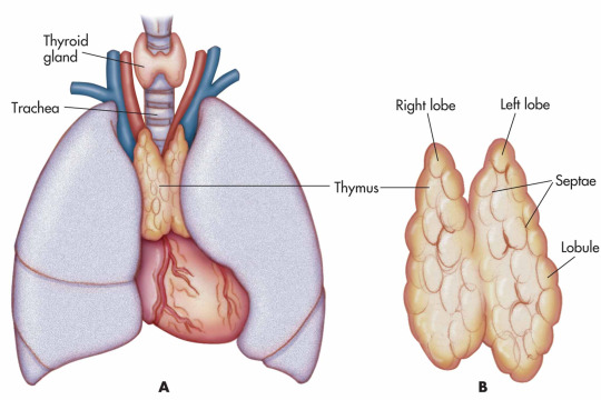
The thymus is composed of two identical lobes and is located in the anterior superior mediastinum, in front of the heart and behind the sternum. Each lobe of the thymus can be divided into a central medulla and a peripheral cortex which is surrounded by an outer capsule.
Function
Facilitates the maturation of T cells - which provide cell-mediated immunity.
T cells begin as hematopoietic precursors from the bone-marrow, and migrate to the thymus, where they are referred to as thymocytes.
In the thymus they undergo a process to ensure the cells react against antigens ("positive selection"), but that they do not react against antigens found on body tissue ("negative selection").
Once mature, T cells emigrate from the thymus to provide vital functions in the immune system.
Each T cell has a distinct T cell receptor, suited to a specific substance, called an antigen.
Most T cell receptors bind to the major histocompatibility complex on cells of the body.
Positive selection
T cells have distinct T cell receptors. These are formed by process recombination gene rearrangement which is error-prone, and some thymocytes fail to make functional T-cell receptors, whereas other thymocytes make T-cell receptors that are autoreactive. The survival and nature of the T cell then depends on its interaction with surrounding thymic epithelial cells.
T cell receptor interacts with the MHC molecules on the surface of epithelial cells.
A T cell with a receptor that doesn't react, or reacts weakly will die by apoptosis.
A T cell that does react will survive and proliferate.
A mature T cell expresses only CD4 or CD8, but not both.
Negative selection
T cells that attack the body's own proteins are eliminated in the thymus. Epithelial cells in the medulla and dendritic cells in the thymus express major proteins from elsewhere in the body. Some CD4 positive T cells exposed to self antigens persist as T regulatory cells.
Pathology
Immunodeficiency - As the thymus is the organ of T-cell development, any congenital defect in thymic genesis or a defect in thymocyte development can lead to a profound T cell deficiency in primary immunodeficiency disease.
Autoimmune disease - Genetic disorders, such as Myasthenia gravis: caused by antibodies that block acetylcholine receptors.
Thymomas - Originate in thymic epithelial cells most often in adults older than 40. Generally detected when they cause symptoms, such as a neck mass or affecting nearby structures such as the superior vena cava. Can be benign; benign but by virtue of expansion, invading beyond the capsule of the thymus ("invasive thyoma"), or malignant (a carcinoma).
Lymphomas - Tumours originating from T cells of the thymus form a subset of acute lymphoblastic leukaemia (ALL)
Thymic cysts - The thymus may contain cysts, usually less than 4 cm in diameter. Thymic cysts are usually detected incidentally and do not generally cause symptoms.
#thymus#thymoma#lymphatic#lymphatic system#immunology#immune system#t cells#medicine#biomed#biomedicine#notes#medblr#studyblr#sciblr#premed#nursing#biology#human biology#med#biomedical science#science#2#anatomy
197 notes
·
View notes
Text
Tonsilitis
Tonsillitis is inflammation of the pharyngeal tonsils.
Inflammation usually extends to the adenoid and the lingual tonsils
Most commonly viral
Most cases of bacterial tonsillitis are caused by group A beta-hemolytic Streptococcus pyogenes (GABHS) - strep throat.
Spread through the air.
Symptoms may include sore throat, fever, enlargement of the tonsils, trouble swallowing, and large lymph nodes around the neck. Complications include peritonsillar abscess.

Recurrent tonsillitis
A polymicrobial flora consisting of both aerobic and anaerobic bacteria
Other competing bacteria are reduced - less interference to GABHS infection.
Streptococcus pneumoniae, Staphylococcus aureus, and Haemophilus influenzae are the most common bacteria isolated in recurrent tonsillitis, and Bacteroides fragilis is the most common anaerobic bacterium isolated in recurrent tonsillitis.
The microbiologies of recurrent tonsillitis in children and adults are different; adults show more bacterial isolates, with a higher recovery rate of Prevotella species, Porphyromonas species, and B fragilis organisms , whereas children show more GABHS. Also, adults more often have bacteria that produce beta-lactamase.
Chronic tonsillitis
Polymicrobial bacterial population present
There is likely a relationship between tonsillar size and chronic bacterial tonsillitis based on both the aerobic bacterial load and the absolute number of B and T lymphocytes.
Fewer dendritic cells on the surface epithelium and more in the crypts and extrafollicular areas during chronic tonsillitis.
Radiation exposure may relate to the development of chronic tonsillitis. A high prevalence of chronic tonsillitis was noted following the Chernobyl nuclear reactor accident in the former Soviet Union.
#medicine#biomed#biomedicine#notes#medblr#studyblr#sciblr#premed#nursing#biology#human biology#med#biomedical science#science#tonsils#tonsilitis#tonsillitis#infection#pathology#2#3#microbiology
71 notes
·
View notes
Text
Tonsils
The tonsils are a set of lymphoid organs consisting of the adenoid tonsil, two tubal tonsils, two palatine tonsils, and the lingual tonsils. These organs play an important role in the immune system.

Palatine tonsil
Main tonsils
Non-keratinized stratified squamous epithelium
Incompletely encapsulated
Long, branched crypts
Located on sides of oropharynx between palatoglossal and palatopharyngeal arches
Reach their largest size in puberty, and they gradually undergo atrophy
Adenoid (also termed "pharyngeal tonsil")
Ciliated pseudostratified columnar epitheium (respiratory epithelium)
Incompletely encapsulated
No crypts, but small folds
Located on roof of pharynx
Tubal tonsils
Ciliated pseudostratified columnar epithelium (respiratory epithelium)
Located on roof of pharynx
Lingual tonsils
Non-keratinized stratified squamous epithelium
Incompletely encapsulated
Long, unbranched crypts
Located behind terminal sulcus (tongue)
The tonsils serve as the immune system's first line of defence against ingested or inhaled foreign pathogens.
The tonsils have specialised antigen capture cells (M cells) on their surface that allow for the uptake of antigens produced by pathogens.
These M cells then alert the underlying B cells and T cells in the tonsil that a pathogen is present and an immune response is stimulated.
B cells are activated and proliferate in areas called germinal centres in the tonsil.
Secretory antibody (IgA) is produced.
Studies suggest that the tonsils also produce T cells themselves, in a manner similar to the thymus.
#tonsil#tonsils#lymphatic#lymph#lymphatic system#anatomy#anp#physiology#medicine#biomed#biomedicine#notes#medblr#studyblr#sciblr#premed#nursing#biology#human biology#med#biomedical science#science
207 notes
·
View notes
Text
Thalidomide
Thalidomide was originally introduced as a non-barbiturate sedative in 1957 and later marketed for the treatment of nausea in pregnant women by the German company Chemie Grünenthal, as antiemetic properties were discovered. In the 1960s however it became apparent that thalidomide treatment resulted in severe teratogenic birth defects and from November 1961 it began to be withdrawn worldwide. It has since been reintroduced into a number of studies and treatments due to its immunosuppresive and anti-angiogenic activity.

Chemical structure of thalidomide. Thalidomide is a stereo-isomer and can exist, depending on the state of the chiral carbon, in two enantiomeric states. Both R and S can interconvert in body fluids and tissues. Thalidomide was distributed as a racemic mix of both enantiomers.
Thalidomide is a piperidinyl isoindole and synthetic derivative of glutamic acid, consisting of two linked glutarimide and pthalimide rings.
The unstable chiral carbon allows two enantiomers to coexist - the S-enantiomer being teratogenic (Franks, 2004.)
The mechanism of action that resulted in these teratogenic effects is still not fully understood.
Leading theories focus on thalidomide's antiangiogenic properties; ability to induce cell death and generate reactive oxygen species; and Cereblon, a thalidomide-binding protein and primary source of teratogenic action (Takumi, 2010).
Thalidomide is hydrolyzed in bodily fluids and metabolized in the liver by cytochrome p450.
When first developed thalidomide was deemed so non-toxic that an LD50 could not be established, but tragically an estimated 25,000 babies were born severely impaired and a further 123,000 miscarried or stillborn (Johnson, 2016).

Antiangiogenic properties
Thalidomide has the ability to inhibit angiogenic vascularization in embryos, thus carrying out teratogenic damage.
The bulk of its damage occurs in days 20-36 of fertilization.
During this time, the primitive vessels formed by vasculoneogensis mature and begin to proliferate and migrate in response to signals and growth factors such as FGF.
It is thought that analogues of thalidomide could block a number of these signals, thus preventing embryonic vascular development outwards towards the limbs and resulting in limb deformities such as phocemelia.
Molecular targets of thalidomide
Cereblon
Forms part of a ubiquitination complex with DNA Binding Protein 1, which in turn selects molecules for destruction.
Thalidomide binds, preventing formation of the complex and proper regulation of developmental signaling molecules, thus initiating teratogenis by inhibiting angiogenesis (Takumi, 2010).
Could also degrade some proteins and prevent breakdown of others.
Tubulin
part of the cytoskeleton and required for cell proliferation and formation of new vessels in the embryo.
When bound by the 5HPP-33 thalidomide analogue, cytoskeletal dynamics are altered preventing cell division.
This could prevent cell proliferation and migration and consequently tissue morphogenesis, the severity of which would be dependent on vascular and tissue maturation at the point of contact with thalidomide (Vergesson, 2015).
#3#thalidomide#drugs#pharmacology#case study#notes#biochemistry#medicine#biomed#biomedicine#medblr#studyblr#sciblr#premed#nursing#biology#human biology#med#biomedical science#science
636 notes
·
View notes
Text
Amphetamines
Amphetamines are sympathomimetic amines which mimic the structures of the catecholamine neurotransmitters, noradrenaline and dopamine.
They are substrates for the neuronal plasma membrane monamine uptake transporters DAT and NET (dopamine and norepinephrine transporters)
Thus act as competitive inhibitors, reducing the reuptake of dopamine and noradrenaline.
In addition, they enter nerve terminals via the uptake processes or by diffusion and interact with the vesicular monoamine pump VMAT-2 to inhibit the uptake into synaptic vesicles of cytoplasmic dopamine and noradrenaline.
The amphetamines are taken up into the storage vesicles by VMAT-2 and displace the endogenous monamines from the vesicles into the cytoplasm.
At high concentrations amphetamines can inhibit monoamine oxidase, which otherwise would break down cytoplasmic monamines.

The cytoplasmic monoamines can then be transported out of the nerve endings via DAT and NET transporters working in reverse, a process thought to be facilitated by amphetamine binding to the transporters. The concentration of extracellular dopamine and noradrenaline is therefore increased, and the reward response generated by increased activation of dopamine receptors can eventually lead to addiction.
The biggest problem facing addiction is the lack of effective medical treatment available. Currently, therapeutic strategies involve giving the patient something similar, but less potent than the drug of addiction, in an attempt to slowly reach abstinence with minimal withdrawal. However, due to memory and learning mechanisms as well as epigenetic and transcriptional changes, relapse is not uncommon.
Pharmacology is less scary than it looks but hard to understand just through reading - here’s a great video
#medicine#biomed#biomedicine#notes#medblr#studyblr#sciblr#premed#nursing#biology#human biology#med#biomedical science#science#pharmacology#amphetamine#amphetamines#drugs#biochemistry#3#addiction
154 notes
·
View notes
Text
My heart skip skips a beat
HEARTBEATS!!
The pause is to allow the atria to fully empty into the ventricle.
Heartbeat on an ECG trace
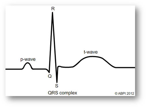
P Interval (Ventricular Diastole)
Atria and ventricles are relaxed
blood is flowing into the atria from the veins.
Atrial pressure increases above that of the ventricle, AV valves open allowing blood to flow into the ventricle
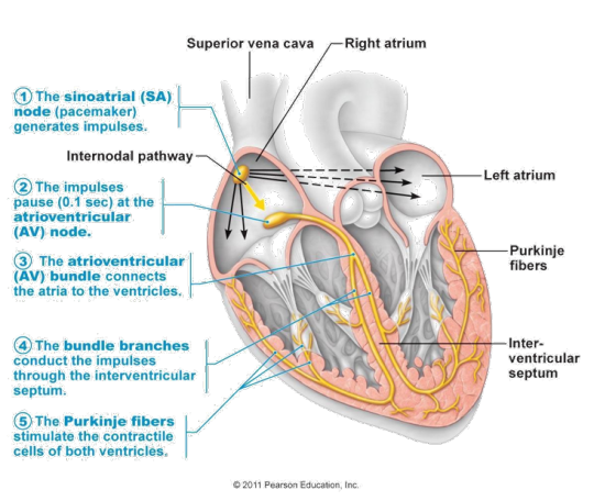
P Wave (Atrial Systole) P-Q
Signal transduction from SA to AV nodes.
SA node fires
Atria contract causing atrial systole
which forces all blood into the ventricles
emptying the atria.
Q Interval (End of Ventricular Diastole)
Depolarisation of interventricular (IV) septum
AV valves remain open - all remaining blood squeezed into the ventricles.
impulse from the SA node reaches the AV node
which spreads the signal throughout the walls of the ventricles via bundles of His and Purkinje fibres
R peak is the end of ventricular diastole and the start of systole.
R Interval (Ventricular Systole)
Ventricular contraction
All blood is now within the ventricles
so pressure is higher than in the atria - AV valves close
ventricles start to contract although pressure is not yet high enough to open the SL (semilunar) valves
ST Segment (Ventricular Systole)
Ventricular contraction
Pressure increases until it equals Aortic pressure,
SL valves open
blood is ejected into the Aorta (and pulmonary artery) as ventricles contract
At this time the atria are in diastole and filling with blood returning from the veins.
plateau in ventricular arterial pressure
T Wave (Ventricular Diastole)
T= moment of Ventricular repolarisation immediately before ventricular relaxation
Ventricles relax
ventricular pressure is once again less than the aortic pressure
so SL valves close
3K notes
·
View notes