Don't wanna be here? Send us removal request.
Statistics
We looked inside some of the posts by cellimagelibrary and here's what we found interesting.
Average Info
Notes Per Post
1K
Likes Per Post
732
Reblog Per Post
331
Reply Per Post
3
Time Between Posts
14 days
Number of Posts By Type
Photo
15
Video
2
Last Seen Tumblr Blogs


Fun Fact
Tumblr is used by 21% of adults online aged 18-29 years.
Photo

Image of the Week – June 22, 2020
CIL:11397 - http://cellimagelibrary.org/images/11397
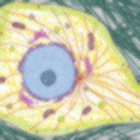
Description: Figure 218 from Chapter 7 (Mitochondria) of 'The Cell, 2nd Ed.' by Don W. Fawcett M.D. The typical structural organization of mitochondria is revealed in this longitudinal section of a mitochondrion and surrounding cytoplasm from pancreas of the bat, Myotis lucifugus. It is bounded by a smooth outer membrane about 7nm thick. An inner membrane has infoldings called cristae that project into the interior of the organelle.
Author: Keith Porter
Licensing: Attribution Non-Commercial No Derivatives: This image is licensed under a Creative Commons Attribution, Non-Commercial, No Derivatives License
43 notes
·
View notes
Photo
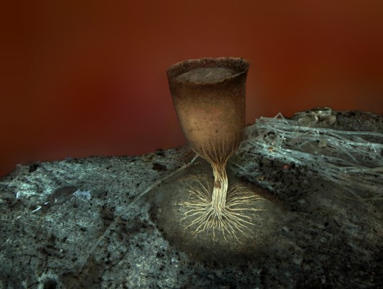
Cheers! Image of the Week – June 15, 2020
CIL:41635 - http://www.cellimagelibrary.org/images/41635
Description: Fluorescent image of the sporangium, an enclosure in which spores are formed, of the slime mold Craterium minutum. Honorable Mention, 2011 Olympus BioScapes Digital Imaging Competition®.
Authors: Dalibor Matýsek and 2011 Olympus BioScapes Digital Imaging Competition®
Licensing: Attribution Non-Commercial No Derivatives: This image is licensed under a Creative Commons Attribution, Non-Commercial, No Derivatives License
15 notes
·
View notes
Video
tumblr
Image of the Week – June 8, 2020
CIL:12375 - http://cellimagelibrary.org/images/12375
Description: Movie showing the dynamics of kinetchore microtubules during meiosis II in primary spermatocytes of the crane-fly Nephrotoma suturalis that were experimentally flattened. Time-lapse polarization microscopy using a Nikon Microphot SA, equipped for liquid crystal polarized light microscopy (LC-PolScope, CRi, Woburn Massachusetts) 60x/1.4 PlanApo oil immersion objective, 1.4 NA oil imm. condenser, with 2.0x zoom lens. Images captured every 15 sec using a QImaging Retigo EXi CCD camera. Raw images were processed using 5-frame algorithm (Shribak and Oldenbourg, 2003). The time series used for the movie is included in this grouped set.
Authors: James R. LaFountain and Rudolf Oldenbourg
Licensing: Public Domain: This image is in the public domain and thus free of any copyright restrictions. However, as is the norm in scientific publishing and as a matter of courtesy, any user should credit the content provider for any public or private use of this image whenever possible.
238 notes
·
View notes
Video
tumblr
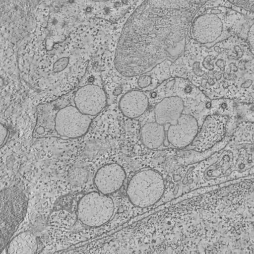
Image of the Week – June 1, 2020
CIL:50519 - http://cellimagelibrary.org/images/50519
Description: We examined the structural organization of cytoskeletal components and membrane systems in neurons of well preserved biopsy material from Alzheimer’s disease (AD). Information was obtained from thin sectioned material using conventional electron microscopy and from thick sections with the high voltage electron microscope. Stereo viewing and computer assisted serial reconstruction techniques were employed to visualize three-dimensional relationships among cytoplasmic components of cortical neurons.
Authors: Mark Ellisman, Rama Ranganathan, Thomas Deerinck, Stephen Young, David Hessler, and Robert Terry
Licensing: Public Domain: This image is in the public domain and thus free of any copyright restrictions. However, as is the norm in scientific publishing and as a matter of courtesy, any user should credit the content provider for any public or private use of this image whenever possible.
34 notes
·
View notes
Photo
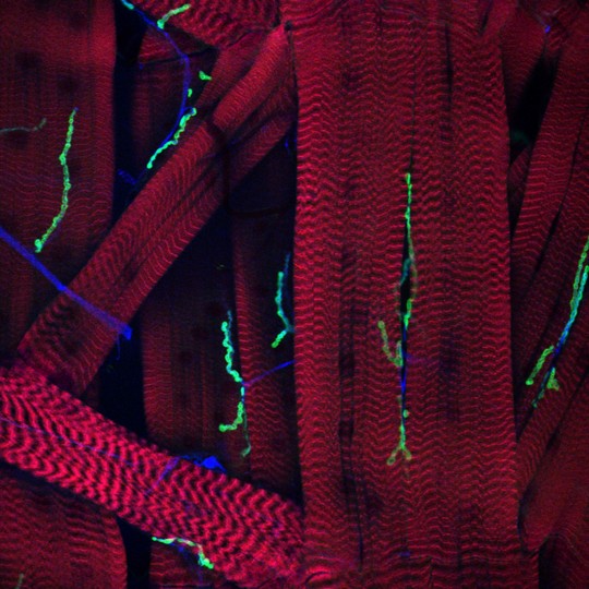
You’ve Got Some Nerve! Image of the Week – February 17, 2020
CIL:41833 - http://www.cellimagelibrary.org/images/41833
Description: Confocal micrograph showing the complex connectivity at the neuromuscular junction of Drosophila (fruit fly). Muscle fibers shown in red and nerve fibers into neuromuscular junction shown in blue and green. Honorable Mention, 2010 Olympus BioScapes Digital Imaging Competition®.
Authors: Timothy Mosca and 2010 Olympus BioScapes Digital Imaging Competition®
Licensing: Attribution Non-Commercial No Derivatives: This image is licensed under a Creative Commons Attribution, Non-Commercial, No Derivatives License
89 notes
·
View notes
Photo

You Dirty Rat! Image of the Week – February 10, 2020
CIL:48107 - http://cellimagelibrary.org/images/48107
Description: Differentiated rat neural stem cells stained for β III-Tubulin (in blue) to reveal neurons and GFAP (in red) to reveal glial cells.
Authors: Natalie Prigozhina
Attribution Only: This image is licensed under a Creative Commons Attribution License.
55 notes
·
View notes
Photo

Dinosaur! Image of the Week – February 3, 2020
CIL:41919 - http://cellimagelibrary.org/images/41919
Description: Polarized light micrograph of Triceratops dinosaur parietal (skull) bone. Honorable Mention, 2009 Olympus BioScapes Digital Imaging Competition®.
Authors: Ellen-Thérèse Lamm and 2009 Olympus BioScapes Digital Imaging Competition®
Licensing: Attribution Non-Commercial No Derivatives: This image is licensed under a Creative Commons Attribution, Non-Commercial, No Derivatives License
47 notes
·
View notes
Photo

Image of the Week – January 27, 2020
CIL:45553 - http://cellimagelibrary.org/images/45553
Description: Imaging an HIV infected dendritic cell reveals numerous viral particles at the tips of filopodia. Actin-based membrane extensions from the dendritic cell are phallodin stained (red). HIV particles (white) decorate the filopodial terminal ends. To label viral particles, a construct containing eGFP at the C-terminus of HIV matrix was expressed. The nucleus of the cell is stained with DAPI and is represented in cyan.
Author: Stuart Turville
Licensing: Public Domain: This image is in the public domain and thus free of any copyright restrictions. However, as is the norm in scientific publishing and as a matter of courtesy, any user should credit the content provider for any public or private use of this image whenever possible.
69 notes
·
View notes
Photo
Coronavirus! - Image of the Week – January 20, 2020
CCDB:6022 - http://www.cellimagelibrary.org/images/CCDB_6022
Project name: Membrane Modifications Induced by Corona Virus
Description: Electron tomography of African Green Monkey epithelial cells infected with SARS-coronavirus
Leaders: Kèvin Knoops, Abraham J. Koster, A. Mieke Mommaas, and Eric J. Snijder
Collaborators: Marjolein Kikkert, Sjoerd H.E. van den Worm, Jessika C. Zevenhoven-Dobbe, and Yvonne van der Meer
Licensing: Attribution Only: This image is licensed under a Creative Commons Attribution License.
41 notes
·
View notes
Photo

Image of the Week - January 6, 2020
CIL:34603 - http://cellimagelibrary.org/images/34603
Description: A cross section of cilium through the cilium-basal body complex extending from the cilium to the transition zone between the cilium and basal body and ending at the proximal tip of the basal body. In cross section the 9 + 2 axoneme of the cilium stands out. The plasma membrane covers the axoneme and is continuous with the plasma membrane covering the cell body. Each doublet tilts inward to the tangent of the axonemal cylinder. Two arms, an inner arm and outer arm, extend from the complete microtubule of each doublet. These arms have ATPase activity and are called dyneins. They cause one doublet to slide against the adjacent doublet. Other protein links, like the spokes, in the axoneme are elastic and induce the characteristic bending of the cilium and prevent excess sliding as the doublets slide past each other.
Author: Richard Allen
Licensing: Attribution Non-Commercial Share Alike: This image is licensed under a Creative Commons Attribution, Non-Commercial, Share Alike License.
69 notes
·
View notes
Photo
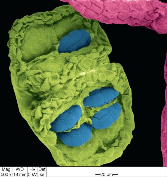
Image of the Week - December 30, 2019
CIL:41488 - http://www.cellimagelibrary.org/images/41488
Description: Colorized scanning electron micrograph of pollen (blue) in anther (green) held by the filament (pink ) from Acacia dealbata grown in Ecuador, Provincia Pichincha.
Authors: Alexis Debut, Sara Guerra, and David Cabrera (graphic designer)
Licensing: Attribution Non-Commercial Share Alike: This image is licensed under a Creative Commons Attribution, Non-Commercial, Share Alike License.
68 notes
·
View notes
Photo

Image of the Week – December 16, 2019
CIL:11397 - http://cellimagelibrary.org/images/11397
Description: Figure 218 from Chapter 7 (Mitochondria) of 'The Cell, 2nd Ed.' by Don W. Fawcett M.D. The typical structural organization of mitochondria is revealed in this longitudinal section of a mitochondrion and surrounding cytoplasm from pancreas of the bat, Myotis lucifugus. It is bounded by a smooth outer membrane about 7nm thick. An inner membrane has infoldings called cristae that project into the interior of the organelle.
Authors: Keith Porter
Licensing: Attribution Non-Commercial No Derivatives: This image is licensed under a Creative Commons Attribution, Non-Commercial, No Derivatives License
74 notes
·
View notes
Photo

Find this and other great images in the Technology Networks new The Spectacular World of Aquatic Organisms Flipbook.
Image of the Week - November 25, 2019
CIL:41017 - http://www.cellimagelibrary.org/images/41017
Description: Laser scanning confocal micrograph of a water flea, Daphnia atkinsoni. This specimen has a "crown of thorns," a defensive trait induced in offspring only when the parents sense chemical cues released by one of their main predators, the tadpole shrimp Triops cancriformis. The water flea's exoskeleton (exterior structure, green) and subcellular details within the organism (nuclei - tiny blue dots) are both visible. First Prize, 2009 Olympus BioScapes Digital Imaging Competition®.
Authors: Jan Michels and the 2009 Olympus BioScapes Digital Imaging Competition
Licensing: Attribution Non-Commercial No Derivatives: This image is licensed under a Creative Commons Attribution, Non-Commercial, No Derivatives License
The Spectacular World of Plants Flipbook -https://go.technologynetworks.com/the-spectacular-world-of-aquatic-organisms-flipbook
42 notes
·
View notes
Photo

Image of the Week – November 18, 2019
CIL:41311 - http://cellimagelibrary.org/images/41311
Description: Scanning electron microscope image of Penta lanceolata stigma (receptive surface for pollen).
Authors: Louisa Howard and Charles Daghlian
Licensing: Public Domain: This image is in the public domain and thus free of any copyright restrictions. However, as is the norm in scientific publishing and as a matter of courtesy, any user should credit the content provider for any public or private use of this image whenever possible.
54 notes
·
View notes
Photo

Image of the Week – November 11, 2019
CIL:50647 - http://www.cellimagelibrary.org/images/50647
Description: Human umbilical vein endothelial cells stained initially for nuclei with DAPI (blue) and for vascular endothelial cadherin (red).
Authors: Ashwin Inala and Eileen Shiuan
Licensing: Public Domain: This image is in the public domain and thus free of any copyright restrictions. However, as is the norm in scientific publishing and as a matter of courtesy, any user should credit the content provider for any public or private use of this image whenever possible.
46 notes
·
View notes
Photo

Spooky Eyes - Happy Halloween - Image of the Week - October 28, 2019
CIL:39022 - http://cellimagelibrary.org/images/39022
Description: Confocal micrograph showing the connections of the visual system in a four-day-old zebrafish embryo. Staining of the neurons, glia and optic nerve illustrate the connections between the retina and the brain. The retina is a multi-layered structure of retinal neurons that processes light information in the eye. The ganglion cell layer is closest to the bulb of the eye moving out towards the layer that contains the light sensitive cells - the rods and cones - towards the back of the retina. Both the retinal ganglion cell layers and photoreceptor cell layers are shown in cyan. The glial cells are shown in purple. The optic nerve (cyan) transmits information from the eye to the brain. The point at which the optic nerves meet is known as the optic chiasm.
Authors: Kara Cerveny and Steve Wilson
Licensing: Attribution-NonCommercial-NoDerivs 2.0 UK: England & Wales (CC BY-NC-ND 2.0 UK)
44 notes
·
View notes
Photo

Image of the Week – October 21, 2019
CIL:48108 - http://cellimagelibrary.org/images/48108
Description: Differentiated rat neural stem cells stained for β III-Tubulin (in green) to reveal neurons, GFAP (in red) to reveal glial cells, and DAPI (in blue) to label nuclei.
Authors: Natalie Prigozhina
Attribution Only: This image is licensed under a Creative Commons Attribution License.
38 notes
·
View notes