#scanning electron microscope
Text
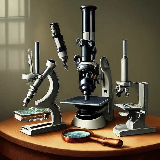
A Journey into the World of Microscopy: From Humble Beginnings to High-Tech Magnification
The science of looking into the hidden invisible Microscopy has transformed our understanding of the world around us. It can explore the universe beyond the reach of our naked eyes, with complex cellular structures, red blood cells, viruses and other viruses and microorganisms taking on amazing perspectives
The history of the microscope is a fascinating story of human curiosity, scientific genius, and relentless exploration. From the humble beginnings of simple magnifying glasses to the sophistication of modern electronic microscopes, the invention of microscopes has shaped our understanding of the microscopic world
In the 1600s, Dutch opticians such as Hans and Zachary Janssen are credited with inventing the first microscope. Known for this hybrid microscope, many lenses were used to magnify objects up to 30 times.At the end of the 17th century, Antony van Leeuwenhoek, Dutch draper some changed our perception of thumbnails. Armed with a well-made single-lens microscope, and explored the hidden reaches of nature. In 1674, Leeuwenhoek discovered microorganisms in lake water, which he aptly named “animalcules”. His discovery laid the foundations of biology and inspired generations of scientists. This incredible feat allowed him to uncover a hidden universe – the first sightings of bacteria, red blood cells, and other microorganisms.
Formation of the scientific environment (17th-19th centuries): Leeuwenhoek’s discoveries boosted scientific research. Robert Hooke, an English scientist, established these developments. In 1665, his book "Micrographia" recorded his observations with a compound microscope. Notably, the term "cell" was coined by Hooke when he examined cork tissue, laying the foundation for cell biology.Microscope systems flourished throughout the 18th and 19th centuries Joseph Lister and other scientists addressed the limitations of the early lenses, introducing improvements that reduced image distortion.
Beyond the Limits of Light: The Beginning of the New Age (19th-20th century): As the 19th century progressed, the limitations of optical microscopy became apparent and scientists yearned for a tool which can go deeper into cells. This research culminated in the development of the electron microscope in the 1930s. The 20th century was revolutionary with the invention of the electron microscope. Unlike light microscopes, which use visible light, electron microscopes use electron beams to achieve much higher magnification.Formation of the scientific environment (17th-19th centuries): Leeuwenhoek’s discoveries boosted scientific research. Robert Hooke, an English scientist, established these developments. In 1665, his book "Micrographia" recorded his observations with a compound microscope. Notably, the term "cell" was coined by Hooke when he examined cork tissue, laying the foundation for cell biology.Microscope systems flourished throughout the 18th and 19th centuries Joseph Lister and other scientists addressed the limitations of the early lenses, introducing improvements that reduced image distortion.
Beyond the Limits of Light: The Beginning of the New Age (19th-20th century): As the 19th century progressed, the limitations of optical microscopy became apparent and scientists yearned for a tool which can go deeper into cells. This research culminated in the development of the electron microscope in the 1930s. The 20th century was revolutionary with the invention of the electron microscope. Unlike light microscopes, which use visible light, electron microscopes use electron beams to achieve much higher magnification.
In the 1930s, German experts Max Knoll and Ernst Ruska made the first electron microscope. This tool let us see tiny things like cells and even atoms by using electron beams, not light, getting images many times bigger. This cool invention showed us the tiny parts inside cells, viruses, and stuff too small to see before. The 1900s brought even more cool microscopes. New kinds like phase-contrast and confocal microscopy let scientists look at live cells without using stuff that could hurt them. Now, the world of looking at tiny things is getting even better. Today, we have high-tech microscopes that use computers and lasers. These let us see and even change tiny things in ways we never could before.
Modern Microscopy's Diverse Arsenal - Today, the field of microscopy boasts a diverse range of specialized instruments, each tailored to address specific scientific needs. Here's a glimpse into some remarkable examples:
Scanning Electron Microscope (SEM): Imagine a high-tech camera that captures images using a beam of electrons instead of light. That's the essence of a SEM. By scanning the surface of a sample with a focused electron beam, SEMs generate detailed information about its topography and composition. This makes them ideal for studying the intricate structures of materials like insect wings, microchips, and even pollen grains.
Transmission Electron Microscope (TEM): While SEMs provide exceptional surface detail, TEMs take us a step further. They function by transmitting a beam of electrons through a very thin sample, allowing us to observe its internal structure. TEMs are the go-to instruments for visualizing the intricate world of viruses, organelles within cells, and macromolecules like proteins.
Confocal Microscopy: Ever wished to focus on a specific layer within a thick biological sample and blur out the rest? Confocal microscopy makes this possible. It utilizes a laser beam to precisely illuminate a chosen plane within the sample, effectively eliminating information from out-of-focus regions. This allows researchers to create sharp, three-dimensional images of cells, tissues, and even small organisms.
Atomic Force Microscopy (AFM): This technique takes a completely different approach, venturing into the realm of physical interaction. AFM employs a tiny cantilever, akin to a microscopic feeler, to physically scan the surface of a sample. By measuring the minute forces between the cantilever and the sample's surface, AFM can map its topography at an atomic level. This provides invaluable insights into the properties of materials at an unimaginable scale, making it crucial for research in fields like nanotechnology and surface science.
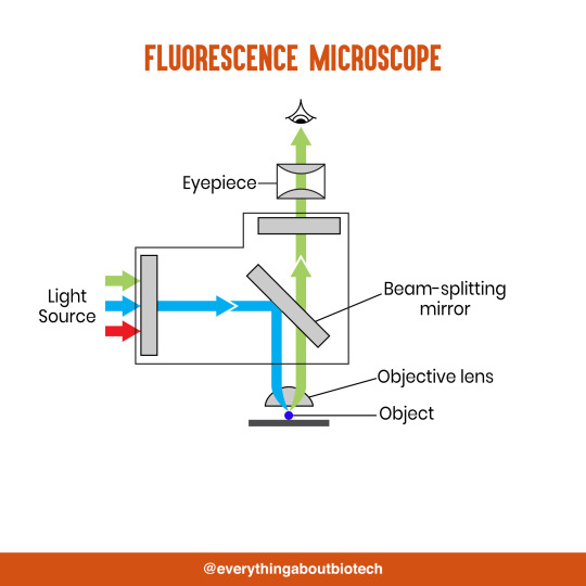
Fluorescence Microscopy: Imagine illuminating a sample with specific wavelengths of light and observing it glowing in response. That's the essence of fluorescence microscopy. This technique utilizes fluorescent molecules or tags that bind to specific structures within a cell or tissue. When excited by light, these tags emit their own light, highlighting the target structures with remarkable clarity. This allows researchers to visualize specific proteins, DNA, or even pathogens within biological samples.
Super-resolution Microscopy (SRM): Overcoming the limitations imposed by the wavelength of light, SRM techniques like STED (Stimulated Emission Depletion) and PALM (Photoactivated Localization Microscopy) achieve resolutions surpassing the diffraction limit. This allows researchers to visualize structures as small as 20 nanometers, enabling the observation of intricate cellular machinery and the dynamics of individual molecules within living cells.
Cryo-Electron Microscopy (Cryo-EM): This powerful technique takes a snapshot of biological samples in their near-life state. Samples are rapidly frozen at ultra-low temperatures, preserving their native structure and minimizing damage caused by traditional fixation methods. Cryo-EM has been instrumental in determining the three-dimensional structures of complex molecules like proteins and viruses, providing crucial insights into their function and potential drug targets.
Correlative Microscopy: Combining the strengths of multiple microscopy techniques, correlative microscopy offers a comprehensive view of biological samples. For instance, researchers can utilize fluorescence microscopy to identify specific structures within a cell and then switch to electron microscopy to examine those structures in high detail. This integrated approach provides a deeper understanding of cellular processes and their underlying mechanisms.
Light Sheet Microscopy (LSM): Imagine illuminating a thin slice of a sample within a living organism. LSM achieves this feat by focusing a laser beam into a thin sheet of light, minimizing photobleaching and phototoxicity – damaging effects caused by prolonged exposure to light. This allows researchers to observe dynamic processes within living organisms over extended periods, providing valuable insights into cellular behavior and development.
Expansion Microscopy (ExM): This innovative technique physically expands biological samples by several folds while preserving their structural integrity. This expansion allows for better resolution and visualization of intricate cellular structures that would otherwise be difficult to distinguish using traditional microscopy methods. ExM holds immense potential for studying the organization and function of organelles within cells.
Scanning Near-Field Optical Microscopy (SNOM): This innovative technique pushes the boundaries of resolution by utilizing a tiny probe that interacts with the sample at an extremely close range. SNOM can not only image the surface features of a sample with exceptional detail but also probe its optical properties at the nanoscale. This opens doors for research in areas like material science and photonics, allowing scientists to study the behavior of light at the interface between materials.
X-ray Microscopy: Stepping outside the realm of light and electrons, X-ray microscopy offers unique capabilities. By utilizing high-energy X-rays, this technique can penetrate deep into samples, making it ideal for studying the internal structure of dense materials like bones and minerals. Additionally, it allows for the visualization of elements within a sample, providing valuable information about their distribution and composition.
From revealing the building blocks of life to aiding in the development of new medicines, the microscope has played an undeniable role in shaping our scientific understanding. As technology continues to evolve, one can only imagine the future breakthroughs this remarkable invention holds in unveiling the secrets of our universe, both seen and unseen. These advancements hold the potential to revolutionize our understanding of biological processes, develop new materials with extraordinary properties, and ultimately pave the way for breakthroughs in medicine, nanotechnology, and countless other fields. As we continue to refine and develop novel microscopy techniques and the future holds immense promise for further groundbreaking discoveries that will undoubtedly revolutionize our perception of the world around us.
#science sculpt#life science#science#molecular biology#biology#biotechnology#artists on tumblr#microscopy#microscope#Scanning Electron Microscope#Transmission Electron Microscope#Confocal Microscopy#Atomic Force Microscopy#Fluorescence Microscopy#Expansion Microscopy#X-ray Microscopy#Super-resolution Microscopy#Light Sheet Microscopy#illustration#illustrator#illustrative art#education#educate yourself#techniques in biotechnology#scientific research#the glass scientists#scientific illustration#scientific advancements
6 notes
·
View notes
Text

Don’t let the purple hue fool you – this image captured by a scanning electron microscope is actually showing us mesoporous gold nanoparticles.
Photograph: Javeria Bashir/AIBN
2023 Australian Institute For Bioengineering And Nanotechnology Image Contest
#javeria bashir#photographer#australian institute for bioengineering and nanotechnology image contest#micro photography#scanning electron microscope#mesoporous gold nanoparticles#nature
3 notes
·
View notes
Text
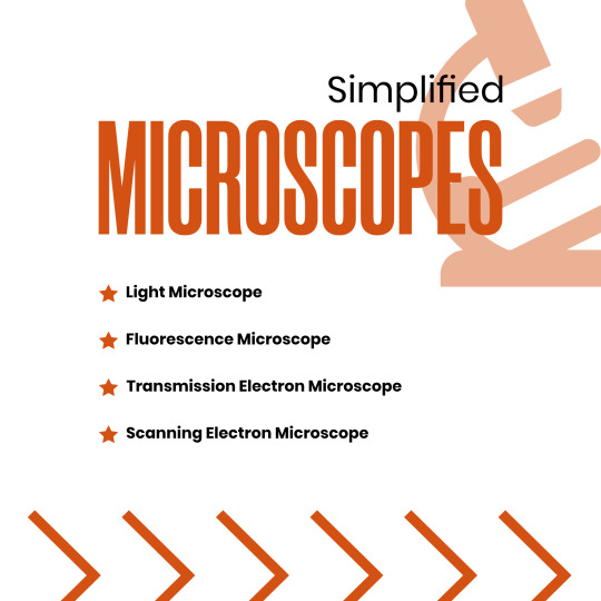


Microscopes Simplified
Light Microscope
Fluorescence Microscope
Transmission Electron Microscope
Scanning Electron Microscope
#microscope#light microscope#Fluorescence Microscope#Transmission Electron Microscope#Scanning Electron Microscope#microscopic#science#microscopy#microscopic life#biotechnology#biotech#biology#biochemistry#studyblr#molecularbiology#notes#class notes#students#science illustration#illustration#illustragram#scientific illustration#graphic design#graphicwork#study notes#study#study motivation#study tips#studygram
81 notes
·
View notes
Text
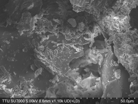


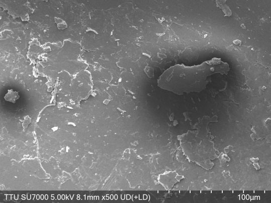
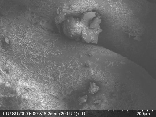


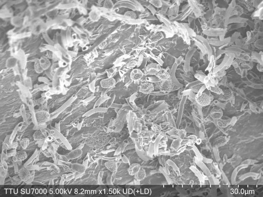

This came to mind, so I'm back to sharing some photos I and a friend took using a scanning electron microscope, this time it's cochineal beetle husks! I think some of these photos really highlight how scanning electron microscopy is so similar to both standard photography and what I view as the opposite end of the spectrum of astrophotography.
SEM -> Electrons are essentially shot at an object to 'scan' it, and then the way they scatter off the surface in a very precise manner is used to reconstruct the image. (I'm certain it's more complicated than this, but that's all you need to know for the basics)
This is why they're kept in vacuum, so that interfering air particles aren't likely to mess with the electron scattering. Plus, with organic materials like these beetle husks and anything else I share photos of (at least at present), they don't actually have enough conductivity intrinsically to properly deflect electrons!
So, they have to be coated with an incredibly thin layer (so as to preserve the actual image still, it's on the order of nanometers thin) of gold and palladium through a process called sputtering to allow for conductivity.
If a region is too "conductive" (It's been a minute, so I don't remember the exact terminology) under the settings with which you're trying to take a photo, the electrons can actually "stick" to the surface in a region, whiting it out and creating what's referred to as 'charging' effects. That's what causes some of the really sharp whites in some of these images! (It can also just be good clarity)
Additionally, with some of the photos (I think 7 and 8 really exemplify this the best with how it sort of looks like a cliff in the image going deeper into the husk), you can see how this can't reconstruct a perfectly in focus image at all 'heights'. The stuff that is put into an SEM if the vacuum doesn't have to be repulled is super tiny and very flat, but even then that very minor amount of height difference is clearly huge.
I just think it's not only really cool to see what stuff like this looks like, but also how similar it can be to standard photography!
5 notes
·
View notes
Text
Uses of Different Types of Microscopes in Forensics
Uses of Different Types of Microscopes in Forensics
The forensic and microbiological labs include a variety of microscopes that may be used. The value of microscopes is increased by how widely they may be used and modified.
#microscope #forensicscience
(more…)

View On WordPress
#comparison microscope#comparison microscope in Forensic Science#dark-field microscope in forensic science#Fluorescent microscope#Forensic science#forensic science notes#microscopy notes#polarising microscope#Scanning Electron Microscope#stereoscopic microscope#stereoscopic microscope uses in forensic science#Transmission Electron Microscope#use of compound microscope in forensic science#use of digital microscope in forensic science#use of fluorescent microscope in forensic science#use of inverted microscope in forensic science#use of phase contrast microscopy in forensic science#use of polarising microscope in forensic science#use of scanning electron microscope#use of scanning electron microscope in forensic science#Uses of Different types of Microscope In Forensics
7 notes
·
View notes
Photo
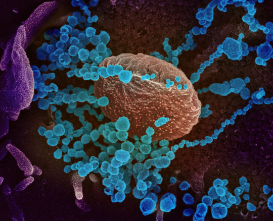
COVID captured under the scanning electron microscope. View more images of Covid under the microscope here.
23 notes
·
View notes
Text
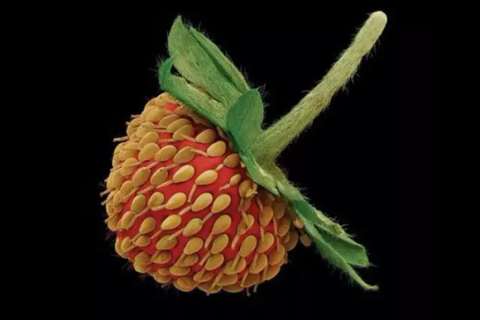
This Nature is Ready for Its Close Up
Micrograph of a strawberry by Rob Kesseler
Fabulous images of nature, really really up close. via DesignBoom:
In order to create the eclectic visual imagery, the original samples are sprayed with a fine coating of gold, then photographed on a scanning electron microscope. Called micrograph artwork, the high resolution and colorful samples represent just a tiny fraction of the unique…
View On WordPress
#magnified nature#microscopic#nature magnified#Rob Kesseler#Scanning Electron Microscope#seeds under microscrope
0 notes
Text

0 notes

Text
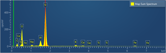
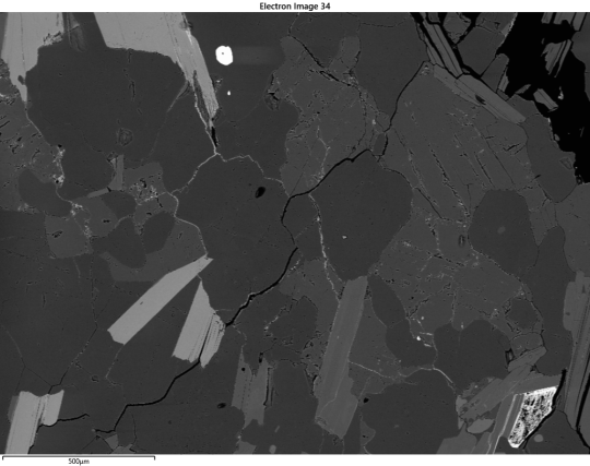


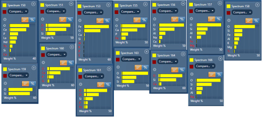
Finally got to look over the SEM data for ec-501 and. HRM.
Like 1, the topography map is surprising because I expected more holes. 501 is a pretty soft sample!
The specimen samples are both suprising and not suprising. Monazite? Annoying but sure, thoigh I didn't expect the La and Nd(neodynium my beloved).
I know the smaller samples are a little fucked, but they are mostly just to help confirm. Like yeah, 151 is very much definitely possibly zircon! But it's so goddamn tiny that Aztec really said "bruh idk???"
We got the two micas and quartz, no surprise. We got plag! And then what the FUCK is going on with 155, 160, 161, and 163? I assume 161 is more zircon. but whay the fuck could the others be??
Anyways, tadah! My new data to interpret. I can pop open aztec later for more details, including individual element maps.
2 notes
·
View notes
Photo
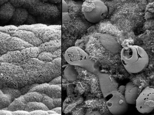
PET Caterpillars
Usually found in hospitals, positron emission tomography (PET) scanners are chamber-sized devices that help to diagnose diseases, spotting changes in how tissues metabolise or break down chemical tracers in a similar way to sugars. The approach helps millions of people during diagnosis, but using PET for research on mammals is expensive with many ethical issues. On the hunt for alternatives, scientists turn to caterpillars. Similar enough to humans in their genetics and large enough that their insides can be inspected by PET, the larvae of the tobacco hornworm (Manduca sexta) make excellent model organisms. Here, using electron microscopy the gut of a healthy caterpillar (left) is compared with cells changed by a bacterial infection (right) – mimicking inflammation seen in colitis – which can be reversed with antibiotics. Caterpillars may have lots to teach us not only about how certain inflammatory diseases progress, but also how researchers might improve and expand on techniques like PET.
Written by John Ankers
Image from work by Anton G. Windfelder and colleagues
Institute of Zoology and Developmental Biology; Cellular Recognition and Defense Processes, Justus Liebig University Giessen and Experimental Cardiovascular Imaging, Molecular Cardiology, Heinrich Heine University Düsseldorf, Germany
Image originally published with a Creative Commons Attribution 4.0 International (CC BY 4.0)
Published in Nature Communications, November 2022
You can also follow BPoD on Instagram, Twitter and Facebook
#science#biomedicine#caterpillars#pet scan#positron emission tomography#electron microscope#colitis#gut#intestine#microbiome
4 notes
·
View notes
Text
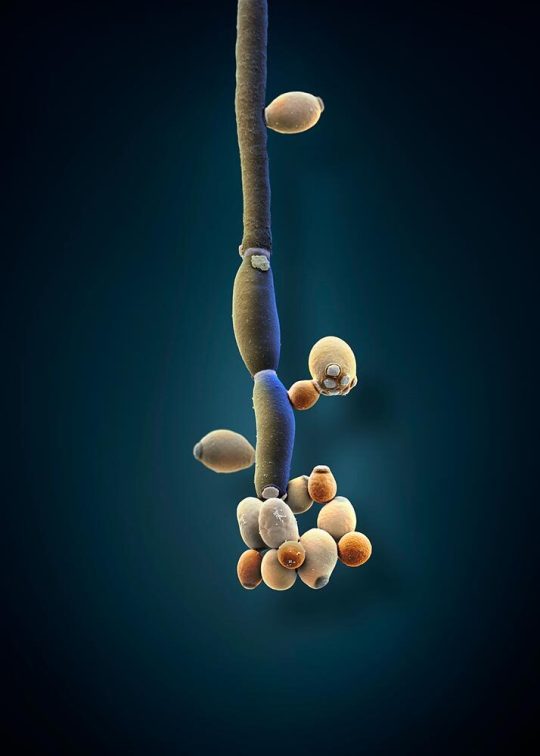
Studies show that patients with irritable bowel syndrome tend to have high levels of the fungus Candida albicans (illustrated here) in their gut. In recent years, scientists have started to take a closer look at how the fungi in your gut microbiome affect your health.
COLORIZED SCANNING ELECTRON MICROSCOPE IMAGE BY MARTIN OEGGERLI
UNIVERSITY HOSPITAL BASEL, SWISS NANOSCIENCE INSTITUTE, BASEL
#martin oeggerli#colorized scanning electron microscope#photographer#micro photography#university hospital basel#swiss nanoscience institute#basel#switzerland#irritable bowel syndrome#candida albicans#fungus#nature#microbiome#national geographic
10 notes
·
View notes
Text
Unlocking the Microscopic World: Exploring the Applications and Functionality of Scanning Electron Microscopes
Delving into the intricate details of the material world requires advanced tools that can unveil the hidden dimensions invisible to the naked eye. Among these, the Scanning Electron Microscope (SEM) stands as a pinnacle of scientific ingenuity, enabling researchers to peer into the microcosm with unparalleled clarity and precision.
Understanding the Mechanics of Scanning Electron Microscopes
At the heart of every SEM lies an electron gun, a marvel of engineering that emits a stream of high-energy electrons. These electrons, directed by sophisticated electromagnetic lenses, form a focused beam aimed at the specimen under study. To ensure an unimpeded path for the electrons, the specimen resides within a vacuum chamber, devoid of air molecules.
As the electron beam traverses the specimen's surface, scanning coils meticulously guide its movements in a predetermined pattern. Through this dance of electrons, a cascade of interactions occurs, generating various signals that capture the essence of the specimen's topography and composition. These signals, including secondary electrons, backscattered electrons, and characteristic X-rays, converge to produce monochrome images teeming with intricate detail and depth.
The Multifaceted Applications of Scanning Electron Microscopes
Materials Science: In the realm of materials science, SEM serves as a beacon illuminating the microstructural landscape of metals, ceramics, and polymers. By scrutinizing grain boundaries, phase distributions, and particle arrangements, researchers glean invaluable insights into material properties and failure mechanisms.
Nanotechnology: SEM's prowess extends to the nanoscale, where it unveils the morphology and distribution of nanoparticles and nanostructures, laying the groundwork for groundbreaking advancements in nanotechnology.
Biology and Life Sciences: Within the realm of biology, SEM unveils a panorama of cellular surfaces, tissues, and biological structures, elucidating their intricate topographies and fostering a deeper understanding of microbial morphology and interactions.
Geology and Earth Sciences: Geologists harness SEM to unravel the mysteries of minerals, rocks, and fossils, unraveling their composition, structure, and geological genesis.
Semiconductor Industry: SEM emerges as a stalwart ally in the semiconductor industry, facilitating quality control, defect identification, and precision measurement of microelectronic components.
Archaeology and Cultural Heritage: In the realm of archaeology, SEM serves as a time machine, offering glimpses into the manufacturing techniques, corrosion processes, and material compositions of ancient artifacts.
Forensic Science: SEM's forensic utility shines bright, aiding investigators in scrutinizing trace evidence and unraveling intricate clues pivotal in criminal investigations.
Material Coating and Surface Modification: SEM serves as a discerning eye in material coating and surface modification studies, enabling researchers to analyze coating thickness, uniformity, and adhesion with unparalleled precision.
Empowering Researchers and Educators Alike
For researchers, SEM embodies a gateway to discovery, offering high-resolution imaging and analytical capabilities crucial in interdisciplinary research endeavors. Likewise, for students, SEM fosters a hands-on learning experience, nurturing a new generation of scientists adept at navigating the intricate realms of materials science, nanotechnology, and beyond.
Exploring the Spectrum of Scanning Electron Microscopes
From conventional SEMs to specialized variants like Environmental SEMs (ESEMs) and Field Emission SEMs (FESEMs), the SEM landscape encompasses a spectrum of instruments tailored to diverse research needs and applications.
Experience Innovation with VBCC Research
At VBCC Research, we catalyze scientific exploration through state-of-the-art facilities and a dedicated team of experts. Our Benchtop Scanning Electron Microscope (SEM) epitomizes versatility and efficiency, boasting intuitive touch-panel operation and rapid image acquisition capabilities. With features such as high and low vacuum modes and optional Energy Dispersive X-ray Spectroscopy (EDS), our SEM empowers researchers to unlock new frontiers in scientific inquiry.
Conclusion
Scanning Electron Microscopes stand as marvels of modern science, transcending the limits of human perception to reveal the hidden intricacies of the microcosm. With their multifaceted applications and transformative capabilities, SEMs herald a new era of discovery, empowering researchers and students alike to unravel the mysteries of the microscopic world.
0 notes
Text
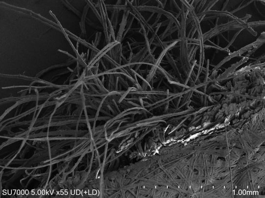


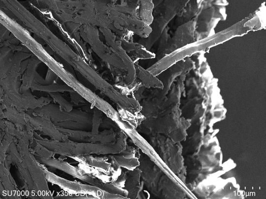
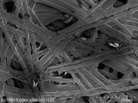
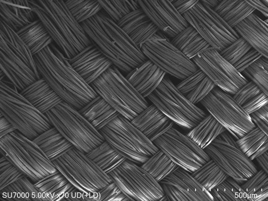
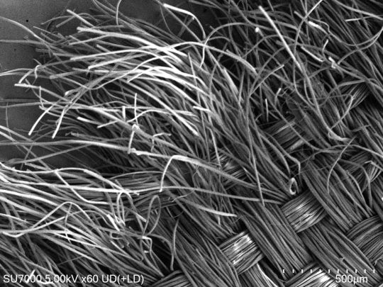
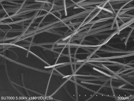
Silkworm cocoon and woven silk fibers under a scanning electron microscope!
I came across these looking for my astrophotography and figured I might as well post them too.
SEMs aren't common in my field, but I was taking a weird topics course which gave me the opportunity to take these photos! Wonderfully cool piece of technology
I had to tear open the cocoon with an x-acto knife myself immediately before this, which was... a choice on my part (how the heck did I forget silkworm cocoons are that tough), but it's also the reason for the rough edges.
Being able to see the difference between the cocoon and woven silk at this level is such a cool comparison.
SEM photos and astrophotography photos are both generally rendered at least initially in black and white, but we have methods that make it much easier to false color astrophotography compared to SEM photos. The technological similarities and differences are actually so cool to me, because it's like yea, big machine for camera for tiny thing and big machine for camera for very big very far thing would sensibly be somewhat similar!
1 note
·
View note
Last Seen Blogs

nishp
Celebrating the National Days

bobo655
Untitled


ramankj
Untitled

breenbowen
The Journaling of Burch 965