#Glia gifs
Text


Arnas Fedaravicius // Glia
Taglist: @sihtricfedaraaahvicius @whumpappreciation @siimonesvensson @gloriouslyalivetoday @melissarose234 @crusader1997 @sivulele @gemini-mama @bathedinheat @vashole @losstboi @fox-bright @whumpybromance @umfood @elwegencyn @keenbagelsharkbanana @the-irish-girl @tinumiel @hb8301 @miss-sparkel-mr-hitch @simpforfictionalaisela25 @alexagirlie @uunotheangel
60 notes
·
View notes
Text


ARNAS FEDARAVICIUS as Alex Summers
Glia (2020)
90 notes
·
View notes
Photo


Drix’s introduction
16 notes
·
View notes
Text
Study: Common pesticide may have made the Zika epidemic worse

- By Pieter Vancamp , Barbara Demeneix , Muséum national d’histoire naturelle (MNHN) , The Conversation -
Before Covid, there was Zika. In 2015, the number of babies born in Brazil with small heads and brains – a condition known as microcephaly – suddenly increased dramatically. The severe deformities left the children disabled for life, and caused intense global concern.
These cases of microcephaly were soon shown to be caused by pregnant women being infected with the mosquito-borne Zika virus. The virus infects and kills cells that form the brain, hampering its proper development.
But the impact was not universal – certain regions in north-eastern Brazil saw far more cases of microcephaly than others.
Scientists began to question whether other factors might be at play that intensified the epidemic in some places. Not long after, they focused their attention on pyriproxyfen, a globally approved pesticide used against household insects in agriculture – including mosquitoes. Pyriproxyfen was used intensively in the regions with the highest numbers of microcephaly cases.
Now, in a new study, we have shown that pyriproxyfen could indeed exacerbate the already severe effects the Zika virus has on foetal brain development.
Pyriproxyfen and Zika
In late 2014, pyriproxyfen was introduced to drinking water throughout Brazil to control the Aedes aegypti mosquito population, which spreads the Dengue and Zika viruses.
But the insecticide is known to accumulate in the environment for years, eventually finding its way into the human body. So to prevent potential side-effects, the World Health Organisation had recommended the daily intake of pyriproxyfen should not exceed 0.3 mg/L for an average adult, and concentrations in drinking water should be lower than 0.01 mg/L.
As babies and foetuses generally absorb or accumulate more chemicals than adults, they can be at greater risk. And because there was a strong geographical overlap between the use of pesticides and microcephaly cases, even these small doses raised issues concerning safety.
Epidemiological and experimental studies have since delivered contradictory results regarding whether or not pyriproxyfen was implicated in the disease outbreak. But in our recently published study, we discovered that pyriproxyfen impairs thyroid hormone signalling in the brain, modifying crucial processes for its proper development.
How pyriproxyfen affects brain development
Thyroid hormone is an important molecule that helps shape the foetal brain. Without it, the brain doesn’t grow as it’s supposed to, leaving affected children with learning difficulties for life.
In the lab, we house genetically modified tadpoles that emit green fluorescence when exposed to thyroid hormone. More thyroid hormone means more green signal, indicating the hormone is active in the tadpoles’ cells.
When we exposed our tadpoles to pyriproxyfen, the green signal dropped dramatically. This demonstrated that the pesticide blocks thyroid hormone action.

Genetically modified tadpoles emit green fluorescence when exposed to thyroid hormone (T3 in the image). When they were simultaneously exposed to pyriproxyfen, the fluorescent signal dropped significantly, showing the insecticide blocks thyroid hormone action. Petra Spirhanzlova, MNHN
One of the most important roles of thyroid hormone is to generate a balanced amount of nerve cells and their supporting cells, known as glia. Together, they form the essential building blocks of the brain.
Since pyriproxyfen blocks normal thyroid hormone action, we thought it might affect the generation of neurons and glial cells too. To investigate this, we used stem cells cultured from mouse brains, and exposed them to increasing doses of the pesticide.
The findings were clear: the higher the dose, the fewer cells were generated, and the more they died, producing an unbalanced proportion of nerve cells and glia.
The brains of the exposed tadpoles in our study did not develop normally, and made them behave unnaturally. Underlying these changes were altered expression patterns of a number of genes.
A key player in this was the gene Msi1, which encodes a protein called Musaschi-1. The Zika virus uses this protein to replicate and infect other cells, and we knew from previous studies that more thyroid hormone resulted in less Musaschi-1.
Not surprisingly, the cells that survived pesticide exposure contained much more of the protein due to blocked thyroid hormone action. That’s why we hypothesised that, through increasing Musaschi-1, pyriproxyfen could allow the virus to replicate faster.
To test this, we infected exposed stem cells with the Zika virus and found that the transcription of key genes was altered compared to when they were infected with the virus alone and not exposed to the pesticide. Although we didn’t observe higher infection rates, it’s still possible that pesticide exposure could aggravate hampered brain development. That can worsen the impact the virus has on a child’s later-life intellectual capacities, and requires further investigation.
Regulating pesticides
This is not the first time a pesticide has been shown to alter the course of a disease.
For example, individuals with higher amounts of another chemical that disrupts the endocrine system, perfluorobutanoic acid, in their blood, are at risk of a more severe course of Covid-19.
But for many other ubiquitous pesticides to which we are continuously exposed in our daily life, we have no clue as to how they affect us, and whether they interact with viral diseases.
We need better testing protocols for pesticides to obtain more solid data to inform health policies and decision makers.
As for pyriproxyfen, Europe is not proposing to use it at concentrations advised by the World Health Organization, but recently reauthorised it.
Our study emphasises again how little we know about the harmful effects of pesticides on human health, notably on brain development, but also the natural environment as a whole.

Pieter Vancamp, Post-doctorant, Muséum national d’histoire naturelle (MNHN) and Barbara Demeneix, Professor Physiology, Endocrinology, Muséum national d’histoire naturelle (MNHN)
This article is republished from The Conversation under a Creative Commons license. Read the original article.
--
Read Also
Mapping dengue hot spots determines risks for Zika and chikungunya
#brain#zika#epidemics#health#brazil#pyriproxyfen#virus#pesticides#microcephaly#pediatrics#neuroscience#medicine#thyroid#genetics#pollution#infectious diseases
0 notes
Photo


Answers to the Friday Brain Tickler
1. Sympathetic Chain Ganglion
The place where pre-synaptic and post-synaptic neuron cell bodies synapse. The neurons here are motor neurons famous for their involvement in the involuntary actions that occur during the fight or flight response: dilate pupil, increase heart rate, dilate bronchioles and blood vessels etc.
How can you tell this from the histology?
Neuron cell bodies are large and have a prominent nucleus and nucleolus that look just like eyes.
Neuron cell bodies in peripheral ganglia are surrounded by satellite cells, small supportive glia.
Neuron cell bodies in sympathetic chain ganglia are separated from each other by axons and their nuclei are what I call ‘shifty eyes’ they won’t look straight at you!
2. Myocardium (cardiac muscle)
The cells that keep you alive by contracting to make your heart beat
How can you tell this from the histology?
Large pink cells - the eosinophilia is due to the high protein (actin and myosin content).
The nucleus is commonly centrally located in the cytoplasm and there is space between the side of the nucleus and the edge the plasma membrane.
There isn’t much connective tissue but plenty of capillaries between cells
3. Dorsal Root (Sensory) Ganglion
Where the cell bodies of all the neurons that carry sensory information back to your spinal cord and brain live.
How can you tell this from the histology?
Same as 1 except the neurons are instead clustered together and their eyes stare right at you! Creepers.
4. Intervertebral Disc
The strong fibrous joint between the vertebral bodies in your spinal cord that resist compressive forces and tensile forces when you move.
How can you this tell from the histology?
This is fibrocartilage. It contains both Type I collagen fiber bundles (pink fibrous material) and scattered chondrocytes within lacunae (the large round cells).
5. Epiphyseal Growth Plate
The region of your bones that allows you to grow in length as a child. It fuses and becomes bone after puberty.
How can you tell this from the histology?
Hyaline cartilage that contains chondrocytes with one distinct area (zone of proliferation) where dividing chondrocytes look like a stack of pancakes.
Hope you enjoyed the histological mentalism!
i♡histo
#histology#pathology#science#med school#med student#vet science#vet school#education#quiz#nursing#nurse#ihearthisto
4 notes
·
View notes
Photo

Na primeira aula de biologia apredemos sobre os tipos de tecidos existentes em seres vertebrados, sendo eles os tecidos epitelial, muscular, conjuntivo e nervoso. Todos esses tecidos são importantes para o funcionamento de nossos corpos, e em alguns casos dependentes.
O tecido epitelial tem a função de absorver e secretar substâncias, de proteger e de sentir as sensações. A proteção é feita por meio do revestimento que cobre tanto a superficie externa (pele), quanto os tratos digestório, respiratório e urogenital, as cavidades corporais, os tubos, os ductos e os vasos sanguinios e linfaticos. Assim vemos a relção do tecido epitelial sobre o tecido conjuntivo. O fato de ter a percepção dos sentidos deve-se aos orgãos do sentido, sendo as células epiteliais especializadas como as células olfatórias que captam os estímos do ambiente.
O tecido muscular apresenta a função de movimento, essas célualas são chamadas de fibras musculares e têm a capacidade de contrair e alongar. Das estruturas ligadas a ele e existem três tipos em que o movimento atua, no esqueletico que são os musculos do sistema locomotor, no cardiaco que são os musculos do coração exclusivamente e o liso que cuida da parte dos vasos e vícera e da derme da pele.
Tecido conjuntivo é o responsavel pela conexão, sustentação e preenchimento. Esse tecido é composto por células e por matriz extracelular. A composição diferenciada da sua matriz extracelular faz com que absorva impactos,resista à tração ou tenha elasticidade. Além de proporcionar suporte estrutural ao tecido, ela regula o comportamento das células, influenciando sua proliferação, diferenciação,migração, morfologia, atividade funcional e sobrevivência. As principais células do tecido conjuntivo são: as células mesenquimais, os fibroblastos, os plasmócitos, os macrófagos, os mastócitos, as células adiposas e os leucócitos.
E o último tecido é o Tecido nervoso, que no meu ver é o mais interessante e o mais importante, sem ele não seriamos capazes de fazer coisas simples, já que ele comanda as ações voluntárias e involutárias do nosso corpo. Sua função é receber informações do meio ambiente atraves dos sentidos e do meio interno, processamento e armazenamento de informações. Esse tecido está interligado ao sistema nevoso. O tecido nervoso é composto pelos neurônios e as céluas da glia. Nos possuimos cerca de 85 milhões de neurônios que se comunica e compreendeinformações por meio dos impulsos nervosos.
Esse foi um breve resumos sobre tecidos que foi dito na primeira aula de biologia celular, onde foi introduzido e apresentado seus significados e funções.
#http://www.ufrgs.br/livrodehisto/#https://pt.slideshare.net/andrezacampos14/tecido-nervoso-54353406?qid=babe5614-e35e-4794-b1e8-bf6e1f985fb5&v=&b=&from_search=8
0 notes

Text
youtube
glia set on lines, 2/24/2024
0 notes
Text

Glia Diversity
Single-cell analysis of fruit fly glia – types of non-neuronal cells of the nervous system – reveals more diversity in structure and shape (morphology) than in signatures of gene expression [activity]
Read the published research paper here
Image from work by Inês Lago-Baldaia and colleagues
Department of Cell and Developmental Biology, University College London, London, UK
Image originally published with a Creative Commons Attribution 4.0 International (CC BY 4.0)
Published in PLOS Biology, October 2023
You can also follow BPoD on Instagram, Twitter and Facebook
6 notes
·
View notes
Text
#glia #shoegaze
3 notes
·
View notes


Text


1 note
·
View note
Text
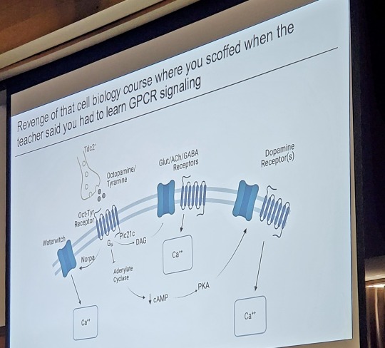
I love it when science talks are funny
#science#science talk#neuroscience#biology#glia#GPCR#laboratory#research#scientist#grad student#phd#phd student#gradblr#public speaking#presentations#ppt#hawtchocolate
79 notes
·
View notes
Text
#glia #shoegaze
10 notes
·
View notes
Last Seen Blogs

deliciousvoidbanana
Senza titolo

giballedrei
Post-Modernism

astrobei
were there boys in lenora?

o-boteco
UM MILHÃO DE FRACASSOS