#Scanning Electron Microscopes
Explore tagged Tumblr posts
Visit Tumblr Blog
Explore Tumblr blogs with no restrictions, modern design and the best experience.
Last Seen Tumblr Blogs


Fun Fact
Tumblr has a 66 index score for customer satisfaction in the US.
Text
Unlocking the Microscopic World: Exploring the Applications and Functionality of Scanning Electron Microscopes
Delving into the intricate details of the material world requires advanced tools that can unveil the hidden dimensions invisible to the naked eye. Among these, the Scanning Electron Microscope (SEM) stands as a pinnacle of scientific ingenuity, enabling researchers to peer into the microcosm with unparalleled clarity and precision.
Understanding the Mechanics of Scanning Electron Microscopes
At the heart of every SEM lies an electron gun, a marvel of engineering that emits a stream of high-energy electrons. These electrons, directed by sophisticated electromagnetic lenses, form a focused beam aimed at the specimen under study. To ensure an unimpeded path for the electrons, the specimen resides within a vacuum chamber, devoid of air molecules.
As the electron beam traverses the specimen's surface, scanning coils meticulously guide its movements in a predetermined pattern. Through this dance of electrons, a cascade of interactions occurs, generating various signals that capture the essence of the specimen's topography and composition. These signals, including secondary electrons, backscattered electrons, and characteristic X-rays, converge to produce monochrome images teeming with intricate detail and depth.
The Multifaceted Applications of Scanning Electron Microscopes
Materials Science: In the realm of materials science, SEM serves as a beacon illuminating the microstructural landscape of metals, ceramics, and polymers. By scrutinizing grain boundaries, phase distributions, and particle arrangements, researchers glean invaluable insights into material properties and failure mechanisms.
Nanotechnology: SEM's prowess extends to the nanoscale, where it unveils the morphology and distribution of nanoparticles and nanostructures, laying the groundwork for groundbreaking advancements in nanotechnology.
Biology and Life Sciences: Within the realm of biology, SEM unveils a panorama of cellular surfaces, tissues, and biological structures, elucidating their intricate topographies and fostering a deeper understanding of microbial morphology and interactions.
Geology and Earth Sciences: Geologists harness SEM to unravel the mysteries of minerals, rocks, and fossils, unraveling their composition, structure, and geological genesis.
Semiconductor Industry: SEM emerges as a stalwart ally in the semiconductor industry, facilitating quality control, defect identification, and precision measurement of microelectronic components.
Archaeology and Cultural Heritage: In the realm of archaeology, SEM serves as a time machine, offering glimpses into the manufacturing techniques, corrosion processes, and material compositions of ancient artifacts.
Forensic Science: SEM's forensic utility shines bright, aiding investigators in scrutinizing trace evidence and unraveling intricate clues pivotal in criminal investigations.
Material Coating and Surface Modification: SEM serves as a discerning eye in material coating and surface modification studies, enabling researchers to analyze coating thickness, uniformity, and adhesion with unparalleled precision.
Empowering Researchers and Educators Alike
For researchers, SEM embodies a gateway to discovery, offering high-resolution imaging and analytical capabilities crucial in interdisciplinary research endeavors. Likewise, for students, SEM fosters a hands-on learning experience, nurturing a new generation of scientists adept at navigating the intricate realms of materials science, nanotechnology, and beyond.
Exploring the Spectrum of Scanning Electron Microscopes
From conventional SEMs to specialized variants like Environmental SEMs (ESEMs) and Field Emission SEMs (FESEMs), the SEM landscape encompasses a spectrum of instruments tailored to diverse research needs and applications.
Experience Innovation with VBCC Research
At VBCC Research, we catalyze scientific exploration through state-of-the-art facilities and a dedicated team of experts. Our Benchtop Scanning Electron Microscope (SEM) epitomizes versatility and efficiency, boasting intuitive touch-panel operation and rapid image acquisition capabilities. With features such as high and low vacuum modes and optional Energy Dispersive X-ray Spectroscopy (EDS), our SEM empowers researchers to unlock new frontiers in scientific inquiry.
Conclusion
Scanning Electron Microscopes stand as marvels of modern science, transcending the limits of human perception to reveal the hidden intricacies of the microcosm. With their multifaceted applications and transformative capabilities, SEMs herald a new era of discovery, empowering researchers and students alike to unravel the mysteries of the microscopic world.
0 notes
Text

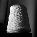
An SEM image of a guitar string held down with carbon tape to a sample mount.
24 notes
·
View notes
Text
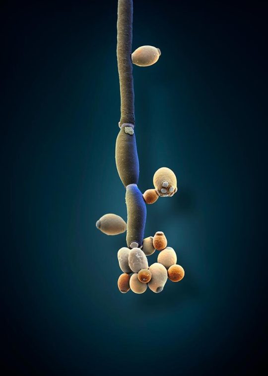
Studies show that patients with irritable bowel syndrome tend to have high levels of the fungus Candida albicans (illustrated here) in their gut. In recent years, scientists have started to take a closer look at how the fungi in your gut microbiome affect your health.
COLORIZED SCANNING ELECTRON MICROSCOPE IMAGE BY MARTIN OEGGERLI
UNIVERSITY HOSPITAL BASEL, SWISS NANOSCIENCE INSTITUTE, BASEL
#martin oeggerli#colorized scanning electron microscope#photographer#micro photography#university hospital basel#swiss nanoscience institute#basel#switzerland#irritable bowel syndrome#candida albicans#fungus#nature#microbiome#national geographic
12 notes
·
View notes
Note
Hello I saw your spider appreciation post. Very cool that their limbs move with hydraulics (I consider physics a form of arcane knowledge).
Would you happen to have a good source for close up spider toe bean pics for less hairy spiders? For art reference, I need details on the translucent jumping spider or similar.
I originality saw them in Biology of spiders by Rainer Foelix. There's a few pics on there. Most spiders are really hairy though, especially the feet. Here's an electron microscope pic of a jumping spider foot

8 notes
·
View notes
Text
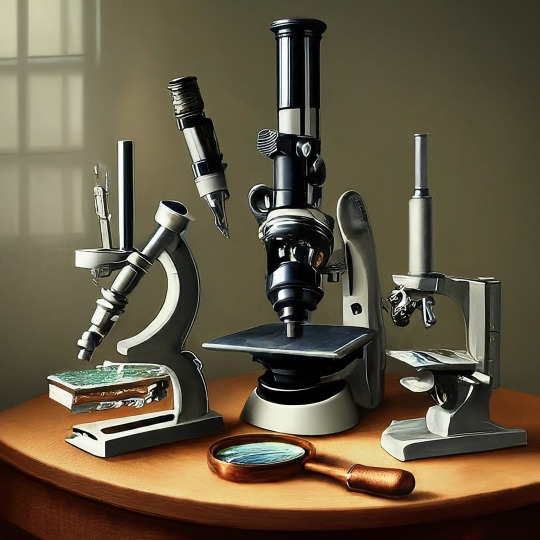
A Journey into the World of Microscopy: From Humble Beginnings to High-Tech Magnification
The science of looking into the hidden invisible Microscopy has transformed our understanding of the world around us. It can explore the universe beyond the reach of our naked eyes, with complex cellular structures, red blood cells, viruses and other viruses and microorganisms taking on amazing perspectives
The history of the microscope is a fascinating story of human curiosity, scientific genius, and relentless exploration. From the humble beginnings of simple magnifying glasses to the sophistication of modern electronic microscopes, the invention of microscopes has shaped our understanding of the microscopic world
In the 1600s, Dutch opticians such as Hans and Zachary Janssen are credited with inventing the first microscope. Known for this hybrid microscope, many lenses were used to magnify objects up to 30 times.At the end of the 17th century, Antony van Leeuwenhoek, Dutch draper some changed our perception of thumbnails. Armed with a well-made single-lens microscope, and explored the hidden reaches of nature. In 1674, Leeuwenhoek discovered microorganisms in lake water, which he aptly named “animalcules”. His discovery laid the foundations of biology and inspired generations of scientists. This incredible feat allowed him to uncover a hidden universe – the first sightings of bacteria, red blood cells, and other microorganisms.
Formation of the scientific environment (17th-19th centuries): Leeuwenhoek’s discoveries boosted scientific research. Robert Hooke, an English scientist, established these developments. In 1665, his book "Micrographia" recorded his observations with a compound microscope. Notably, the term "cell" was coined by Hooke when he examined cork tissue, laying the foundation for cell biology.Microscope systems flourished throughout the 18th and 19th centuries Joseph Lister and other scientists addressed the limitations of the early lenses, introducing improvements that reduced image distortion.
Beyond the Limits of Light: The Beginning of the New Age (19th-20th century): As the 19th century progressed, the limitations of optical microscopy became apparent and scientists yearned for a tool which can go deeper into cells. This research culminated in the development of the electron microscope in the 1930s. The 20th century was revolutionary with the invention of the electron microscope. Unlike light microscopes, which use visible light, electron microscopes use electron beams to achieve much higher magnification.Formation of the scientific environment (17th-19th centuries): Leeuwenhoek’s discoveries boosted scientific research. Robert Hooke, an English scientist, established these developments. In 1665, his book "Micrographia" recorded his observations with a compound microscope. Notably, the term "cell" was coined by Hooke when he examined cork tissue, laying the foundation for cell biology.Microscope systems flourished throughout the 18th and 19th centuries Joseph Lister and other scientists addressed the limitations of the early lenses, introducing improvements that reduced image distortion.
Beyond the Limits of Light: The Beginning of the New Age (19th-20th century): As the 19th century progressed, the limitations of optical microscopy became apparent and scientists yearned for a tool which can go deeper into cells. This research culminated in the development of the electron microscope in the 1930s. The 20th century was revolutionary with the invention of the electron microscope. Unlike light microscopes, which use visible light, electron microscopes use electron beams to achieve much higher magnification.
In the 1930s, German experts Max Knoll and Ernst Ruska made the first electron microscope. This tool let us see tiny things like cells and even atoms by using electron beams, not light, getting images many times bigger. This cool invention showed us the tiny parts inside cells, viruses, and stuff too small to see before. The 1900s brought even more cool microscopes. New kinds like phase-contrast and confocal microscopy let scientists look at live cells without using stuff that could hurt them. Now, the world of looking at tiny things is getting even better. Today, we have high-tech microscopes that use computers and lasers. These let us see and even change tiny things in ways we never could before.
Modern Microscopy's Diverse Arsenal - Today, the field of microscopy boasts a diverse range of specialized instruments, each tailored to address specific scientific needs. Here's a glimpse into some remarkable examples:
Scanning Electron Microscope (SEM): Imagine a high-tech camera that captures images using a beam of electrons instead of light. That's the essence of a SEM. By scanning the surface of a sample with a focused electron beam, SEMs generate detailed information about its topography and composition. This makes them ideal for studying the intricate structures of materials like insect wings, microchips, and even pollen grains.
Transmission Electron Microscope (TEM): While SEMs provide exceptional surface detail, TEMs take us a step further. They function by transmitting a beam of electrons through a very thin sample, allowing us to observe its internal structure. TEMs are the go-to instruments for visualizing the intricate world of viruses, organelles within cells, and macromolecules like proteins.
Confocal Microscopy: Ever wished to focus on a specific layer within a thick biological sample and blur out the rest? Confocal microscopy makes this possible. It utilizes a laser beam to precisely illuminate a chosen plane within the sample, effectively eliminating information from out-of-focus regions. This allows researchers to create sharp, three-dimensional images of cells, tissues, and even small organisms.
Atomic Force Microscopy (AFM): This technique takes a completely different approach, venturing into the realm of physical interaction. AFM employs a tiny cantilever, akin to a microscopic feeler, to physically scan the surface of a sample. By measuring the minute forces between the cantilever and the sample's surface, AFM can map its topography at an atomic level. This provides invaluable insights into the properties of materials at an unimaginable scale, making it crucial for research in fields like nanotechnology and surface science.
Fluorescence Microscopy: Imagine illuminating a sample with specific wavelengths of light and observing it glowing in response. That's the essence of fluorescence microscopy. This technique utilizes fluorescent molecules or tags that bind to specific structures within a cell or tissue. When excited by light, these tags emit their own light, highlighting the target structures with remarkable clarity. This allows researchers to visualize specific proteins, DNA, or even pathogens within biological samples.
Super-resolution Microscopy (SRM): Overcoming the limitations imposed by the wavelength of light, SRM techniques like STED (Stimulated Emission Depletion) and PALM (Photoactivated Localization Microscopy) achieve resolutions surpassing the diffraction limit. This allows researchers to visualize structures as small as 20 nanometers, enabling the observation of intricate cellular machinery and the dynamics of individual molecules within living cells.
Cryo-Electron Microscopy (Cryo-EM): This powerful technique takes a snapshot of biological samples in their near-life state. Samples are rapidly frozen at ultra-low temperatures, preserving their native structure and minimizing damage caused by traditional fixation methods. Cryo-EM has been instrumental in determining the three-dimensional structures of complex molecules like proteins and viruses, providing crucial insights into their function and potential drug targets.
Correlative Microscopy: Combining the strengths of multiple microscopy techniques, correlative microscopy offers a comprehensive view of biological samples. For instance, researchers can utilize fluorescence microscopy to identify specific structures within a cell and then switch to electron microscopy to examine those structures in high detail. This integrated approach provides a deeper understanding of cellular processes and their underlying mechanisms.
Light Sheet Microscopy (LSM): Imagine illuminating a thin slice of a sample within a living organism. LSM achieves this feat by focusing a laser beam into a thin sheet of light, minimizing photobleaching and phototoxicity – damaging effects caused by prolonged exposure to light. This allows researchers to observe dynamic processes within living organisms over extended periods, providing valuable insights into cellular behavior and development.
Expansion Microscopy (ExM): This innovative technique physically expands biological samples by several folds while preserving their structural integrity. This expansion allows for better resolution and visualization of intricate cellular structures that would otherwise be difficult to distinguish using traditional microscopy methods. ExM holds immense potential for studying the organization and function of organelles within cells.
Scanning Near-Field Optical Microscopy (SNOM): This innovative technique pushes the boundaries of resolution by utilizing a tiny probe that interacts with the sample at an extremely close range. SNOM can not only image the surface features of a sample with exceptional detail but also probe its optical properties at the nanoscale. This opens doors for research in areas like material science and photonics, allowing scientists to study the behavior of light at the interface between materials.
X-ray Microscopy: Stepping outside the realm of light and electrons, X-ray microscopy offers unique capabilities. By utilizing high-energy X-rays, this technique can penetrate deep into samples, making it ideal for studying the internal structure of dense materials like bones and minerals. Additionally, it allows for the visualization of elements within a sample, providing valuable information about their distribution and composition.
From revealing the building blocks of life to aiding in the development of new medicines, the microscope has played an undeniable role in shaping our scientific understanding. As technology continues to evolve, one can only imagine the future breakthroughs this remarkable invention holds in unveiling the secrets of our universe, both seen and unseen. These advancements hold the potential to revolutionize our understanding of biological processes, develop new materials with extraordinary properties, and ultimately pave the way for breakthroughs in medicine, nanotechnology, and countless other fields. As we continue to refine and develop novel microscopy techniques and the future holds immense promise for further groundbreaking discoveries that will undoubtedly revolutionize our perception of the world around us.
#science sculpt#life science#science#molecular biology#biology#biotechnology#artists on tumblr#microscopy#microscope#Scanning Electron Microscope#Transmission Electron Microscope#Confocal Microscopy#Atomic Force Microscopy#Fluorescence Microscopy#Expansion Microscopy#X-ray Microscopy#Super-resolution Microscopy#Light Sheet Microscopy#illustration#illustrator#illustrative art#education#educate yourself#techniques in biotechnology#scientific research#the glass scientists#scientific illustration#scientific advancements
7 notes
·
View notes
Text
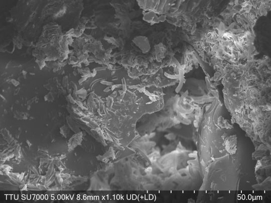


This came to mind, so I'm back to sharing some photos I and a friend took using a scanning electron microscope, this time it's cochineal beetle husks! I think some of these photos really highlight how scanning electron microscopy is so similar to both standard photography and what I view as the opposite end of the spectrum of astrophotography. SEM -> Electrons are essentially shot at an object to 'scan' it, and then the way they scatter off the surface in a very precise manner is used to reconstruct the image. (I'm certain it's more complicated than this, but that's all you need to know for the basics) This is why they're kept in vacuum, so that interfering air particles aren't likely to mess with the electron scattering. Plus, with organic materials like these beetle husks and anything else I share photos of (at least at present), they don't actually have enough conductivity intrinsically to properly deflect electrons! So, they have to be coated with an incredibly thin layer (so as to preserve the actual image still, it's on the order of nanometers thin) of gold and palladium through a process called sputtering to allow for conductivity. If a region is too "conductive" (It's been a minute, so I don't remember the exact terminology) under the settings with which you're trying to take a photo, the electrons can actually "stick" to the surface in a region, whiting it out and creating what's referred to as 'charging' effects. That's what causes some of the really sharp whites in some of these images! (It can also just be good clarity) Additionally, with some of the photos (I think 7 and 8 really exemplify this the best with how it sort of looks like a cliff in the image going deeper into the husk), you can see how this can't reconstruct a perfectly in focus image at all 'heights'. The stuff that is put into an SEM if the vacuum doesn't have to be repulled is super tiny and very flat, but even then that very minor amount of height difference is clearly huge. I just think it's not only really cool to see what stuff like this looks like, but also how similar it can be to standard photography!
10 notes
·
View notes
Text
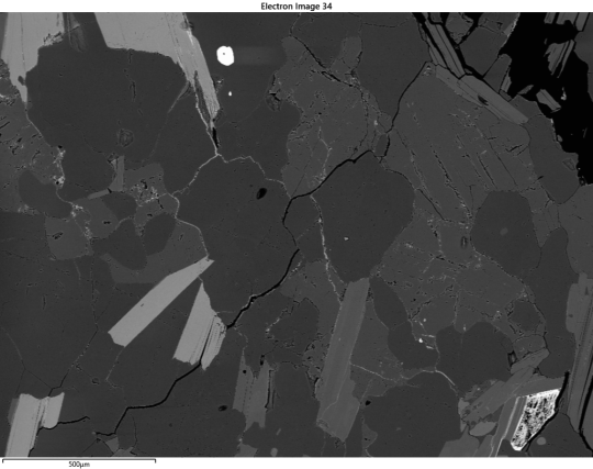


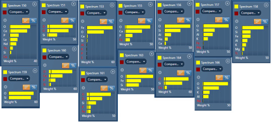
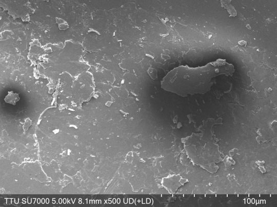
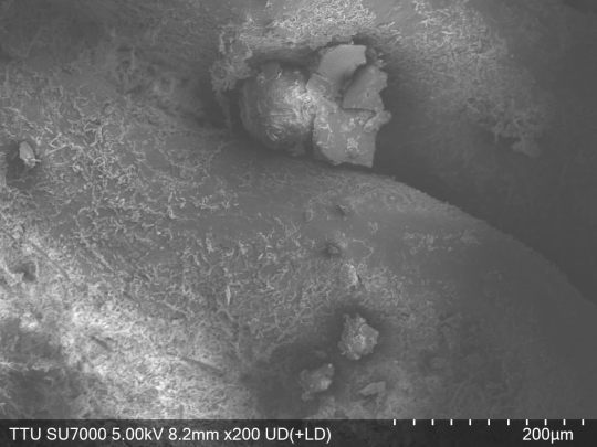
Finally got to look over the SEM data for ec-501 and. HRM.
Like 1, the topography map is surprising because I expected more holes. 501 is a pretty soft sample!
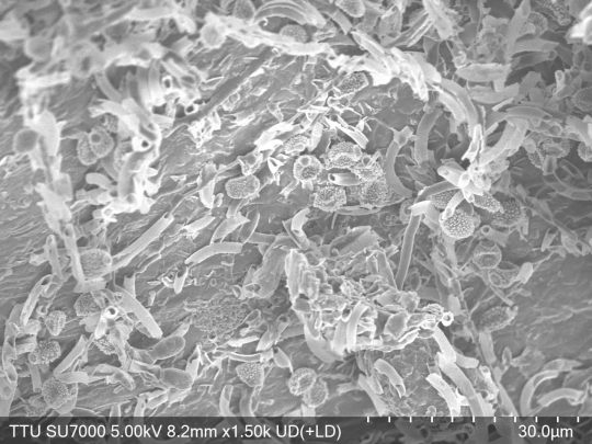
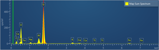
The specimen samples are both suprising and not suprising. Monazite? Annoying but sure, thoigh I didn't expect the La and Nd(neodynium my beloved).
I know the smaller samples are a little fucked, but they are mostly just to help confirm. Like yeah, 151 is very much definitely possibly zircon! But it's so goddamn tiny that Aztec really said "bruh idk???"
We got the two micas and quartz, no surprise. We got plag! And then what the FUCK is going on with 155, 160, 161, and 163? I assume 161 is more zircon. but whay the fuck could the others be??
Anyways, tadah! My new data to interpret. I can pop open aztec later for more details, including individual element maps.
2 notes
·
View notes
Text
#Scanning Electron Microscopes Market#Scanning Electron Microscopes Market Share#Scanning Electron Microscopes Market Size#Scanning Electron Microscopes Market Trends#Scanning Electron Microscopes Market Growth
0 notes
Text
Scanning Electron Microscopes Market set to hit $13.9 billion by 2035
Industry revenue for Scanning Electron Microscopes is estimated to rise to $13.9 billion by 2035 from $5.3 billion of 2024. The revenue growth of market players is expected to average at 9.2% annually for the period 2024 to 2035.
Scanning Electron Microscopes is critical across several key applications including material science, semiconductor manufacturing, biological research and failure analysis. The report unwinds growth & revenue expansion opportunities at Scanning Electron Microscopes’s End User, Technology and Application including industry revenue forecast.
Industry Leadership and Competitive Landscape
The Scanning Electron Microscopes market is characterized by intense competition, with a number of leading players such as Thermo Fisher Scientific Inc, Carl Zeiss AG, JEOL Ltd, Hitachi High-Technologies Corporation, FEI Company, Danish Micro Engineering, Tescan Orsay Holding, Keysight Technologies Inc, Nikon Metrology NV, Leica Microsystems, Olympus Corporation and Bruker Corporation.
The Scanning Electron Microscopes market is projected to expand substantially, driven by technological advancements in sem and increased demand from the nanotechnology industry. This growth is expected to be further supported by Industry trends like SEM Applications in Life Sciences and Healthcare.
Detailed Analysis - https://datastringconsulting.com/industry-analysis/scanning-electron-microscopes-market-research-report
Moreover, the key opportunities, such as opportunity in nanomaterials analysis, opportunity in forensic investigations and growth via digital transformation, are anticipated to create revenue pockets in major demand hubs including U.S., Japan, Germany, China and UK.
Regional Shifts and Evolving Supply Chains
North America and Europe are the two most active and leading regions in the market. With challenges like high cost of acquisition and extensive training requirements, Scanning Electron Microscopes market’s supply chain from raw material procurement / components manufacturing / sem assembly & testing to end user applications is expected to evolve & expand further; and industry players will make strategic advancement in emerging markets including India, Brazil and South Africa for revenue diversification and TAM expansion.
About DataString Consulting
DataString Consulting offers a complete range of market research and business intelligence solutions for both B2C and B2B markets all under one roof. We offer bespoke market research projects designed to meet the specific strategic objectives of the business. DataString’s leadership team has more than 30 years of combined experience in Market & business research and strategy advisory across the world. DataString Consulting’s data aggregators and Industry experts monitor high growth segments within more than 15 industries on an ongoing basis.
DataString Consulting is a professional market research company which aims at providing all the market & business research solutions under one roof. Get the right insights for your goals with our unique approach to market research and precisely tailored solutions. We offer services in strategy consulting, comprehensive opportunity assessment across various sectors, and solution-oriented approaches to solve business problems.
0 notes
Text
shaking myself rn......... i rlly wanna finish all my icons so i can rp the jellybean immediately
#my scanning electron microscope assignment can Wait until i'm done!!!!!!!!!!!!! all that matters is jb from xod <3 💖💖🥰#i swearrrrr her blog is literally ready i just need all my goddamn icons 😭😭 SHAKING MYSELF MORE#🧸 ––– ✧・゚: *✧・゚:* 002. OOC.
1 note
·
View note
Text

Labnic Digital Microscope features Siedentop trinocular head type with head inclined at 30 degrees and the headlight path fixed to 50:50. The digital microscope features a working stage with two mechanical layers. The digital microscope is Wi-Fi and USB-enabled.
0 notes
Text

Just Watching
1 note
·
View note
Text

Don’t let the purple hue fool you – this image captured by a scanning electron microscope is actually showing us mesoporous gold nanoparticles.
Photograph: Javeria Bashir/AIBN
2023 Australian Institute For Bioengineering And Nanotechnology Image Contest
#javeria bashir#photographer#australian institute for bioengineering and nanotechnology image contest#micro photography#scanning electron microscope#mesoporous gold nanoparticles#nature
4 notes
·
View notes
Text

After initial tests created a series of large holes in the wall of the lab, the higher-power Scanning Tunneling Tennis Ball Microscope project was quickly shut down.
Tennis Balls [Explained]
Transcript Under the Cut
[Cueball fires eight tennis ball at decreasing heights using a tennis ball machine, making four "thunk" noises. Megan is standing behind him.]
[Ten noises come from the right side of the panel.] Bonk Bonk Bonk Bonk Bonk OW! Bonk OW! Bonk Bonk
[Megan has her hand to her chin.] Megan: Ok, there's definitely a person over there. Let's do one more pass to try to measure their height.
[Caption below the panel:] Electrons are small and hard to work with, so some scientists have developed a scanning tennis ball microscope instead.
2K notes
·
View notes
Text
youtube
This video shows how a Scanning Electron Microscope works? And how to use a virtual scanning electron microscope to acquire a good image. It is a great learning and teaching tool. You can access the virtual TEM at https://myscope-explore.org/virtualSE... developed by Microscopy Australia and Thermofisher Scientific.
#scanning#electronmicroscope#electronmicroscopy#electronbeam#image#images#nano#lotus#butterfly#lens#electrons#education#digitalliteracy#onlinelearning#microscope#microscopy#teaching#fiber#forensics#hair#arthropods#wool#superhydrophobic#waterrepellent#e-learning#virtuallearning#digitaltools#digitaleducation#openeducationalresources#openeducation
1 note
·
View note