#colorized scanning electron microscope
Explore tagged Tumblr posts
Visit Tumblr Blog
Explore Tumblr blogs with no restrictions, modern design and the best experience.
Last Seen Tumblr Blogs


Fun Fact
Hackers stole 65M passwords from Tumblr in 2013.
Text
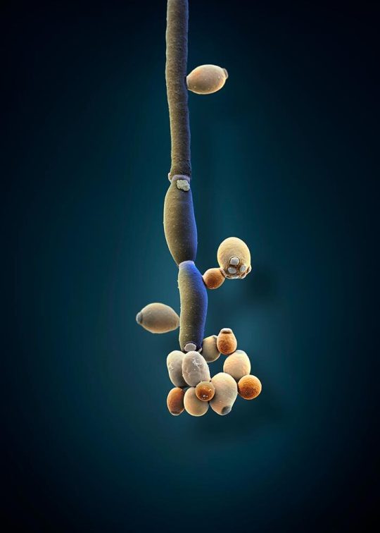
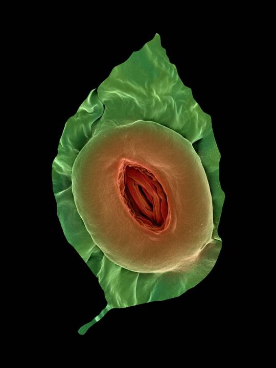
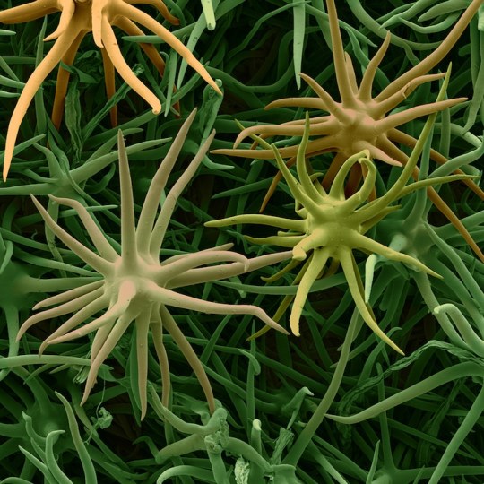
Studies show that patients with irritable bowel syndrome tend to have high levels of the fungus Candida albicans (illustrated here) in their gut. In recent years, scientists have started to take a closer look at how the fungi in your gut microbiome affect your health.
COLORIZED SCANNING ELECTRON MICROSCOPE IMAGE BY MARTIN OEGGERLI
UNIVERSITY HOSPITAL BASEL, SWISS NANOSCIENCE INSTITUTE, BASEL
#martin oeggerli#colorized scanning electron microscope#photographer#micro photography#university hospital basel#swiss nanoscience institute#basel#switzerland#irritable bowel syndrome#candida albicans#fungus#nature#microbiome#national geographic
12 notes
·
View notes
Text
The Madagascan Sunset Moth: these moths are often mistaken for butterflies, thanks to their colorful, iridescent appearance

The scientific name of this species is Chrysiridia rhipheus, but it's commonly known as the Madagascan sunset moth. These gorgeous day-flying moths can be found only in Madagascar.

Above: a dorsal view of the same species
The Madagascan sunset moth has a strikingly colorful appearance, especially when its underwings are exposed, as a rainbow-like effect is produced by the iridescent scales that cover the underwings (and appear in smaller sections on the dorsal side of the wing).

Above: some of the iridescent scales on the underwings of Chrysiridia ripheus, as seen through a scanning electron microscope
The markings on the dorsal side are primarily black, with some patches of iridescent green, blue, and red. A "fringe" of white scales can also be seen along the edge of each wing; these are especially prominent on the hindwings.

Above: the dorsal patterns are visible only when the wings are left open
Like most day-flying moths, the adults of this species will often feed on nectar. Their caterpillars are known to consume Omphalea plants, which are toxic; those toxins are sequestered by the caterpillar and then retained through the pupal and adult stages of development, which means that the adult moth is toxic. Their colorful appearance is likely aposomatic, deterring predators by signaling that the moth itself is toxic.

Above: a magnified view of the white scales that outline the hindwings
Sources & More Info:
Navsari Agricultural University: Sunset Moth: the most beautiful insect
Oxford University Museum of Natural History: Madagascar
Moth Identification Guide: Madagascan Sunset Moth
California Academy of Sciences: Sunset Moth
Optica: Polarization-Sensitive Color Mixing in the Wing of the Madagascan Sunset Moth
Wikipedia: Chrysiridia rhipheus
#entomology#lepidoptera#madagascan sunset moth#chrysiridia rhipheus#arthropods#moths#insects#urania rhipheus#animal facts#sunset moth#bugs#cool animals#colorful moths#madagascar#aposomatic coloration#moths are amazing#and beautiful#don't let anyone tell you otherwise
639 notes
·
View notes
Text

Close-Up of the Large Red Damselfly (Pyrrhosoma nymphula)!
Scanning electron microscope image reveals the stunning complexity of the Large Red Damselfly. From its vibrant, multi-colored compound eyes to the intricate hair-like structures covering its face, this insect is a true masterpiece of evolution.
✨ Fun Fact: Damselflies have near-360° vision, thanks to their massive compound eyes, allowing them to spot prey and predators with incredible accuracy!
📸: @eyeonscience
67 notes
·
View notes
Text
Wet Beast Wednesday: tardigrades
Last week on Wet Beast Wednesday I covered the largest animals to ever exist on our planet. This week I'm going to pull a full 180 and cover the smallest animals yet on this series. Meet the tardigrade, the internet's favorite micro-animal the is said to be basically immortal. How true is that? Let's see.

(Image: an electron microscope image of a tardigrade. It looks a lot like a potato with eight stubby legs tipped with long claws. At the front is a small, circular mouth. It has no other discernable features. In the background are bits of plant matter that look like seaweed at this scale. End ID)
The tardigrades are 1,300 known species (and probably a lot of unknown ones too) in the phylum Tardigrada. They are also part of the superphylum Ecdysozoa, which are animals that grow by molting their outer cuticles or exoskeletons. In particular, the tardigrades are believed to be a sister group of the arthropods, the group that contains crustaceans, insects, isopods, and a lot of other things. Tardigrades are truly tiny, the largest species reaching a whopping 1.5 millimeters in length, though most species reach no more than 0.5 mm. They have round, segmented bodies with four pairs of legs that end in either claws or suction discs. The body segments consist of a head, three body segments with a pair of legs each, and a caudal segment with the final pair of legs. The first three legs are used for movement while the final pair points backwards and is used for grabbing onto substrate. All of the body segments except for the final one correspond to segments found in the head section of insects. Tardigrades are missing many hox genes, genes that direct the body plan during development. Their ancestors may have had a body plan more similar to insects, but the loss of the hox genes has compressed them into walking heads with a bit of butt. The mouth is tubular and sucks in food. In the mouth are stylets, needle-like structures used to pierce food objects. Once food is drawn into the mouth, a structure called the buccopharyngeal apparatus activates. This is a combination of spines and muscle that acts like an inner jaw that pulls food into the digestive tract. The buccopharyngeal apparatus is distinct enough to be used as a major identifying feature between species. Tardigrades are translucent and many images you've seen of them have false color to show the details or are 3D models based on scanning electron microscope imagery of them. Tardigrades molt their exoskeletons multiple times (up to 12) during their lifecycle. Some species are unable to poop normally and instead all their waste is discarded during the molt. It was formerly believed that tardigrades could exchange genes with each other without mating, a process called horizontal gene transfer that is seen in bacteria, archaea, and other micro-organisms. It has since been discovered that while still capable of horizontal gene transfer, it is quite a bit rarer in tardigrades than we thought.

(Image: an electron microscope image of a tardigrade standing on a bit of plant matter. This one has a closed mouth with a ring of triangular tooth-like structures. It also has two simple eyes that look like black dots. End ID)
The name "tardigrade" means "slow walker", which is fitting as, despite their eight legs, tardigrades have a slow and awkward gait. This is the result of their legs being unjointed, only able to pivot at their connection to the body. Their gait has been compared to that of bears, hence why they are often called water bears and their discoverer, Johann August Ephraim Goeze, called them "kleiner wasserbär", meaning "little water bear". Tardigrades are found worldwide and have inhabited virtually every habitat, from the tops of mountains to the deep sea, from hot springs to the antarctic, from freshwater to saltwater. The one thing they have in common is a need to stay wet. Tardigrades can survive out of water as long as they can stay moist and are often found in mosses, hence another common name: moss piglets. The majority either eat plants or bacteria, but some will feed on smaller tardigrades or other micro-animals. Their famous survivability makes it easy for tardigrades or their eggs to be carried to new habitats by larger animals or other phenomena. Tardigrades are one of the first micro-animals to colonize a new habitat and they are a pioneer species, the first species to colonize a new environment and whose presence makes that environment fore suitable for other species to follow. Tardigrades are a major food source to other micro-animals and larger organisms. Most species have distinct males and females, though a few reproduce through parthenogenesis. In most cases, molting female will lay her eggs in her shed cuticle and males will them fertilize them. Other species have a form of internal reproduction. Males and females will court each other before mating and females will usually allow multiple males to fertilize her eggs. Female tardigrades are typically larger and more abundant than males. Eggs can take up to 14 days (species dependent) before hatching. All tardigrades of the same species have the exact same number of cells as each other. They are also born with the same number of cells they will have as an adult. Their growth is driven by enlargement of the existing cells rather than cellular reproduction making new cells. The lifespan ranges between a few months to a few years, depending on species.
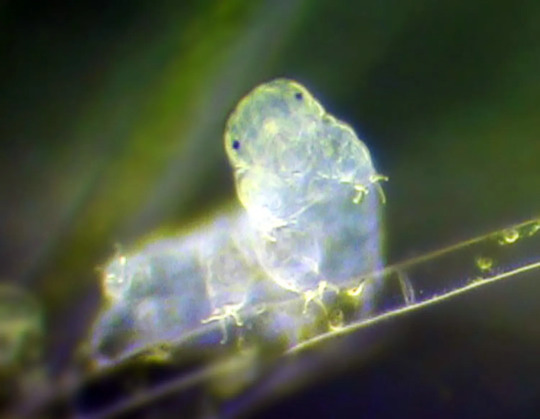
(Image: a color photo of a tardigrade. It is a pale, translucent white, making it hard to make out details. Its body is curved, with the front end pointing at the camera. It has two simple eyes. End ID)
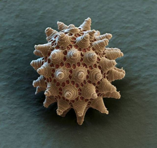
(Image: an electron microscope image of a tardigrade egg. It is round but covered in small pores and conical structures. End ID)
The most famous feature of tardigrades is their legendary durability. It is commonly said that tardigrades can survive just about anything (except for the things that are actually trying to kill them. They are prey to a lot of species after all). Among the things they can survive is extreme heat, extreme cold, dehydration, extremely high and low pressure, exposure to ionizing radiation (that's the scary kind), low oxygen environments, environmental toxins, heavy impacts, and the vacuum of fucking space. While the can survive in extreme conditions, tardigrades are not considered extremophiles. True extremophiles thrive in extreme environments and are negatively impacted by leaving them. Tardigrades can survive in extreme environments, but are negatively impacted and can't survive as well there as they can in less extreme places. The main trait that has allowed tardigrades to survive all five mass extinctions in history is cryptobiosis. Cryptobiosis is the rare ability for an animal to enter a state of dormancy where their metabolic processes come to an almost complete stop. While in cryptobiosis, metabolic activity drops to 0.01% normal and water content drops to 1% normal. In this state, the tardigrade is called a tun. Tardigrades usually enter cryptobiosis in response to arid conditions. One experiment showed that a species of tardigrade could last for at least 30 years in this state and return to normal lifestyle functions when exposed to water. Tardigrades will also enter cryptobiosis in response to low oxygen, toxic chemical exposure, increased or decreased temperature, and excessive salt content in the water. Tardigrades also show extreme resistance to both high and low pressure. They can live in 0 atmospheres of pressure and some species can survive up to 6,000 atmospheres, more than double the pressure at the bottom of the Marianas trench. More interesting is their ability to survive dangerous radiation. They can survive 1,000 times the dose of gamma radiation that humans can. Early tests focused on tardigrades in cryptobiosis and concluded that the extremely low water content of a cryptobiotic tardigrade doesn't leave much opportunity for the radiation to react with the animal. However it was later found that active and fully hydrated tardigrades are still considerably resistant to radiation. Studies into this resistance indicate that tardigrades can very efficiently repair damaged DNA and have unique proteins called Dsup that provides additional protection. Dsup introduced to human cells has provided additional protection against x-rays.
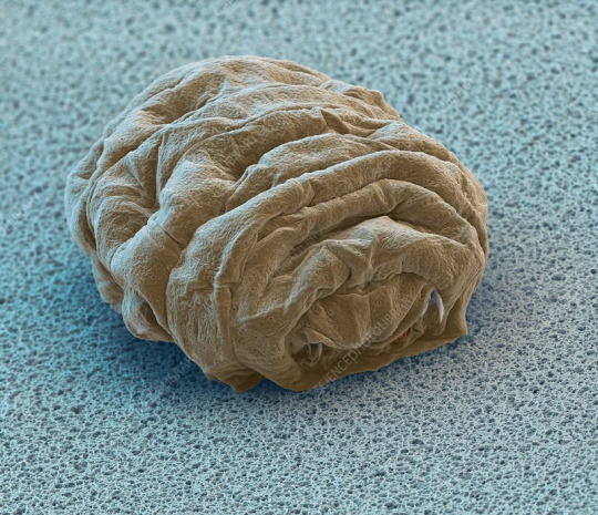
(Image: an electron microscope image of a tun - a tardigrade in cryptobiosis. It is smaller and very wrinkly, with the legs and mouth retracted into the body. End ID)
Tardigrades were the first animals to be exposed to the vacuum of space. They were exposed for 10 days, some in a state of cryptobiosis at the time of exposure and some still active. It was found that they were able to survive the vacuum when shielded from the sun's ultraviolet radiation, with those already in cryptobiosis doing better. Upon being rehydrated, many were able to resume normal life functions and successfully reproduce, though others died after being rehydrated. Those that were exposed to UV radiation fared much worse, with only a few hydrated individuals surviving. The individuals in cryptobiosis had a lower survival rate when exposed to UV than those not exposed to UV and were less successful at reproducing afterwards. Studies of tardigrade's space survival abilities and resistance to radiation could go a long way in helping human space travel. One of the largest dangers of space travel is that space is full of nasty radiation from the sun that Earth's magnetic field protects us from. Some scientists speculate about the possibility of accidentally seeding other planets or moons with tardigrades or other space-resistant organisms. This is a problem because introducing Earth life to other world has the potential to damage any native ecosystems and if we find life in space in the future we don't want to have to figure out if it's something we accidentally put there. While tardigrades could likely survive on other planets, they would eventually die without a food source. Some sources reported that tardigrades may have colonized the moon after an experiment with them crashed. Unfortunately, the moon is not crawling with tardigrades now. It's way too dry for them to exit cryptobiosis even if they survived the crash, which they probably didn't.

(Image: art of a tardigrade floating in the vacuum of space. End ID. Source: University of California - Santa Barbara)
#wet beast wednesday#tardigrade#water bear#moss piglet#micro animal#microbiology#marine biology#biology#zoology#ecology#animal facts#informative#science#space#astrobiology#radiation#cryptobiosis#tun#image described
202 notes
·
View notes
Text
Modern technology and dinosaur fossils
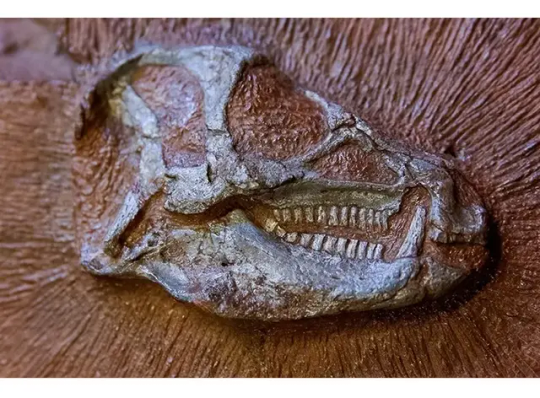
There isn't any credible scientific evidence that supports the mainstream mineralization theory but on the contrary modern technology proves that it can't happen the way the theory suggests. Modern technology, especially microscopy, has dramatically deepened our understanding of dinosaur fossils, revealing details that were unimaginable just a few decades ago. Here’s a concise overview of what microscopy and related tools have uncovered: 1. Discovery of Soft Tissues Using scanning electron microscopy (SEM) and transmission electron microscopy (TEM), scientists have found: • Blood vessel-like structures • Red blood cell-shaped objects • Bone cells (osteocytes) with preserved structures These were first reported in a T. rex femur by Dr. Mary Schweitzer in the early 2000s, challenging the idea that soft tissue couldn’t survive millions of years.
2. Protein and Molecular Structures Microscopy, combined with mass spectrometry and immunohistochemistry, has revealed: • Preserved collagen fibers, confirming a link between dinosaurs and birds. • Reactions with antibodies that only bind to certain proteins, suggesting that original molecules survived in some fossils.
3. Pigmentation and Feather Color With electron microscopes, researchers have identified melanosomes (pigment-containing structures) in fossilized feathers. These led to: • Reconstructions of actual feather colors in dinosaurs like Anchiornis and Microraptor (e.g., iridescent black, reddish tones).
4. Growth and Disease Evidence Bone cross-sections viewed under light and electron microscopes show: • Growth rings, like tree rings, indicating growth rates. • Signs of bone cancer, arthritis, and injuries helping us understand dinosaur health. 5. Cellular and Nanostructural Detail Advanced imaging reveals: • Nano-scale preservation of original tissue structure. • Differences between fossil material and possible contamination (ruling out modern biofilm or bacteria in some studies). In Summary: Microscopy and modern tech have turned dinosaur bones into biological time capsules, revealing: • Cellular structure • Protein preservation • Pigment patterns • Disease evidence This new level of insight has reshaped our view of dinosaurs not just as skeletons, but as once-living, dynamic creatures with feathers, color, disease, and growth cycles. Here’s a list of modern technologies that have been used to analyze and reveal new facts about dinosaur fossils especially those related to soft tissue, cellular structures, and ancient biology:
Scanning Electron Microscopy (SEM) • Produces high-resolution images of fossil surfaces. • Used to identify blood vessel-like structures, osteocytes, and melanosomes (pigment organelles).
Transmission Electron Microscopy (TEM) • Offers ultra-high magnification to see internal structures of cells and fibers. • Helped confirm collagen fibers and sub-cellular features in fossils.
Mass Spectrometry (LC-MS/MS) • Detects and sequences ancient proteins like collagen. • Confirmed molecular similarities between dinosaurs and birds.
Immunohistochemistry • Uses antibodies to test for specific proteins. • Proved some structures in fossils react like real biological tissue, not just mineralized look-alikes.
Synchrotron X-ray Imaging • High-energy, non-destructive imaging of fossil interiors. • Maps the chemical composition of fossils, including traces of blood, soft tissues, or pigments.
Raman Spectroscopy • Identifies molecular bonds and organic compounds. • Confirms presence of preserved proteins and pigments.
Fourier-Transform Infrared Spectroscopy (FTIR) • Detects organic molecules in fossils. • Supports claims of soft tissue preservation by matching spectral signatures of known proteins.
Confocal Laser Scanning Microscopy • Produces 3D images of soft tissue structures inside fossils. • Used to observe elasticity and fine internal details of preserved vessels.
9. Micro-CT (Computed Tomography) Scanning • Creates detailed 3D models of fossil interiors. • Reveals hidden bone structures, growth rings, and sometimes trapped soft tissue.
Stable Isotope Analysis • Measures carbon, oxygen, and other isotopes. • Reveals insights about dinosaur diets, environments, and metabolism. These technologies are revolutionizing paleontology by uncovering molecular and cellular data that were never thought possible in such ancient remains. Here’s a summary with examples of specific dinosaur species where modern technology has revealed extraordinary fossil details:
Tyrannosaurus rex • Discovery: Soft tissue structures including blood vessels, osteocytes, and collagen fragments. • Technology used: Scanning Electron Microscopy (SEM), Mass Spectrometry (LC-MS/MS), Immunohistochemistry. • Significance: First confirmed protein sequences from a dinosaur; supports a close evolutionary link to birds.
2. Anchiornis huxleyi • Discovery: Microscopic pigment structures called melanosomes in fossilized feathers. • Technology used: Transmission Electron Microscopy (TEM), Synchrotron X-ray imaging. • Significance: Revealed feather colors — likely black, gray, and reddish hues — making it one of the first dinosaurs reconstructed with accurate coloration.
Brachylophosaurus canadensis (a hadrosaur) • Discovery: Preserved blood vessels, cells, and possible nuclei in bone tissue. • Technology used: Confocal Laser Scanning Microscopy (CLSM), FTIR spectroscopy, SEM. • Significance: Demonstrated that even delicate cellular structures can persist under rare conditions.
Microraptor gui • Discovery: Feather structure and iridescent coloration patterns. • Technology used: Electron Microscopy, Melanosome analysis. • Significance: Showed it had shiny, bird-like plumage linking flight-related traits with non-avian dinosaurs. These breakthroughs have turned dinosaur bones into molecular time capsules, and they continue to reshape how we imagine these ancient creatures.
#dinosaurs#paleontology#dinosaur extinction#science#geology#cosmology#cosmos#mary schweitzer#mary schweizer#space
12 notes
·
View notes
Text

Lily, you're never going to make tardigrade an insult, this is never going to be a thing. People LOVE tardigrades! Just look at these little fellas go:

Credit: My Microscopic World
Besides, you saying that Twitter's full of tardigrades only makes the tardigrades sound bad.

The original image from Steve Gschmeissner/Science Photo Library is a colorized scanning electron micrograph of a tardigrade; microscopic sized Twitter/X.com paraphernalia added separately.
14 notes
·
View notes
Text

Why the moon shimmers with shiny glass beads
The Apollo astronauts didn’t know what they’d find when they explored the surface of the moon, but they certainly didn’t expect to see drifts of tiny, bright orange glass beads glistening among the otherwise monochrome piles of rocks and dust.
The beads, each less than 1 mm across, formed some 3.3 to 3.6 billion years ago during volcanic eruptions on the surface of the then-young satellite. “They’re some of the most amazing extraterrestrial samples we have,” said Ryan Ogliore, an associate professor of physics in Arts & Sciences at Washington University in St. Louis, home to a large repository of lunar samples that were returned to Earth. “The beads are tiny, pristine capsules of the lunar interior.”
Using a variety of microscopic analysis techniques not available when the Apollo astronauts first returned samples from the moon, Ogliore and a team of researchers have been able to take a close look at the microscopic mineral deposits on the outside of lunar beads. The unprecedented view of the ancient lunar artifacts was published in Icarus. The investigation was led by Thomas Williams, Stephen Parman and Alberto Saal from Brown University.
The study relied, in part, on the NanoSIMS 50, an instrument at WashU that uses a high-energy ion beam to break apart small samples of material for analysis. WashU researchers have used the device for decades to study interplanetary dust particles, presolar grains in meteorites, and other small bits of debris from our solar system.
The study combined a variety of techniques — atom probe tomography, scanning electron microscopy, transmission electron microscopy and energy dispersive X-ray spectroscopy — at other institutions to get a closer look at the surface of the beads. “We’ve had these samples for 50 years, but we now have the technology to fully understand them,” Ogliore said. “Many of these instruments would have been unimaginable when the beads were first collected.”
As Ogliore explained, each glass bead tells its own story of the moon’s past. The beads — some shiny orange, some glossy black — formed when lunar volcanoes shot material from the interior to the surface, where each drop of lava solidified instantly in the cold vacuum that surrounds the moon. “The very existence of these beads tells us the moon had explosive eruptions, something like the fire fountains you can see in Hawaii today,” he said. Because of their origins, the beads have a color, shape and chemical composition unlike anything found on Earth.
Tiny minerals on the surface of the beads could react with oxygen and other components of Earth’s atmosphere. To avoid this possibility, the researchers extracted beads from deep within samples and kept them protected from air exposure through every step of the analysis. “Even with the advanced techniques we used, these were very difficult measurements to make,” Ogliore said.
The minerals (including zinc sulfides) and isotopic composition of the bead surfaces serve as probes into the different pressure, temperature and chemical environment of lunar eruptions 3.5 billion years ago. Analyses of orange and black lunar beads have shown that the style of volcanic eruptions changed over time. “It’s like reading the journal of an ancient lunar volcanologist,” Ogliore said.
IMAGE: Microscopic views of lunar volcanic glass. (Image: Katharine Robinson and G. Jeffrey Taylor, Nature Geoscience, 2014)
3 notes
·
View notes
Text
Book Review: In Search of Stardust
In Search of Stardust: amazing micrometeorites and their terrestrial imposters, by Jon Larsen. I’ll give this a three out of five stars. It has information, just not at all the info I was looking for. In short: I wanted information on micrometeorites. I got, mostly, a photo gallery instead.
I mean, you would think from the title that this book would have not just info on what micrometeorites look like, but how to go about finding them, and maybe even a story or two on why one would get into such a pursuit in the first place. That’s what I was looking for, for the best reason of all. Writing material!
After all, if objects fallen from the sky are the stuff of folklore and fantasy, wouldn’t it be interesting to have a modern mage or cunning woman taking advantage of scientific knowledge and microscopes to pluck up grains of stardust for their spellwork? Wouldn’t it be cool to get in types of magic that can ordinarily only be cast under a shooting star - but a clever wizard has figured out the key factor is really micrometeorite dust in the air. And then weaponizes that knowledge to lay a smackdown no one saw coming.
All of this would be awesome. But it’s not info you’ll get in this book.
What is in this book? A bare minimum of info on how the author got his photos. (Seriously, there’s not enough to even try to replicate the procedure.) The fact that this was a several-year project with samples from all seven continents. A page on how to verify a micrometeorite. (Highly technical, and yet still too sparse in scientific detail.) A few interesting pages on how where the meteorite hits, going with or against Earth’s rotation, determines if it has a soft landing as an unmelted lump or a hard one and melts into a glassy sphere.
And lots, lots of photographs. Scanning electron micrographs to show internal structure. Color pics of all kinds of micrometeorites, from cryptocrystalline to olivine to glass. Some extraterrestrial spherules that are not, technically, micrometeorites. Nearly forty pages of anthropogenic spherules; things that look like micrometeorites but are mostly caused by human activity. Sparks from grinding wheels, welding drops, urban road dust, fireworks, and more. After that come some terrestrial lookalikes (mineral grains and fulgurites are interesting), microtektites, and some oddities that include Darwin glass, volchovites, ooids, pisoids, and Pele’s tears.
All told, if you want tiny scraps of info and a lot of visual examples, check this book out of a library. If you want real meat on things that fall from the sky... better luck elsewhere.
2 notes
·
View notes
Text
Get to Know Me Tag :)
I was tagged by @telomeke in their post here. Thanks for the tag! 🥰
do you make your bed?
Yes. To be fair though, I am not a messy sleeper 😅. Let's just say that, in less than a minute, I can make the bed I slept in look like it hadn't been slept in lol 🤣🤣🤣
what's your favourite number?
I was born on the 9th day of the 9th month. Imagine how obnoxious I was back in '99 lol. 😁
what is your job?
Let's just say I'm fortunate enough to have an occupation that lets me do experiments, mix nefarious things, obtain spectacular (fail) results, and get paid while I'm at it 😅
if you could go back to school, would you?
I often describe myself as an infinite learner. So, while it's true that I can learn things outside of uni, there are still some things that I want to comprehend in an academic setting. I mean, how else can I get my hands on a Scanning Electron Microscope, spectrophotometer, and the like?? 😅
can you parallel park?
Yes. Curiously, my parallel park is better than my reverse parking lol.
a job you had that would surprise people?
Not a job, but most people are surprised when I tell them that I used to be a competitive cheerleader/dancer 😅😅😅 Apparently, I don't look "perky" enough lol. Another thing that also surprises other people is when I tell them I used to do theatre and even played the Virgin Mary in a school play back in high school. Again, my resting bitch face is working against me 🤣🤣🤣
do you think aliens are real?
I'll believe when there is definitive proof that there are. And before I get dragged into the mud, the same goes with religious stuff. I might have played the role of the Virgin Mary in a school theatre, but I am agnostic, at best lol.
can you drive a manual car?
Yes. I actually prefer driving manual cars to automatic because I tend to get bored easily and shifting gears helps me focus lol.
what's your guilty pleasure?
I usually drink unlimited amounts of long black coffee during the day and drink beer mixed with vodka before bedtime. But I will prolly live happily ever after if somebody gives me unlimited access to some tasty chicken nuggets lol
tattoos?
I would want some but I don't like needles lol
favorite color?
I like reds, yellows, and blues.
favorite type of music?
Depends on my mood. There are times I like heavy metal. Other times I would prefer classical music. However, I do like The Beatles so their music is always somewhere in my Spotify playlists.
do you like puzzles?
Yes. I used to do cryptograms and logic puzzles while commuting to my uni.
any phobias?
Does fear itself count? 😅 No, the worst that I feel is anxiety. However, I could always convince myself to face whatever it is that is giving me anxiety. For example, I might not like needles, but I can still get my vaccination with little fuss lol.
favorite childhood sport?
Running. I just like getting from one place to another as fast as my legs could carry.
do you talk to yourself?
If I am alone, yes. I even address myself in the third person 😅
what movies do you adore?
I listed some of my faves in a different tag game here.
coffee or tea?
Definitely coffee but my GP is on my ass for having low blood pressure so I am slowly migrating to green tea 😅 😅 😅
first thing you wanted to be growing up?
Pretty sure I wanted to be a librarian because (1) I'll be surrounded by books, (2) I will be able to hear myself think since libraries are quiet, and (3) it's almost guaranteed that I will be (mostly) left alone 😅 😅 😅
Whew. Tagging @dribs-and-drabbles, @lost-my-sanity1, @imlivingformyselfdontmindme, @waitmyturtles and everybody who sees this and wants to play this game 😊
15 notes
·
View notes
Text
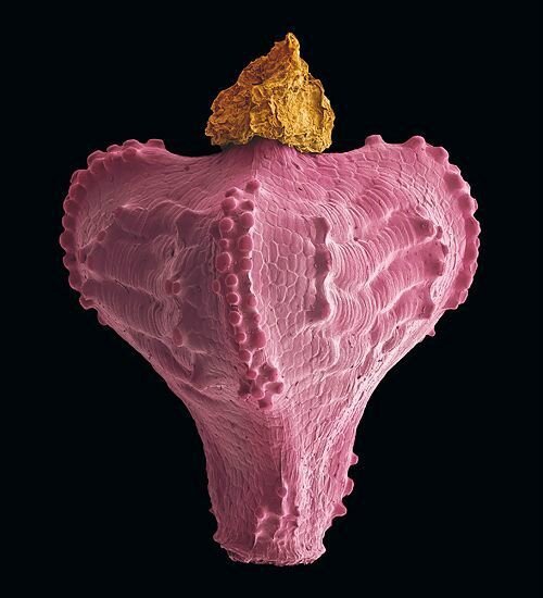

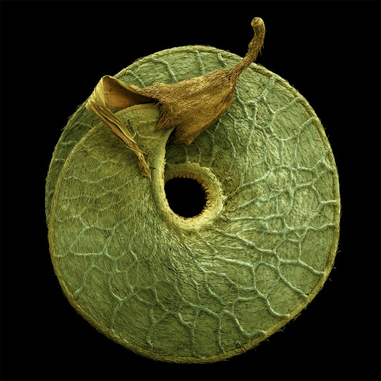
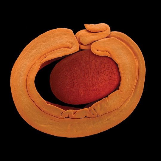
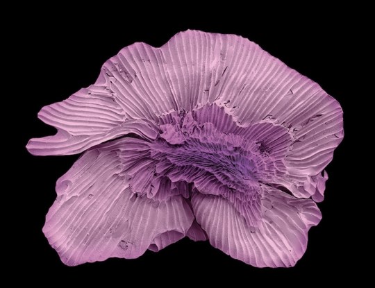
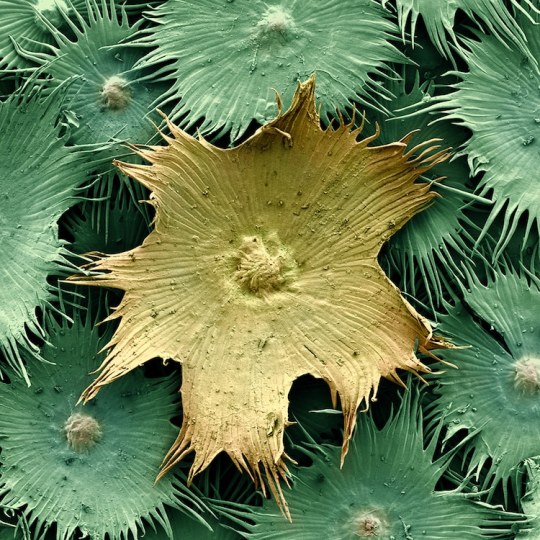
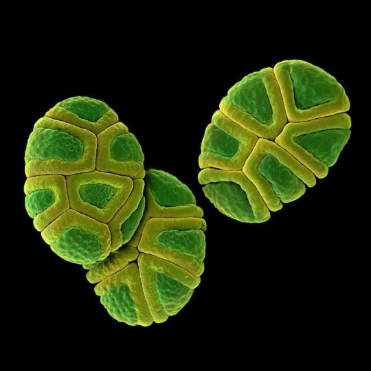

Rob Kesseler is a visual artist and Emeritus Professor of Arts, Design & Science at Central Saint Martins, London. For the past twenty years he has worked with botanical scientists and molecular biologists around the world to explore the living world at a microscopic level. Using a range of complex microscopy processes he creates multi-frame composite images of plant organs.
Using scanning electron microscopy and a mix of microscopic, scientific, digital, and manual processes, artist Rob Kesseler develops coloured micrographs of the intricate patterns within pollen and seed grains, plant cells, and leaf structures. The photographs feature specifics of cellular composition that are undetectable without magnification.
Kesseler tells that as a child, his father gifted him a microscope, marking a pivotal moment in his creative career. “What the microscope gave me was an unprecedented view of nature, a second vision,” he writes, “and awareness that there existed another world of forms, colours and patterns beyond what I could normally see.” The artist says his use of color is inspired by the time he spends researching and observing, and that just like nature, he employs it to attract attention.
36 notes
·
View notes
Text

Hello, EDS maps.
A bunch of thin sections imaged using under the SEM (scanning electron microscope) and then overlain with elemental EDS data in false color composites.
The dark blues are olivines, being FeO (red) /MgO (blue) rich; cyan crystals are diopside pyroxenes, being CaO (green) and MgO (blue) rich, and the pink crystals are Fe-Ti oxides, being FeO (red) and TiO2 (pink) rich. The very green groundmass in some samples is probably plagioclase, which is compositionally quite different from the cyan groundmass of other presumably more pyroxene rich samples. (These are foidites)
Time to do some analysis...
#cannot wait to get into the nitty gritty of these#eds mapping I love you I love you#phdee#geology#volcanology
3 notes
·
View notes
Text

Finally had our ideas for what the construction nanites look like come together.
We'll be refining this look. So, this image will be an "artist's visualization of the construction nanites", while some later image will be an electron microscope scan of them and there will be more detail but no color.
3 notes
·
View notes
Text
How a Water Analysis Lab Near Me Helps Identify Microplastics in Water?
Water pollution is a growing concern worldwide, and microplastics have emerged as a significant threat to water quality. These tiny plastic particles, often invisible to the naked eye, have infiltrated water bodies, posing risks to both human health and the environment. If you are searching for a "Water Analysis Lab near me," it is crucial to choose one that specializes in detecting microplastics in water. This blog explores how water analysis labs help identify microplastics and why testing is essential for safe drinking water.
What Are Microplastics?
Microplastics are small plastic particles less than 5 millimeters in size. They come from various sources, including:
Primary microplastics: Manufactured small plastics found in personal care products, industrial abrasives, and microbeads.
Secondary microplastics: Resulting from the breakdown of larger plastic debris due to environmental factors like sunlight and wave action.
These tiny particles enter water systems through wastewater discharge, stormwater runoff, and even atmospheric deposition, making their way into drinking water sources.
Why Are Microplastics a Concern?
Microplastics pose multiple risks, including:
Health Hazards: Studies suggest that microplastics may carry harmful chemicals and toxins, potentially leading to endocrine disruption, reproductive issues, and other health complications.
Marine and Aquatic Life: Fish and other aquatic organisms consume microplastics, which can lead to bioaccumulation, affecting the entire food chain.
Water Quality: The presence of microplastics compromises water purity and safety, making it essential to conduct regular testing.
How a Water Analysis Lab Near Me Detects Microplastics
1. Sample Collection and Preparation
The first step in microplastic detection is water sample collection. Labs follow strict protocols to prevent contamination during sample collection, using specialized glass or metal containers. Once collected, the sample undergoes preparation, which involves filtration and concentration techniques.
2. Filtration and Separation
To isolate microplastics from water, labs use:
Membrane Filtration: Water is passed through fine-mesh filters to trap microplastic particles.
Density Separation: Since plastics have different densities, solutions like sodium chloride or zinc chloride are used to separate plastic particles from organic matter.
3. Microscopic and Spectroscopic Analysis
After isolation, microplastics are analyzed using advanced techniques such as:
Optical Microscopy: A microscope helps identify microplastic size, shape, and color.
Fourier Transform Infrared Spectroscopy (FTIR): This technique detects plastic polymer composition, differentiating microplastics from other particles.
Raman Spectroscopy: Used to identify molecular structures and confirm plastic types.
Scanning Electron Microscopy (SEM): Provides high-resolution images for detailed structural analysis.
4. Polymer Identification
Once identified, the plastic polymers are classified into types such as polyethylene, polypropylene, polystyrene, and polyester. Understanding the polymer type helps in assessing potential sources of contamination.
5. Data Interpretation and Reporting
After analysis, the lab provides a comprehensive report detailing the presence, concentration, and type of microplastics in the sample. Businesses, government agencies, and individuals use these reports to make informed decisions on water treatment and environmental policies.
Who Needs Microplastic Testing?
1. Households and Residential Communities
Individuals seeking safe drinking water can benefit from testing services, ensuring their water sources are free from microplastics.
2. Water Treatment Plants
Testing is crucial for municipal water suppliers to monitor microplastic levels and improve filtration methods.
3. Industries and Manufacturing Units
Factories and production units discharge wastewater, which may contain plastic waste. Regular testing helps in compliance with environmental regulations.
4. Environmental Organizations
NGOs and research institutions analyze microplastic pollution trends, aiding in awareness and policy development.
5. Hotels and Restaurants
Food and beverage businesses must ensure the water they use is free from contaminants, including microplastics, to maintain high safety standards.
How to Choose the Best Water Analysis Lab Near Me
When looking for a reliable water testing facility, consider the following factors:
Accreditation and Certification: Ensure the lab is accredited by relevant regulatory bodies.
Advanced Testing Techniques: Opt for a lab that uses modern spectroscopy and microscopy methods.
Experience and Expertise: A lab with a history of conducting microplastic analysis is preferable.
Comprehensive Testing Services: Choose a lab that offers a full spectrum of water quality tests beyond microplastic detection.
Fast and Accurate Reporting: Timely results with precise data interpretation are essential.
Conclusion
Microplastics are an emerging environmental threat that necessitates proper monitoring and control. A "Water Analysis Lab near me" specializing in microplastic detection can help individuals, industries, and governments ensure clean and safe water. Regular testing helps prevent health risks, supports sustainability efforts, and ensures compliance with water safety standards. By choosing a reputable water analysis lab, you can take a proactive step toward protecting your health and the environment from microplastic contamination.
0 notes
Text
Petrographic Analysis: Revealing the Hidden Insights of Rocks and Minerals
Petrographic analysis is a crucial scientific technique used to study rocks and minerals, providing valuable insights into their composition, texture, and origin. This article explores the methods, applications, and significance of petrographic analysis in various fields such as geology, engineering, and environmental science.
What is Petrographic Analysis?
Petrographic analysis involves examining thin sections of rocks or minerals under a polarizing microscope. The primary goal is to identify mineral constituents, textures, and relationships within the rock. This analysis can reveal essential information about the rock’s history, formation conditions, and potential uses. If you are looking for a Petrographic Analysis then you may visit this website https://c3sinc.com/petrographic-analysis-scanning-electron-microscopy/.
!https://miro.medium.com/v2/resize:fit:875/0*QgBsT_l7ry6Su9Ks.jpg
Key Components of Petrographic Analysis
Thin Section Preparation: Samples are cut into thin slices, usually around 30 micrometers thick, allowing light to pass through for microscopic examination.
Microscopic Examination: A polarizing microscope is used to analyze the optical properties of minerals, which helps in their identification.
Data Interpretation: The collected data is interpreted to understand the mineralogy, petrology, and geological history of the sample.
Methods of Petrographic Analysis
Petrographic analysis employs several methods to obtain and interpret data:
1. Optical Microscopy
Description: Uses light to examine thin sections of rocks.
Applications: Identifies mineral types and textures based on their optical properties, such as birefringence and interference colors.
2. Scanning Electron Microscopy (SEM)
Description: Provides high-resolution images of mineral surfaces.
Applications: Offers detailed information about mineral morphology and composition.
3. X-ray Diffraction (XRD)
Description: Analyzes crystal structures by measuring the diffraction of X-rays.
Applications: Identifies mineral phases present in the sample and quantifies their abundance.
4. Chemical Analysis
Description: Determines the chemical composition of minerals using techniques like Energy Dispersive X-ray Spectroscopy (EDX).
Applications: Provides insights into the geochemical processes that formed the rock.
Applications of Petrographic Analysis
Petrographic analysis has wide-ranging applications across various fields:
1. Geology
Stratigraphy: Helps in understanding the layering of rocks and geological history.
Petrology: Aids in classifying rocks based on their mineral composition and origin.
2. Engineering
Construction Materials: Evaluates the suitability of rocks and aggregates for construction projects.
Foundation Studies: Assesses the stability and integrity of geological formations for infrastructure development.
3. Environmental Science
Contamination Studies: Analyzes soil and rock samples to identify pollutants and assess environmental impact.
Resource Exploration: Assists in the exploration of natural resources, such as minerals and fossil fuels.
Importance of Petrographic Analysis
Petrographic analysis is essential for several reasons:
Understanding Geological History: Provides insights into the formation and evolution of the Earth’s crust.
Resource Management: Helps in the efficient extraction and use of natural resources.
Infrastructure Safety: Ensures that construction projects are built on stable geological foundations, reducing risks associated with landslides and other geological hazards.
Environmental Protection: Aids in identifying and mitigating the impacts of human activities on geological formations.
Conclusion
Petrographic analysis is a powerful tool in the study of rocks and minerals, offering valuable insights into their properties and origins. Its applications in geology, engineering, and environmental science make it an indispensable part of earth sciences.
As technology advances, petrographic analysis will continue to evolve, enhancing our understanding of the Earth’s complex systems and contributing to sustainable resource management and environmental protection. Whether in academia, industry, or environmental studies, petrographic analysis remains a cornerstone of geological investigation.
0 notes
Text
Evolution and Impact of the Laboratory Microscope
The laboratory microscope stands as one of the most revolutionary instruments in the history of science. From the early rudimentary versions that merely magnified objects to today’s sophisticated digital and electron microscopes, this tool has significantly advanced our understanding of the natural world. It has not only enabled the exploration of the microscopic realm but has also catalyzed groundbreaking discoveries across various scientific fields. This article delves into the evolution, significance, and future of the laboratory microscope, underscoring its unparalleled role in scientific progress.
A Brief History of the Laboratory Microscope
The concept of magnification dates back to ancient civilizations, but it was not until the late 16th century that the first true microscope was invented. Hans and Zacharias Janssen, Dutch spectacle makers, are credited with creating the first compound microscope around 1590. This early model, which combined multiple lenses, allowed for greater magnification than simple magnifying glasses.
However, the microscope as we know it today owes much to Antonie van Leeuwenhoek, often referred to as the "Father of Microbiology." In the 1670s, van Leeuwenhoek built simple microscopes with lenses capable of magnifying objects up to 300 times their size. With these instruments, he made the first observations of bacteria, protozoa, and other microorganisms, opening an entirely new world to scientific inquiry.
The 19th and 20th centuries saw significant advancements in microscope technology. Achromatic lenses, developed by Joseph Jackson Lister in the 1830s, eliminated color distortions, enabling clearer and more accurate observations. The invention of the electron microscope in the 1930s, which uses beams of electrons instead of light, allowed scientists to observe structures at the atomic level. This marked a monumental leap in our ability to study the building blocks of life.
The Modern Laboratory Microscope: A Multidisciplinary Tool
Today’s laboratory microscopes are sophisticated, highly specialized instruments used in various scientific disciplines. In biology, they are indispensable for studying cell structures, bacteria, viruses, and other microorganisms. In chemistry and materials science, microscopes help researchers analyze the composition and structure of materials at a molecular level. In medicine, they are critical for diagnosing diseases, studying tissue samples, and developing new treatments.
One of the most significant advancements in recent years has been the development of digital microscopes. These devices combine traditional optical technology with digital imaging, allowing for real-time observation and the ability to capture, store, and share images and videos. Digital microscopes have made microscopy more accessible, enabling remote collaboration among scientists and educators worldwide.
Confocal microscopes, another modern innovation, use laser scanning to produce high-resolution images of thick specimens. This technology is particularly useful in biological research, where it allows for the detailed study of complex structures within cells and tissues.
The Impact of the Laboratory Microscope on Scientific Discoveries
The laboratory microscope has been at the heart of countless scientific breakthroughs. In microbiology, it enabled the discovery of microorganisms, leading to the development of the germ theory of disease and the advent of modern medicine. The ability to observe pathogens and understand their behavior was instrumental in developing vaccines, antibiotics, and other life-saving treatments.
In cell biology, the microscope has allowed scientists to explore the intricate workings of cells, leading to discoveries such as the structure of DNA, the process of mitosis, and the mechanisms of cellular communication. These insights have been crucial in understanding genetic diseases, cancer, and other medical conditions.
The microscope has also played a pivotal role in materials science. By enabling the examination of materials at the atomic level, it has facilitated the development of new materials with unique properties, such as superconductors, nanomaterials, and advanced composites. These materials have a wide range of applications, from electronics to aerospace to renewable energy.
The Future of Laboratory Microscopy
As technology continues to advance, the future of laboratory microscopy looks promising. One area of exciting development is super-resolution microscopy, which breaks the diffraction limit of light to achieve even higher levels of detail than traditional optical microscopes. This technology has the potential to revolutionize fields such as neurobiology, where it could provide unprecedented insights into the functioning of the brain.
Another emerging trend is the integration of artificial intelligence (AI) with microscopy. AI-powered microscopes can automate the analysis of images, allowing for faster and more accurate data interpretation. This capability is particularly valuable in fields such as pathology, where AI can assist in the early detection of diseases such as cancer.
Moreover, the ongoing miniaturization of microscopes is making them more portable and affordable, broadening their accessibility. Handheld and smartphone-based microscopes are already being used in educational settings and in remote areas for medical diagnostics. These portable devices have the potential to democratize science, enabling more people to engage in scientific research and discovery.
Conclusion
The laboratory microscope is more than just an instrument; it is a window into the hidden world that exists beyond the limits of human vision. From its humble beginnings to its current state as a highly advanced tool, the microscope has been instrumental in expanding our understanding of the natural world. Its impact on science and medicine cannot be overstated, as it has paved the way for countless discoveries and innovations.
As we look to the future, the laboratory microscope will undoubtedly continue to evolve, pushing the boundaries of what we can see and understand. Whether in the hands of a student, a researcher, or a medical professional, the microscope remains an essential tool for exploration and discovery. Its legacy is one of curiosity, innovation, and the relentless pursuit of knowledge—a legacy that will continue to inspire future generations of scientists.
1 note
·
View note
Text
Unveiling the Polaritonic Cathode Ray Tube: UPCRT

The UPCRT (Ultra-Thin Polaritonic Cathode Ray Tube) is a revolutionary display concept that combines the responsiveness of classic CRTs with the advanced capabilities of polaritonic materials. Let's explore each layer of the UPCRT to understand how it works and what makes it special.
Layer 1: The Electron Gun
Function: The electron gun generates a focused beam of electrons, which is essential for creating the display image.
Technology: The UPCRT uses a Field Emission Gun (FEG) instead of the traditional thermionic cathode.
How it Works:
Sharp Emission Tip: Made from materials like carbon nanotubes or metallic nanowires, the tip is extremely sharp, enhancing the electric field.
Strong Electric Field: A high voltage is applied, causing electrons to be emitted from the tip through quantum tunneling.
Beam Formation: The emitted electrons form a concentrated beam, which is then accelerated towards the display screen.
Benefits:
Compact Design: FEGs are smaller than traditional CRT guns, allowing for a slimmer display.
Power Efficiency: FEGs operate at room temperature, reducing power consumption.
Immediate Response: High electron emission rates ensure near-instantaneous response times.
Precision: A micro-focusing system ensures the electron beam is tightly focused for sharp visuals.
Layer 2: Electron Beam Deflection
Function: This layer controls the movement of the electron beam across the display, painting the picture one pixel at a time.
Technology:
Electrostatic Deflection Plates: Lightweight plates with varying electric charges deflect the electron beam efficiently.
MEMS-Based Beam Steering (Future): Microscopic mechanical structures (MEMS) could offer even faster and more precise deflection.
Benefits:
Efficiency: Electrostatic deflection is more efficient and allows for a thinner display.
Precision: MEMS-based steering can provide ultra-fast scanning speeds and pinpoint accuracy.
Layer 3: The Polaritonic Layer
Function: This layer emits light when struck by the electron beam, creating the image.
Materials:
Quantum Dots/Wells: Made from advanced materials like 2D perovskites or graphene, these structures emit specific colors when excited by electrons.
Microcavity: Reflective mirrors trap light within the polaritonic layer, enhancing light output.
How it Works:
Excitation: The electron beam excites the quantum dots/wells to a higher energy state.
Light Emission: As they relax back to their ground state, they emit light, creating a pixel on the screen.
Benefits:
Brightness: Polaritonic materials offer exceptional light emission capabilities.
Color Gamut: Precisely engineered quantum dots/wells provide a broader range of colors, leading to richer visuals.
Layer 4: Display Structure
Function: This layer houses all the components and ensures efficient light transmission to the viewer.
Components:
Thin-Film Assembly: Integrates the FEG and deflection system into a single, ultra-thin film.
Flat Panel Design: The polaritonic layer rests on top of the microcavity structures, creating a sleek display profile.
Transparent Electrodes: A transparent conducting oxide (TCO) like Indium Tin Oxide (ITO) allows light to pass through efficiently.
Vacuum Seal: All components are housed within a thin, vacuum-sealed panel to maintain optimal performance.
The UPCRT in Action: A Symphony of Electrons and Light
Electron Generation: The FEG generates a focused beam of electrons.
Beam Deflection: Electrostatic deflection plates or MEMS precisely control the movement of the electron beam.
Light Emission: The electron beam strikes the polaritonic layer, exciting the quantum dots/wells.
Color Creation: The excited quantum dots/wells emit light of specific colors.
Light Amplification: The microcavity structure traps and amplifies the emitted light, enhancing brightness and color vibrancy.
Electron Gun Configuration
The UPCRT uses only one electron gun per display. This is a significant improvement over traditional shadow mask CRTs, which required three electron guns for red, green, and blue subpixels.
Why a Single Electron Gun Works:
Fast Scanning: The electron beam scans the display rapidly, visiting each pixel in quick succession.
Polaritonic Layer: The quantum dots/wells in the polaritonic layer emit different colors based on their material properties, allowing a single electron gun to create a full-color image.
Conclusion
By harnessing the power of field emission guns, MEMS-based deflection, and advanced polaritonic materials, the UPCRT promises unmatched brightness, energy efficiency, and a compact design. It retains the immediate response and analog-like control of classic CRTs, making it a compelling choice for next-generation displays.
0 notes