#Skin Treatment & Platelet Rich Fibrin Matrix.
Text
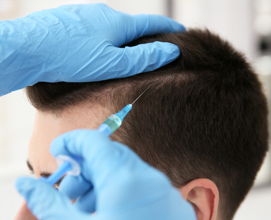
PRP Hair-Regrowth (Roller) Treatment - Twacha Aesthetic Skin Treatmemt Clinic

For a long time, PRP has been the gold standard for plasma therapy used to cure hair growth. However, continued research and clinical trials have led to the development of a new and significantly improved treatment, the next-generation plasma therapy known as PRF or Platelet Rich Fibrin.
Our blood contains fibrin, a protein that prevents clotting and provides strength and structure.
PRF can also be obtained by doing a simple blood test on your own blood. But compared to PRP, the outcomes and treatment experience are significantly better.
No Anti-coagulants: In PRP tubes, an anti-coagulant is typically used to help stop the retrieved plasma from thickening and clotting. Anticoagulants disrupt the fibrin matrix and stop the protein from functioning as it ought to. Since PRF doesn’t contain any additional anti-coagulants, the fibrin matrix is left unharmed and can immediately begin to function to restore cellular rejuvenation.
Virtually Painless : Simply injecting platelets into the scalp is significantly less uncomfortable than doing so while also injecting anticoagulants. The majority of the discomfort and stinging at the injection site is really brought on by anticoagulants. By removing anti-coagulants from PRF, the entire treatment becomes almost painless.
Platelet Count: PRF extraction can generate a platelet count at the injection site that is almost ten times greater than what the body naturally produces. This is far more than what a PRP procedure can retrieve.
Long-term benefits from increased growth factors: Growth factors are our body’s natural “healing element,” helping all cells, including hair cells, to grow and mend. They do a lot of things inside the scalp, including promoting stem cell rejuvenation, re-oxygenating and repairing damaged cells, increasing cell turnover, stimulating blood circulation, and more. In contrast to PRF, which releases these growth factors over a period of 7 to 10 days, PRP releases these growth factors instantly but over a short period of time (the same day). Introducing a healing cascade over the following several months (over days and weeks), this slower release of growth factors ensures that both the number and quality of growth factors are significantly higher, which contributes to the longer-term effects of the treatment.
https://twachaskin.com/hair-treatments/prp-hair-regrowth-roller-treatment/
#skincare#bodycare#skinluxury#WellnessSolutions#HealthyLifestyle#MindBodySoul#SelfCare#HolisticHealth#NaturalHealth#AlternativeMedicine#HealthyHabits#WellnessJourney#HealthyLiving#FitnessMotivation#MentalHealthAwareness#HealthyEating#WellnessGoals#WellnessCommunity#WellnessInspiration#SelfLove#PersonalGrowth#StressManagement#HealthyMindset#PositiveVibes#Mindfulness#HealthyBody#HealthyMind#HealthySpirit
2 notes
·
View notes
Text
Under Eye Restoration For Dark Circles: How It Can Help Brighten Your Appearance?
Dark circles can cast a shadow on your face, affecting your overall appearance and confidence. Imagine a solution that can naturally brighten your appearance and boost your confidence. Say goodbye to under-eye woes with under-eye restoration!
Together, we will discover the power of PRP (Platelet-Rich Plasma) and PRFM (Platelet-Rich Fibrin Matrix) in rejuvenating the delicate under-eye area. We will explore the benefits of this advanced treatment, how it works, and what you can expect throughout the process. By the end of this guide, you will have a comprehensive understanding of under-eye restoration and how it can help you achieve a brighter, more youthful appearance.
What is Under Eye Restoration Treatment?
A minimally invasive procedure called under eye restoration using PRP and PRFM uses your body’s inherent capacity for healing. Similar to a standard blood test, a tiny blood sample is extracted. The platelet-rich plasma (PRP) and platelet-rich fibrin matrix (PRFM) are then separated from other blood components during the centrifugation processing of the blood.
The PRP and PRFM contain concentrated growth factors and regenerative proteins that promote skin rejuvenation. The extracted PRP and PRFM are carefully injected into the targeted areas under the eyes, stimulating collagen production and improving the appearance of dark circles, sagging, and crepiness.
The procedure is performed using fine needles to ensure precision and minimize discomfort. The treatment typically takes about 30 minutes to an hour, depending on individual needs and desired outcomes. You may return to your regular routine with little downtime following the procedure.
How does the treatment work?
PRP and PRFM are rich in growth factors and bioactive proteins crucial in tissue repair and regeneration. When injected into the under-eye area, PRP and PRFM stimulate collagen synthesis and cellular turnover.
By promoting collagen production, PRP and PRFM help improve the skin’s texture, tone, and firmness. The growth factors and proteins in PRP and PRFM also support blood vessel formation and enhance the skin’s natural healing response, improving skin quality and a more vibrant appearance. Additionally, it has been shown that PRP and PRFM have anti-inflammatory and antioxidant capabilities, which can further aid in the regeneration of the under-eye region.
How does the treatment target sagging and crepiness under the eyes?
Sagging and crepiness under the eyes can be caused by a loss of collagen and elastin fibers and decreased skin thickness. Under-eye restoration with PRP and PRFM addresses these concerns by promoting collagen production and improving the overall health and quality of the skin.
The growth factors and proteins in PRP and PRFM stimulate the regeneration of collagen and elastin fibers, leading to tighter and smoother skin. As collagen production increases, the under-eye area becomes firmer and more resilient, reducing sagging and crepiness. By targeting these specific concerns, under eye restoration with PRP and PRFM can help you achieve a more refreshed and youthful appearance.
What is the recommended treatment plan for undereye restoration?
Undereye restoration using PRP and PRFM is typically performed as a series of treatments to achieve optimal results. The exact treatment plan may vary depending on individual needs and the assessment of the licensed professional. It is crucial to have an in-depth consultation with an experienced CONTŌR specialist who can assess your issues and provide a custom treatment plan.
A series of three treatments is often recommended to maximize the benefits of undereye restoration. The interval between treatments allows the skin to respond to the PRP and PRFM injections and gradually improve over time.
What are the benefits of Under Eye Restoration treatment?
Under eye restoration, using PRP and PRFM offers several benefits in addressing dark circles.
Dark circles can be caused by various factors, including thinning skin, blood vessels showing through the skin, or hyperpigmentation. PRP and PRFM treatments target these underlying causes to help reduce the appearance of dark circles and improve the overall look of the under-eye area. The therapy can help achieve a brighter and more youthful appearance by stimulating collagen production and promoting skin rejuvenation.
Undereye restoration treatments with PRP and PRFM can significantly improve the skin’s texture, tone, and elasticity.
The regenerative properties of PRP and PRFM help stimulate collagen and elastin production, which are crucial for maintaining skin firmness and elasticity. Patients may notice a smoother, tighter, and more supple skin texture in the under-eye area as the treatment progresses. Restoring healthy skin attributes can contribute to a more rejuvenated and refreshed appearance.
The treatment helps brighten the overall appearance.
One of the primary goals of undereye restoration is to brighten the overall appearance by reducing the prominence of dark circles. The combination of PRP and PRFM helps improve blood flow and circulation in the under-eye area, reducing the appearance of blood vessels or shadows contributing to dark circles. Additionally, the treatment promotes the production of new, healthier skin cells, resulting in a brighter and more radiant look.
What is the recovery process after under-eye restoration treatment?
After undergoing under-eye restoration with PRP and PRFM, it’s essential to understand the recovery process to ensure optimal results. The recovery period for this procedure is typically minimal, with most individuals able to resume their daily activities immediately. However, following the post-treatment instructions is essential for a smooth recovery.
To optimize the results of your under-eye restoration and minimize any potential side effects, consider the following tips for post-treatment care:
Keep the treated area clean.
Apply cold compresses.
Avoid excessive sun exposure.
Avoid strenuous activities.
Follow the recommended skincare routine.
Properly caring for your under-eye area during recovery and following the aftercare instructions can support the healing process and maximize the benefits of under-eye restoration with PRP and PRFM.
Explore Under Eye Restoration Treatment with PRP and PRFM
Under eye restoration, using PRP and PRFM is a powerful and natural approach to improving the appearance of dark circles. The treatment harnesses the regenerative properties of PRP and PRFM to address sagging and crepiness under the eyes and promote skin rejuvenation.
Under eye restoration with PRP and PRFM could be an excellent option if you struggle with dark circles and seek an efficient treatment. To determine if you’re a good candidate for this procedure, speak with us at CONTŌR.
Embrace the opportunity to take control of your appearance and feel more confident and ready to face the world. Contact us today to learn more about our under-eye restoration treatment!
0 notes
Text
Best PRF Treatment in Hyderabad
Hair Loss and Hair Thinning are common hair-related complaints in recent times. Hair fall has multiple causes which include nutritional, hormonal or genetic conditions. Platelet-rich fibrin (PRF) is the popular alternative for Platelets Rich Plasma (PRP) Therapy.
FMS Skin and Hair Clinics is one of the best hair clinics for PRF treatment in Kondapur, Hyderabad. PRF is considered as an advanced version of PRP with far more superior effects. PRF injections are made from a person's own blood and a protein matrix called fibrin. Once harvested, we get a high concentration of white blood cells, fibrin and platelets containing growth factors.
PRF is prepared according to the low-speed centrifugation concept and is highly enriched with platelets and leukocytes that provides an increased concentration of growth factors. These growth factors assist in tissue regrowth. PRF treatment for hair is more effective in treating hair loss issues.
At FMS Skin and Hair Clinics, PRF is prepared using a specialized device called Arthrex Autologous Conditioned Plasma (ACP) Double Syringe System. PRF treatment is done by a trained and certified dermatologist.
Know more about PRF Treatment at https://www.fmsskin.com/best-prp-platelet-rich-plasma-prf-in-hyderabad/

0 notes
Text
PRFM for Hair Loss - Hairline International Hair & Skin Clinic-

Androgentic alopecia affects up to 30% of men over the age of 30 and 50% of men over the age of 50 and women as well. Several modalities of treatment have been promoted for hair regrowth. Only 2 medications minoxidil and finesteride and 655nm laser treatment have been approved by the FDA.
Platelet rich fibrin matrix (PRFM) is a new breakthrough in hair regrowth.
Advantages of PRFM Treatment
PRFM presents more viable and intact and activated platelet in fibrin matrix which produces a more prolonged exposure to growth factors in a more natural time course. It is believed that this natural kinetics will yield more sustained hair growth.

Studies show that PRFM is more effective in grade 3, 4, and 5 in men and grade 2 in female.
All patients tolerated the procedure well. No patients noted any significant ecchymosis.
Hair shedding didn’t worsen in any case.
Significant hair regrowth and improvement in thickness seen in 3 sessions of treatment.
The procedure takes only 20 mins. Its sustain release up to 7 days after the procedure allows cell angiogenesis and hair re growth in the scalp. It increases hair density and thickness almost 2 times in 6 months.
Platelet rich fibrin matrix is extensively used by surgeons worldwide to treat chronic lower extremity ulcers. It promotes wound healing via cell proliferation and new cell re growth.
PRFM is very effective in combination with hair transplantation. It stimulates dermal angiogenesis and wound healing which helps in better survival of the transplanted graft. Moreover, it also improves the density of the thinning hair by stimulating cell proliferation.
Q. What is platelet rich fibrin matrix treatment for hair loss?
A. Platelet rich fibrin matrix treatment is considered to be one of the latest inventions in hair treatment. In this treatment a small amount of blood is collected from the patient and centrifuged. This separates and concentrates the patient’s own platelets and fibrin into a matrix. The matrix is then injected into the thinning or bald scalp which stimulates cell proliferation and regrowth of hair through targeted tissue regeneration.
Q. How is it better than the current line of treatments advised for hair loss?
A. Platelet rich fibrin matrix releases growth factors into the scalp through a sustained mechanism. In vitro studies have shown that growth factors are released up to 7 days. This stimulates new hair growth. Pronounced results are seen after 3 weeks of the treatment.
Q. Why is it superior to platelet rich plasma?
A. PRFM is superior to PRP. The procedure takes only 20 mins. PRFM requires only 4 sessions to see visible result. Its sustain release up to 7 days after the procedure allows cell angiogenesis and hair regrowth in the scalp. It increases hair density and thickness almost 2 times in 6 months.
Q. What cases should opt for this treatment?
A. A dermatologist decides the cases after thorough history taking and blood investigations but any individual with hair thinning and balding can opt for the treatment.
Q. Is there a scientific evidence for such a treatment?
A. Platelet rich fibrin matrix is extensively used by surgeons worldwide to treat chronic lower extremity ulcers. It promotes wound healing via cell proliferation and new cell regrowth.
Q. What results are visible?
A. Results are visible after 4 sessions of therapy in hair loss patients.
Q. How is it more effective in combination with hair transplantation?
A. PRFM is very effective in combination with hair transplantation. It stimulates dermal angiogenesis and wound healing which helps in better survival of the transplanted graft. Moreover, it also improves the density of the thinning hair by stimulating cell proliferation.
#PRFM for Hair Loss in Bangalore#Hair Regrowth Treatment Bangalore#regenera activa treatment cost in india#best re-genera hair treatment in bangalore
0 notes
Text
What Are the Reasons to Try the Platelet-Rich Fibrin Face Treatment for Your Face?

The PRF or platelet-rich fibrin is an enhanced version of PRP or platelet-rich plasma. PRF is a natural treatment that uses an individual’s blood cells to enhance skin texture, collagen production, and hyaluronic acid.
The platelet-rich fibrin face treatment in Fort Lauderdale can also be used to restore hair, manage wounds, and repair knee and skin regrowth. Though PRF and PRP have the same principle and goal, they are not the same. In this blog, you will learn some reasons to try the PRF treatment for your face.
Reasons to Consider the PRF Treatment for Your Face
Some of the top reasons why you need to consider PRF treatment for your face are as follows:
It can eliminate wrinkles and facial folds
PRF promises and helps to deliver an enhanced skin complexion with fewer fine lines and wrinkles. While the blood in the PRP is made using anticoagulants and at a faster speed to destroy all red blood, PRF is made by spinning the tubes at a lesser speed and isolating the white blood cells and platelets from the red blood cells.
The method used in platelet-rich fibrin face treatment in Fort Lauderdale allows for a higher platelet concentration and can maintain the fibrin matrix without anticoagulants. This is why this treatment can help to lessen wrinkles and facial folds; 48 hours after this treatment, you can notice the changes.
It can treat under-eye loose skin and dark circles.
The skin beneath your eye is considered the thinnest skin on your body, which is why it is natural to be more vulnerable to wrinkles and fine lines. PRF can easily treat the effects of under-eye aging by reducing the wrinkle’s visibility and filling in hollows. Dermal fillers are usually considered the best treatment for this sensitive area, but now PRF is considered the best option.
It can enhance skin texture and treat acne scars.
The skin cells in our body can be exposed to various damaging factors like smoking, pollution, blood flow, less blood flow, and poor skin maintenance. All these can lead to a more dreaded and dull skin tone that will make you look older.
But due to the healing power of platelets, platelet-rich fibrin face treatment in Fort Lauderdale can not only improve moisture to your skin but also improve blood flow to improve the appearance of your complexion and refine your skin tone.
The results can last for around a year.
One of the benefits of this PRF procedure is that the patient can enjoy the results of this treatment for around a year. This is a great thing considering that the procedure lasts for around 45 minutes, and you will only require two to five appointments for a complete treatment. There are also fewer side effects risks compared to other treatment forms available on the market.
Bottom Line
If you want to enhance the look of your age and remove dark spots and other aging spots, you can opt for platelet-rich fibrin face treatment in Fort Lauderdale. This is one of the treatments you can find on the market; you must ensure that you undergo this procedure from a reputed clinic or medical spa.
0 notes
Text
PRP每月科技新知 (2022/06/01)- PRP可用於異位性皮膚炎和其他類型的濕疹
---這是一篇個案報告---
前言: 異位性皮膚炎是一種慢性皮膚發炎疾病,最常見於兒童時期,但也影響許多成年人。大多數異位性皮膚炎患者使用保濕劑和外用類固醇進行治療。然而,這些治療並不總是能夠良好控制疾病。富含血小板血漿 (PRP) 用於治療許多皮膚病。PRP是經由血小板釋放各種物質(例如生長因子)發揮作用,從而誘導傷口癒合��一系列反應,包括抗發炎和組織再生,從而促進皮膚健康。這篇文章報導了一例異位性皮膚炎患者,使用 PRP 成功控制皮膚症狀。
個案報告: 一名 44 歲的女性於 2016 年 5 月就診,有長期反覆面部濕疹病史。患者使用類固醇乳膏六個月後,效果不佳,並且也詢問眼眶下黑眼圈和皺紋的治療方法。在討論治療方式後,決定進行 PRP 注射。 使用 4-cc 富含血小板的纖維蛋白基質試劑盒 (Selphyl, Aesthetic Factors, Inc) 進行了兩次 PRP 針劑注射治療,每次間隔 6 週。 除了在眼睛下方施用外,PRP 也使用在鼻唇溝中以達到年輕化的目的。
圖 1:面部濕疹對 PRP 針劑注射治療的效果。
結論: 根據這篇文章和其他研究的結果,PRP 可用於治療異位性皮膚炎和其他類型的濕疹。這位病人追蹤觀察兩年,症狀緩解是前所未有的。
# 防疫新選擇,免出門安全購物:
KimClaire網頁購買: https://kimclaire.1shop.tw/prp?tag=LINE
原文摘要:
Atopic dermatitis (AD) is a chronic inflammatory skin condition that most often presents in childhood but also affects many adults. Most patients with AD are treated with moisturizers and topical corticosteroids. However, these treatments do not always allow for an adequate control of the disease. Platelet-rich plasma (PRP) is used in treating many dermatologic conditions. It works by releasing bioagents, such as growth factors, from platelets, which induce a wound-healing cascade involving inflammation and tissue regeneration that promotes skin health. Here, we report a case of AD in which the patient successfully achieved an extended control of skin symptoms with PRP therapy.
A 44-year-old woman presented in May 2016 with a long-standing history of recurrent facial eczema. Six months later, the patient returned for a refill of the hydrocortisone cream and inquired about treatments for infraorbital dark circles and wrinkles. After discussing the treatment options, we decided to proceed with PRP injections, and informed consent was obtained. Two PRP therapy sessions were performed, spaced 6 weeks apart, using a 4-cc platelet-rich fibrin matrix kit (Selphyl, Aesthetic Factors, Inc). In addition to administration beneath the eyes, PRP was also administered into nasolabial folds for rejuvenation purposes.
Fig 1: Response of facial eczema to PRP treatment.
Based on our results and those of other studies, there is an indication for the potential use of PRP in the management of AD and other types of eczema. Two years of symptom relief is unprecedented. A randomized clinical trial with a greater number of participants is warranted to rigorously evaluate the seemingly positive effects of PRP in treating skin eczema, including AD. Further research may also shed more light on the pathophysiology of AD as well as the biochemical pathways through which PRP can exert its therapeutic effects.
文章出處:
Saba Vafaei-Nodeh, BSc, and Shadan Kabiri-Abyaneh, MD, CCFP. Long-term control of atopic dermatitis with platelet-rich plasma. JAAD Case Rep. 2020 Nov 5;7:54-56.

0 notes
Text
The Vampire Facelift Treatment Aids in the Rejuvenation of Your Face
Looking for a Prp vampire facial near me? Have a look at this.
People go to great lengths to rejuvenate their faces to appear beautiful and alive. Face-lifting, also known as a vampire facelift, is a non-surgical cosmetic procedure that removes excess skin and makes the person appear younger and more alive. This PRP treatment rejuvenates the skin and gives you a younger, fresher, and more youthful appearance.
This procedure was created for those who want to look younger and wrinkle-free. Using a mixture of hyaluronic acid and platelet-rich fibrin matrix, this method helps to increase the volume in the face (PRFM). The key element in this process is PRFM, which is usually found in the patient's blood and produces unique substances, platelet-rich plasma (PRP), which is injected into the patient's wrinkled skin.
The Vampire facelift formula reduces muscle and fat, making the under-eye area and cheeks appear flat and hollow. This vampire facelift procedure can treat the following areas: corners of the mouth, wrinkles on the nose, and lines on the corners of one's eyes, wrinkles on the forehead, frown lines, smile lines, and cheek plumping. Not only does it reduce wrinkles, but it also improves the appearance of the skin.
The Advantages of a Vampire Facelift Treatment
● Improves the skin's quality
● Improves skin clarity
● Combating the effects of ageing
● Blood flow has improved.
● A simple procedure
● It increases collagen production.
● Skin folds are reduced.
● The skin softens.
● The sagging skin returns to normal.
● Scars and spots are completely removed.
● The skin becomes constricted.
This method promotes the formation of new blood vessels, which increases blood flow to the skin, resulting in healthier and better skin. Following the treatment, new cells begin to form, resulting in the formation of new fatty tissue and the appearance of brighter, younger, and fresher skin.
0 notes
Text
Is PLATELET-RICH FIBRIN better than fillers: The Ultimate Guide
We may age in years, but it does not mean we have to enjoy it! Additionally, the aging process might hit prematurely, which can be pretty unpleasant. Numerous variables, including sunlight exposure, lifestyle choices, and even heredity, can influence how we age.
How frequently do you find yourself evaluating your looks in the mirror? Perhaps you’ve begun to see fine wrinkles or a loss of volume in your face. Additionally, you may have started to notice skin coloring or texture alterations in various places of your body. While you may not be able to halt time, you still can turn back the hands of time!
If you want to rejuvenate your appearance, your blood may be the key. You see, Platelet-Rich Fibrin is a blood solution. Blood will be taken from a vein in the arm and processed in a centrifuge to extract the red blood cells from the white blood cells. What remains is plasma with growth factor-rich platelets, white blood cells, and mesenchymal stem cells. After that, the Platelet-Rich Fibrin is injected into various parts of the face to focus regeneration precisely where it is wanted.
Due to the absence of a blood thinner in Platelet-Rich Fibrin (which distinguishes it from Natural Growth Factor Injections) clumps and creates a fibrin matrix when injected. For 7-10 days, the fibrin clot steadily distributes growth factors into the surrounding area.
The Platelet-Rich Fibrin Process
Fibrin is a biological framework that arises in the body in response to damage. Platelets flowing in the blood will adhere to a fibrin scaffold. When platelets adhere to the fibrin scaffold, they trigger and produce growth factors that initiate wound healing by forming new skin cells and blood vessels. A regulated inflammatory response is triggered by injecting Platelet-Rich Fibrin into a targeted location on the face or scalp, stimulating new cell development and collagen formation.
What Is the Purpose of Platelet-Rich Fibrin?
Platelet-Rich Fibrin is used to treat many of the same conditions as Natural Growth Factor Injections; however, Platelet-Rich Fibrin releases compounds for a significantly more extended time, up to one week, compared to Natural Growth Factor Injections, which only releases growth factors for a few hours. This indicates that Platelet-Rich Fibrin may provide superior long-term advantages.
Growth factors are responsible for stimulating stem cells to produce more collagen and elastin. The platelets in Platelet-Rich Fibrin are more resilient than those in Platelet-Rich Fibrin, resulting in a speedier healing process.
The produced Platelet-Rich Fibrin is instantly available for injection into the skin to plump the hollows beneath the eyes and dark circles and enhance skin tone and texture. Additionally, Platelet-Rich Fibrin can be injected directly into the scalp to help with hair regeneration and growth.
How Does Platelet-Rich Fibrin Improve Poor Skin And Black Circles Around The Eyes?
The skin under our eyes is among the thinnest on our bodies. It is frequently one of the first regions to exhibit symptoms of aging. Platelet-Rich Fibrin is an excellent treatment choice for this area and may be preferable to heat-based skin tightening devices.
Platelet-Rich Fibrin can also be used to improve the effects of fillers placed beneath the eyes. Injecting fillers beneath the eyes can rapidly address hollowness beneath the eyes and is an excellent way to revitalize this region. However, if fillers are injected superficially beneath the eyes, they might induce puffiness or a blue hue.
Additionally, it might be challenging to address dark circles only with fillers. By compressing the loose skin around the eyes and reducing the dark circles, administering Platelet-Rich Fibrin and the fillers can considerably boost the outcomes of the fillers.
Reasons To Give Platelet-Rich Fibrin a Try
1. PLATELET-RICH FIBRIN IS COMPLETELY NATURAL.
As previously mentioned, Platelet-Rich Fibrin is a treatment that utilizes your platelets, which is one of the treatment’s most distinguishing characteristics.
2. PLATELET-RICH FIBRIN GROWTH FACTORS ARE YOUR ANTI-AGING MEDICATION.
Platelet-Rich Fibrin’s growth factors are critical for reversing the unmistakable symptoms of aging. It has many platelets, white blood cells, and a trace amount of stem cells. Platelets adhere to the fibrin matrix and produce growth factors that stimulate new skin cells, collagen, and blood vessels. Additionally, the growth factors in Platelet-Rich Fibrin are delivered more slowly than those in Natural Growth Factor Injections, providing an extra benefit of a longer-lasting anti-aging procedure!
3. PLATELET-RICH FIBRIN MAY BE APPLIED TO ANY PART OF THE BODY OR FACE.
It has been utilized to facilitate quicker recovery following surgical operations such as bone transplants and dental implants in the medical profession. Additionally, it can aid in the healing process following cosmetic surgical treatments. Platelet-Rich Fibrin can be carefully injected anywhere around the face or body, improving the skin’s texture, tone, discoloration, and general health.
4. PLATELET-RICH FIBRIN CAN BE CONSOLIDATED WITH OTHER TREATMENTS TO ACHIEVE EVEN BETTER RESULTS.
Some clinics provide various treatments targeted at enhancing your skin’s look and assisting you in achieving the youthful skin you’ve been dreaming of.
Even various fillers have distinct qualities. Platelet-Rich Fibrin can be used with any of these therapies to maximize progress and assist you in achieving your personal goals.

After Care Recommendations for any Platelet-Rich Fibrin treatment:
AVOID ALL NSAIDS for 2-4 weeks before the treatment! These drugs interfere with the platelet coagulation process in Platelet-Rich Fibrin and may reduce the effectiveness of the treatment. Tylenol does not contain aspirin and is thus safe to consume before your therapy.
Hydration is critical for collecting an adequate amount of high-quality Platelet-Rich Fibrin during therapy and will improve your treatment outcomes. Drink at least 64 ounces of water on the days preceding and the day of your session, and moisten the injection region before your visit.
Before your therapy, take 2000 mg of Vitamin C every day. The sooner you begin, the better.
For 2-4 weeks before your therapy, abstain from omega-3 fatty acids, fish oil, turmeric, garlic supplements, and aspirin. It’s also recommended that you refrain from alcoholic beverages for seven days before your therapy (including red wine)
If you’re interested to learn more and experiencing skin restoration, the PLATELET-RICH FIBRIN treatment may be ideal for you! You may receive this treatment with the assistance of clinics such as Spirited Aesthetics and Wellness! Age like great wine, and be prepared to slay every single day.
0 notes
Text
Is vampire facial worth it?
People make infinite attempts to rejuvenate their faces to seem beautiful and exist. Face-lifting is a corrective non-surgical process that aids in eliminating additional skin and Shows the person looks more youthful and nice, commonly known as Vampire Facial Boston. This PRP therapy helps in strengthening the skin and presents you with younger, fresh, and facial features. If you want to have the beautiful skin you need to take care of this, you need to require facials and massage for making them nice. Take your best facial spa in Boston to have the most attractive skin.
Vampire Facial In Boston was acquired for people who want to look young and wrinkle-free. This best facial spa in Boston helps in improving the volume in the surface using a mixture of hyaluronic acid and platelet-rich fibrin matrix (PRFM). The key element during this process is PRFM, which is normally found in the family of the patient and designs unique substances, platelet-rich plasma (PRP), which is inserted into the wrinkled skin of the person. The dermatologists guarantee that this procedure is secure and there are no side effects. The symptom of aging is restored completely and making skin healthy.
📷
Best facial spa in Boston - The vampire facelift method decreases the muscle and fat making your under-eye area and cheeks looking bad. The spaces which can be handled by this vampire facelift for facial spa Boston ma procedure are sides of the mouth, wrinkles on the nose, lines on the corner of your eyes, wrinkles on the temples, frown lines, smile lines, plumping of the cheeks. It not only betters the texture and other different things.
Benefits of vampire facelift procedure - facial spa Boston MA.
Promote enhanced skin quality
Enhances the skin clarity
Fight with the factors of aging
Correcting in the blood flow
A painless method
It incites collagen production
Decreases the skin folds
Skin converts soft
The sagging skin gets normal
Complete removal of scars and spots
The skin gets tight
This facial spa Boston ma helps in enhancing the creation of new blood vessels, so increasing the blood flow to your skin appearing in more youthful and healthier skin. After the treatment, new cells begin to form, which generates new fatty tissue to look brighter, younger, and fresh - facial spa Boston ma.
The whole process requires an hour of blood draw, construction of the PRP, platelet-rich fibrin form, and surely the vampire facelift procedure. Since revival and restoration of the skinning method are created using patients’ actual compensation, there is no major problem, and the system lasts for one year. Consult the best facial spa Boston ma provider to get this procedure done effectively.
If you are not aware of the face lifting process, you should consult your dermatologist - the best medical spa and seek his view on the same before recommending the system. In extension, the vampire facelift is not the only efficient way but also correct. You can find this aesthetic style at an affordable rate. It is also based on your facial skin and the part of your face handled.
Article Source :- https://evolutionmedspaboston.blogspot.com/2021/06/is-vampire-facial-worth-it.html
0 notes
Text
Clinical Review about the Role of Platelet Rich Plasma for the Treatment of Traumatic and Degenerative Musculoskeletal Disorders

Abstract
The use of orthobiologics compounds is rapidly expanding in the field of orthopedics and sports medicine. Platelet rich plasma (PRP) represents the second generation of ortobiologics that has numerous advantages as an autologous blood derivate for the treatment of traumatic and degenerative musculoskeletal diseases. Platelet is naturally involved in haemostasis and tissue healing processes due to their content in growth factor and other bioactive molecules. Basic science and preclinical evidence supports the use of platelet derived growth factors as well as of PRP for enhancing reparatory processes in musculoskeletal tissues. Clinical results about the use of PRP for bone, tendon, cartilage or muscle healing are encouraging and continue to accumulate in the recent years. Proteomic profiling and biomarker based PRP characterization have the potential of advancing the field of PRP application. High quality studies are awaited in order to enable clear cut therapeutic indications
Keywords: Platelet rich plasma; Orthopedics; Tendon; Osteoarthritis; Bone; Muscle
Abbreviations: PRP: Platelet Rich Plasma; BMP: Bone Morphogenetic Protein; HA: Hyaluronic Acid; WBC: White Blood Cells; RBCs: Red Blood Cells; ADP: Adenosine Diphosphate; ATP: Adenosine Triphosphate; TGF: Transphorming Growth Factors; PDGF: Platelet Derived Growth Factor; ECM: Cell-Extracellular Matrix; FGF: Fibroblast Growth Factors; IL-1: Interleukin -1; MSCs: Mesenchymal Stem Cells; TNF-α: Tumor Necrosis Factor α; NFκβ: Nuclear Factor Kappa-Beta; 3D: Three Dimensional; ADSC: Adipose Derived Stem Cells; ACT: Autologus Chondrocyte Implantation Techniques; GF: Growth Factors; PRF: Platelet Rich Fibrin Products; OA: OsteoArthritis; KL: Kellgren-Lawrence; IA: IntraArticular; IKDC: International Knee Documentation Committee; KOOS: Knee Injury and Osteoarthritis Outcome Score; MRI: Magnetic Resonance Imaging; HHS: Harris Hip Score; VAS: Visual Pain Analogue Score; OCL: OsteoChondral Lesions; PCL-TCP: PolyCaproLactone-20% TriCalcium Phosphate; TLIF: Trans-foraminal Lumbar Inter-foraminal Fusion; VEGF: Vascular Endothelial Growth Factor; HGF: Hepatocyte Growth Factor; RCT: Rotator Cuff Tear; ACL: Anterior Cruciate Ligament Reconstruction; DASH: Disabilities of the Arm; Shoulder and Hand; CRAT: Chronic Recalcitrant Achilles Tendinopathies; MRSA: Methicillin-Resistive Staphylococcus Aureus
Introduction
The use of orthobiologics is expanding at a rapid pace in the field of bone and joint surgery, tendon and wound healing [1]. While a precise definition has not been elaborated, orthobiologics are considered to be the naturally occurring elements that are used in order to initiate, augment or modulate healing of bone, joints, tendons, ligaments, muscles and/or cutaneous defects. Among the biological compounds currently considered as orthobiologics are included the bone grafts of various origins, autologous blood and conditioned serum, platelet rich plasma (PRP), growth factors and stem cells. Some of these factors such as bone grafts or autologous blood have a long history of use in orthopaedic and/or rheumatologic settings. The use of platelet rich plasma (PRP) or stem cells has been initiated with the beginning of the third millennium and is currently in different stages of penetrating clinical practice. Taking advantage of cutting edge research and using advanced technologies, orthobiologics are processed or engineered to respond to a certain clinical need.
The era of orthobiologics is considered to originate in the pioneering discovery of bone morphogenetic protein (BMP), the first growth factor to be described. Marshal Urist [2] an orthopedic surgeon, isolated BMP from demineralized bone matrix demonstrating its role in bone healing of fractures and nonunion. The modern use of orthobiologics has been stratified by some authors in three stages of increasing complexity as referring to the intrinsic mechanism of action. The first generation is represented by viscosupplementation with hyaluronic acid (HA), the second stage involves the use of PRP while the third and most advanced stage consists in cell based therapies and the use of growth factors [3,4]. In the following we will introduce basic science motivating the use of PRP further presenting the current status of the use of PRP in orthopaedic practice in the field of cartilage, tendon and bone healing.
Platelet rich plasma for musculoskeletal healing
PRP is a plasma suspension derived from whole blood containing variable amounts of platelets [5] Depending on the preparation process PRP might contain as well white blood cells (WBC) and red blood cells (RBCs). Platelet content ranges from 2 to 6 fold above baseline, making PRP a valuable source of concentrated autologous platelets. PRP is usually prepared from autologous blood using extracorporeal blood processing methods such as cell savers/separators, centrifugation or filtration [6]. The large variability of blood processing methods result in plasma samples with variable composition and platelet content that inevitable influences the biological effect [7].
PRP was used for the first time in 1987 as a blood substitute during open heart surgery [8]. In 1990 an autologous fibrin sealant (fibrin glue) obtained by polymerization of fibrinogen with thrombin or calcium chloride [9] was introduced as a topical hemostatic while the first preparation of an autologous PRP product from a small quantity of blood was described in 1999 [10]. Initially used in dental and oral and maxillofacial surgery, PRP use has spread in various fields from sports medicine to cosmetics, orthopedic surgery and ophthalmology. The relatively low cost, easiness in use as well as massive commercial involvement has facilitated PRP rapid expansion in medical practice. As with every relatively new method, the use of PRP has opponents and advocates. There is a strong basic science motivation for the use of platelet concentrate as a healing promoter and/or enhancer, however, evidence from welldesigned clinical trials to support specific clinical indications are only beginning to accumulate.
Basic science- platelets and their role in hemostasis and tissue healing
Platelets are the smallest cellular components of blood. With a diameter ranging from 2-6 μm, platelets are a-nucleated but do have, however, cellular organelles such as mitochondria, a contractile cytoskeleton and intracellular vesicles. Platelets are formed in the bone marrow representing fragmented parts of cytoplasm from megakaryocytes differentiated from a myeloid precursor. Platelets contain among intracellular vesicles dense and alpha granules. Dense granules content consists in calcium, serotonin as well as Adenosine diphosphate (ADP) and Adenosine triphosphate (ATP) molecules. Alpha (α) granules are formed during megakaryocyte stage; contain clotting factors as well as more than 30 types of growth factors, cytokines and other proteins [11]. Platelet membrane is folded and contains an interconnected network of canaliculi. In normal resting state, platelets have a round shape and are not thrombogenic. Upon activation platelets spread their membrane forming pseudopodia, aggregate and release their granular content through canaliculi system exerting their role in haemostasis and wound healing.
Haemostasis involves the balanced action of local vasculature, plasma factors as well as platelets. After an injury, blood vessel walls contracts, the exposed sub endothelial collagen binds the plasmatic Von Willebrand factor facilitating platelet adhesion and activation. Other two mechanisms are the Thromboxane A2 from arachidonic acid within the phospholipidic layer of cellular membrane and thrombin activation. Upon activation platelets release their granular content resulting in the formation of initial clot plug. The second haemostasis stage involves the formation of fibrin from blood fibrinogen by activation of the coagulation factors cascade. Fibrin network stabilizes the platelet plug consolidating the clot. The third haemostasis step involves the activation of WBCs that release fibrinolytic cytokines that will produce clot lysis and blood vessel re-permeabilization after healing [12].
Wound healing is a complex event that involves intercellular, cell-extracellular matrix (ECM) interaction as well as growth factors and cytokines. The type of healing response and efficiency depends on the extent of injury and wound type. In this process, platelets and platelet released growth factors such as platelet derived growth factor (PDGF) have a significant role. Basically wound healing begins with blood clotting process and local haemostasis. Further on, in the following 2-3 days, inflammation is produced by migration of blood neutrofils and subsequently of tissue resident macrophages. Activated macrophages release growth factors such as members of transphorming growth factors (TGF) family, fibroblast growth factors (FGF), PDGF, interleukin -1(IL-1). After third day, local angiogenetic processes as well as fibroblast proliferation begins, followed by ECM collagen deposition after day 5. Wound epithelization in the case of skin injuries and tissue remodelling concludes the healing process that can last 10-14 days depedingly on anatomic location and host dependent parameters [13]. Platelet derived growth factors are therefore involved in multiple stages of wound healing starting with degranulation process and inflamation, to matrix deposition, colagen production and reepitelization. It is important to note that an important part of the growth factors contained by the α granules have receptors on various musculoskeletal tissues justifying their use for enhancing healing of these structures.
In the process of fracture repair and calus formation (bone healing) platelet derived growth factors exert a stimulatory action on bone cells. Bone growth, turnover and repair after fracture or in surgically induced fusion processes represents an interplay between the activity of cellular elements and numerous biochemical and biomechanical factors. Cells (osteoblasts, osteoclasts, osteocytes, osteoprogenitor cells, and the hematopoietic component in the bone marrow) cooperate in matrix deposition, resorbtion and remodeling [14]. Similar with the wound healing process, fracture repair and calus formation incorporates an innitial stage of clot formation, followed by inflamation, proliferation and remodelling. At fracture sites, platelet degranulation release PDGF, members of TGF-β family, EGF, that are present as well in bone and cartilage. Chondrocytes and osteocytes are enriched in TGFβ1 receptors [15] while a combination of PGF, TGF- , FGF, and EGF has been found to stimulate osteoblast differentiation to mature osteocytes [16]. Platelet derived growth factors are involved in bone healing in by three mechanisms: during osteogenesis induce the presence and proliferation of osteoprogenitor cells within the fracture area, participate to osteoinductive process by stimulating progenitor differentiation to mature osteocytes being involved as well in osteoconduction. Osteoconduction requires the presence of a natural or synthetic scaffold acting as a ECM (a natural autologus or allogeneic bone graft of a syntethic cone substitute). Platelet derived growth factors, especially PDGF was shown to be involved in chemotaxis of stem cells, mitogenesis and differentiation, contributing to graft population and de novo bone formation [17]. This supports the use of PRP for enhancing bone repair in fractures, in combination with bone grafts in non or delayed unions and bone fusion procedures.
Cartilage repair and regeneration Cartilage lesions, traumatic or degenerative, are challenging to treat due to the inherent tissue structure with a poor cellularity and lack of vascularity that does not allow for innitiation of classical wound healing processes [18]. In vitro and in vivo studies on the effect of different PRP formulation or platelet derived growth factors are available (for a systematic review of basic science of cartilage repair using PRP [19]. PRP was found to increase chondrocyte and mesenchymal stem cell (MSCs) proliferation [20] and to increase cartilage ECM compound synthesys (proteoglycan, glycosaminoglycan, and type II collagen deposition) [21]. In inflmatory conditions, in the presence of IL-1β, tumor necrosis factor α (TNF-α) or nuclear factor kappa-beta (NFκβ), PRP partially decreased the inhibitory effect of inflamation on collagen II and aggregan gene expression [22] with strong restoration of type II collagen and proteoglycan from the inhibition of IL-1β+TNF-α in a three dimensional (3D) model in the presence of collagen matrix [23].
Evidence from animal studies using PRP formulation as adjunct therapy in focal cartilage repair procedures reported histological improvment of repair tissue [24] while others reported worsening gross apearance and histological scores compared to untreated group [25]. ECM matrix deposition proteoglycan [26] or collagen II content of repair tissue [27] increased in the PRP treated groups compared to control. PRP was found to increase gross and histologic appearance of focal defects treated with PRP conditioned adipose derived stem cells (ADSC) pointing toward a method for enhancing chondrogenesis [28]. In vivo studies using PRP for treating osteoarthritis or inflamatory arthritis reported the increase of proteoglycan mRNA levels, cartilage macroscopic and histologic appearance as well as attenutation of synovial and cartilage inflamation. The pro inflamatory environment of arthritic joints could be modulated by platelet growth factor release and PRP administration [29]. It has been proposed that PRP application could improve cartilage repair after bone marrow stimulation techniques by improving subchondral plate derived MSCs chondrogenesis [30]. PRP could be used as well in combination with scaffolds when repairing chondral or osteochondral defects or in combination with autologus chondrocyte implantation (ACT) techniques [31].
Clinical results regarding PRP application
The main rationale for using PRP in clinical practice is to deliver a concentrate of platelet derived proteins including growth factors (GF) that assist and enhance the reparative processes. The ease of preparation of an autologus blood derivative at the time of surgery or application is appealing. In an appropriate laboratory, operating theatre or even in an appropriate room of an outpatient clinic facility, PRP can be prepared in the extent of couple of minutes using commercially available equipments from blood collected by venous puncture using an anticoagulant. Non coagulated blood is used mostly for preparation of fibrin and/or platelet rich fibrin products (PRF) [32]. PRP can be delivered via open or arthroscopic surgery during various orthopedic procedures as a step of a ligament, meniscal, tendon or muscle repair. PRP can be mixed with bone or ligament grafts, and is usually activated in order to form a gelatinous mass that is easier to handle during open surgery. Minimally intervention procedures in the form of injection therapy using fluid PRP can be performed by a sports medicine, rheumatologist, physical therapist or orthopedist. Injectional therapy is preferably performed under ultrasound guidance to maximize results [6,33]. PRP prepared from blood collected on anticoagulant can be activated at the preparation time. For activation, calcium chloride, autologus prepared thrombin or soluble collagen type I are preffered to bovine thrombin products due to risck of inducing coagulopathy [34]. Collagen activation might be prefferable for preserving growth factor avaialbility than thrombin [35]. Other oppinions advocate the use of inactivated PRP since platelets can be activated by the contact with the tissue to be treated. From platelet granules 95% of growth factors are relased during the first hour post preparation. In the following 5-7 days platellets secrete and release additional growth factors. Different types of PRP preparation exist and a working classification based on platellet and fibrin content is currently accepted and validated [36,37] (Table 1).
Clinical application of PRP in joint healing
Joint environment requires a delicate balance of catabolic and anabolic factors that promote development, turnover and repair. The currently definition and treatment orientation focused mainly on cartilage pathology is giving way to a more integrative approach conssidering joint as a complex organ componed of subchondral bone, synovial tissue, fatty sinovium, subcutaneous fat as well as cartilage, intraarticular tendon and menisci and periarticular ligaments [38]. The intraarticular use of PRP products could act simoultaneously in a concerted manner to restore protein synthesis to rebalance metabolic pathways that are disturbed in post traumatic, degenerative or inflamatory joints. It has been used to treat cartilage lesions, to prevent posttraumatic arthritis and to retard progression in osteoarthritis and rheumatoid or psoriasic arthritis. In a systematicc review including 59 papers of which 22 clinical studies, Filardo et al. [39] concluded that the existent clinical evidence denotes overall good outcomes and no adverse effects. Instalation of results as well as the reported clinical benefits are more likelly be the result less of cartilage restauration but more of overall joint metabolic balancing. Thus, tissue regeneration in itself might not be the predominat mechanism of PRP action thac could rather induce reduction of inflamatory mechanisms. Reduced inflamatory cell chemotaxis toward symovium and periarticular tissue has as result decreased pain and increased mobility [39]. In a case series of 50 active patients with knee osteoarthritis (OA) grade 1-3 Kellgren-Lawrence (KL) were treated with 2 intraarticular (IA) injections at 1 month interval of autologous PRP were followed up to 1 year using International Knee Documentation Committee (IKDC) subjective and objective score, Knee injury and Osteoarthritis Outcome Score (KOOS) and magnetic resonance imaging (MRI).
All patients significantly improved in terms of pain and reported quality of life [34]. In a prospective, randomized, comparative clinical trial enrolling 104 patients with unilateral hip OA followed up over 12 months, autologous PRP was delivered in three doses over two weeks interval under ultrasound guidance. Harris hip score (HHS) and visual pain analogue score (VAS). Compared to the use of hyaluronic acid (HA), PRP was proved to be as safe and efficacious as HA at 12-month follow-up in terms of functional improvement and pain reduction [33]. A non-randomized, prospective study on 312 patients with knee OA and Outerbridge I-IV chondropathy were treated with three IA PRP doses at 2 weeks interval. Significant improvement in pain and functional parameters were recorded at 6 months post last injection [40,41]. A prospective study compared the use of PRP versus high and low molecular weight HA in 150 patients with knee OA. At 2 months interval, similar improvements in terms of pain and function was recorded in PRP and high molecular weight HA groups, however PRP group showed significant improvement at same parameters at 6 months follow up [42]. It has been argued that to date the power as well as quality of the studies being limited the role of PRP injections in the treatment of OA is still unclear [43,44]. However, high level evidence studies are beginning to accumulate supporting the use of PRP formulations for OA treatment. In a FDA sanctioned, double blind, placebo controlled randomized study, PRP administration improved WOMAC scores by78% from the baseline score versus only 7% for the placebo control group after 1 year with no adverse effect. Study concluded PRP is safe and benefits patients with knee OA [45].
The presence or absence of leucocyte fraction within the PRP preparation is a factor that influences results. A metaa-analysis including 6 randomized controlled trials and 3 prospective comparative studies compared clinical outcomes and rates of adverse reactions between LP-PRP and LR-PRP for the treatment of knee OA. The study concluded that there is sufficient evidence to state LP-PRP improves functional outcome scores compared with HA and placebo, both LR-PRP and LP-PRP being safe [46]. PRP has been used as well for the treatment of cartilage defects. A randomized controlled trial evaluated the safety and efficacy of IA injections of PRP compared to HA for the treatment of osteochondral lesions of the talus (OCL). Pain reduction and functional improvement at short time follow up (6 months) was significant higher for the PRP group, recommending the procedure for the treatment of OCL with this location [47].
When used as an adjunct therapy, in combination with microfractures for the treatment of OCL, PRP resulted in resulted in improved functional score status in the follow up time (medium 16, 5 months) The study concluded that further investigations will be required to determine the long-term efficacy of this approach [48]. In a randomized prospective controlled study the effect of PRP versus HA as adjunct therapy for microfracture in OCL was investigated with a medium 15,3 months follow up. Both PRP and HA injections improved the clinical outcomes and can be used as adjunct therapies to treating OCL with microfracture. Because a single dose of PRP provided better results, PRP was recommended as the primary adjunct treatment option in the talar OCL in the postoperative period [49] (Table 2).
PRP in bone regeneration
PRP is used predominantly in maxillofacial surgery as an additive to autologus or synthetic bone grafting. For the orthopaedic practice, its use remains limited mainly due to the current lack of well documented evidence based medicine as well as of clinical treatment algorithms. PRP has been used as a co-adjuvant method for enhancing union of long bones (acute fractures, pseudoarthrosis) and in bone defect grafting. Results from animal studies are controversial. One study investigating the healing 8 mm femoral non unions in rats using polycaprolactone-20% tricalcium phosphate (PCL-TCP) composite scaffolds, mixed with PRP reported accelerated early vascular ingrowth and improved longer-term functional graft integration compared to PCL-PCT only [50]. Other studies are reporting no beneficial efects when using PRP combined with collagen sponge for the healing of calvarial defects in rats [51] or limited regenerative potential when mixed with xenogeic bone grafts for treating mandibular defects in dogs [52].
Two randomized prospective clinical trials with a total of 148 cases, published before December 2011 were evaluated. One of the studies compared recombinant human BMP-7 (rh- BMP-7) versus PRP for the treatment of pseudoarthrosis, the other compared the union of valgising tibial osteotomies in three conditions (PRP, PRP plus mesenchymal stem cells and no adjuvant therapy). The evaluation concluded that the studies had low power and moderate to high risk of bias not being able to support the use of PRP as an adjuvant therapy for these indications [53].
A prospective review of 23 patients who underwent transforaminal lumbar inter-foraminal fusion (TLIF) with PRP with a minimum 2-year follow-up concluded non-significant differences between PRP treated group compared to historical non-treated lot, however, faster healing and bony fusion could be reported in the PRP group [54]. A study using PRP as adjuvant modality to prevent syndesmosis non-union during total ankle reconstruction using DePuy Agility system, reported statistically significant improvement in the 8- and 12-week fusion rates as well as significant reduction in delayed unions and non-union in the PRP group [55]. PRP has been investigated as a method for percutaneous treatment of enhancing long bone healing for clinical applications. It is proposed to be an efficient method to address delayed union, however only limited results can be obtained for nonunion and only in selected cases [56]. The essential factor is reported to be the average time from the initial surgery to PRP injection for non-union, less than 11 months seems to be critical for good outcomes. A prospective study investigating the role of fluoroscopic guided percutaneous injection of PRP for selected cases of delayed unions or nonunions of long bones (femur or tibia) concluded that sufficient union could not be induced by PRP administration in the case of non unions. However, in selected patients with delayed unions of long bones PRP can be reccomended to augment the preexistent fracture fixation methods (intramedular nail or plate fixation) [57,58].
In a prospective randomized study the efficacy of PRP was compared to the use of rhBMP-7 in combination with autologus bone graft in 120 patients with tibial, femoral, humeral radial and ulnar non unions with a maximum 9 months follow up. The study concluded that the application of rhBMP-7 as a bone-stimulating agent is superior compared to that of PRP with regard to their clinical and radiological efficacy. Evidence accumulated in the recent years point toward a necessary effort to standardize PRP procurement protocols, therapeutic formulations, dosage, and timing of application as well as modalities of reporting clinical outcomes. This will derive in accumulation of high quality clinical evidence required for establishing if there is a role for PRP use as bone healing stimulator in orthopedic applications (Table 3).
PRP for tendon healing
PRP treatment for tendon and ligament injuries and degeneration was one of its earliest use for musculoskeletal applications. In vitro studies support the mitogenic activity of PRP on tenocytes, the stimulatory effect on their ECM protein production. Moreover, PRP promotes expression of angiogenetic factors such as vascular endothelial growth factor (VEGF) or hepatocyte growth factor (HGF) by tenocytes contributing to healing process [6]. Growth factors in PRP cocktail were proven to exert anabolic effects, increased chemotaxis of bone marrow cells, improved histologic organization, and increased force at failure in vitro as well as in animal models [59,60]. The anticatabolic effect of TGF-β known to inhibit expression of potent catabolic factors such as IL-1β and TNF-α as well as of matrix degradative enzymes might have a role in protecting tendons from degradative processes.
Several clinical studies report about the use of different PRP formulations as injection therapy or as tendon repair augmentation procedure. Revising the results from 2 randomized and 3 non-randomized with comparative control studies investigating the role of PRP as augmentation procedure for complete rotator cuff tear (RCT), Cahhal et al. [61] concluded that PRP does not have an effect on overall re-tear rates or shoulder-specific outcomes after arthroscopic rotator cuff repair [61]. A meta-analysis including sevens studies on 379 patients undergoing arthroscopic RCT procedures with and without PRP application found no benefits on the overall clinical outcomes and re-tear rate. There was, however, a decrease rate of re-tears among patients treated with PRP for small- and medium-sized rotator cuff tears but not for large- and massive-sized tears [62].
In a multicenter retrospective review on 180 cases investigated the role of ultrasound guided injections in treating tendinopathies (most common sites lateral epicondyle, Achilles, and patellar tendons). Majority of the patients reported moderate pain improvement, 95% of patients having no pain at rest 68% reported no pain during activities, and 85% of patients were satisfied with the procedure [63]. Another study investigated the effect of PRP application at the site of patellar tendon harvest for anterior cruciate ligament reconstruction (ACL). PRP was reported to increase healing of patellar tendon harvest site as assessed by MRI after 6 months and reduced pain in the immediate postoperative period. However, isokinetic testing results were not different between the PRP treated and non-treated groups at 6 months [64]. In a systematic review, the role of PRP in treating tendon injuries and tendinopathies was investigated. PRP was used for patellar (2 studies) and elbow tendinosis, (3 studies) Achilles tendon injuries (3 studies) rotator cuff repair (2 studies) and for augmenting ACL reconstruction procedures (3 studies).
The type of the studies investigated were 3 prospective, randomized, double-blind, 3 were prospective cohort studies and 7 were case reports or case-control studies. Eight of the studies investigated reported favorable outcomes after the use of PRP as augmentation in rotator cuff surgery, injection in elbow tendinosis, patella tendinosis, and Achilles tendon injuries (repair after acute tear and revision surgery), one prospective randomized controlled study showed no significant improvment in PRP application as injection therapy in Achiles tendonopathy.
A large variability in the modality of obtaining PRP, the volume of blood collected, the activation methods as well as modalities of application (injection, gel, fibrin membrane scaffold) making the results difficult to compare. The meta– analysis concluded that PRP application has advantages such as faster recovery, possible reduction of recurence and no adverse effects, however, more randomized controlled comparative studies are needed in order to ascertain the clinical efficiency in tendon healing. The optimal dosage, number and interval in the case of injection therapy needs to be further clarified. Special investigation are requiered in order to compare the use of liquid PRP to gel or scaffold/matrix basedd formulation relative to their potential additive effect [65].
Whenever the use of PRP for chronic overuse tendinopaties is more efficient than other existent treatment methods and whenever a certain anatomic location is more prone to be responsive, is still a question of investigation. To date, results from clinical studies report a moderate to medium effects in the treatment of elbow or Achiles tendinopathies. A multicentric randomized controlled trial compared the use of PRP and needling under local anesthesia compared to needling only for lateral epicondilitis in 230 patients (in 12 centers over 5 years). Even thought no significant differences could be detected at 12 weeks , at 24 weeks, clinically meaningful improvements regarding pain were reported for the PRP group [66]. A randomized controlled trial compared the use of PRP versus corticosteroids in 100 patients with elbow epicondilitis. Pain and functionality as assesed by Disabilities of the Arm, Shoulder and Hand (DASH) was found to be significantly improved in the PRP group exceeding the cortocosteroid effect even at 2 years interval. The authors concluded that in order to establish a clinical therapeutic algorythm, further investigation and follow up of the study are needed [67]. In a randomized controled trial comparing the efect of PRP to whole blood injection in 76 patients with lateral epicondilitis for maximum 12 months follow up concluded that no significant evidence could be detected between groups regarding pain and functionality [68].
In a retrospective study, intra-tendon administration of a single PRP injection was found to have significant role in improving pain and function in mid-portion Chronic Recalcitrant Achilles Tendinopathies (CRAT) over a median 50 months follow up with no adverse effects and significant lower tear rate [69]. In another retrospective study on 26 patients with Achilles tendinopathy that have undergone surgery with PRP administration or injection PRP treatment alone, showed significant degrees of improvement in pre-MRI and post-MRI imaging studies with no significant differences between the groups [70].
A systematic review included all clinical evidences on the use of PRP as a method for biological augmentation of ACL repair, Andriolo et al. [71] included 15 clinical trials, 1 randomized controlled, 3 prospective comparative studies, and 1 retrospective comparative trial. In the studies investigated PRP was used either to improve healing of patellar tendon (in bone patellar bone BPB procedures), to coat the intraarticular portion of the graft or administered within the bonny tunnels in hamstring procedures to enhance bone tendon healing. No adverse effects and even reduced surgical morbidity in two of the studies, better healing response of patellar tendon with BTB procedures as assessed radiologically or functionally. PRP might enhance graft maturation with no significant evidence on osteoligamentous healing or prevention of tunnel enlargement [71] (Table 4).
A preclinical study reports on PRP delivered in gelatin hydrogen efficient in improving avascular zone meniscal tears healling in rabbits [72]. Currently no study has been published on clinical results using PRP for meniscal repair while one registered clinical trial has been wihtdrawn prior to enrollment [73].
Antimicrobial activity of PRP
PRP posses antimicrobial activity due to WBC content, to intrinsic microbiostatic and microbiocidal effect of platelet α granules, as well as of complement or other heat-sensitive components within plasmatic fraction [5]. An in vitro study tested the antimicrobial activity of pooled PRP samples finding antimicrobial activity against Methicillin-resistive Staphylococcus aureus (MRSA) and E Coli [74]. Preclinical evidence suggest that use of PRP might be efficient in addressing surgical wound or even MRSA infections. In a rabbit model of MRSA osteomielitis, local application of PRP gel exterted antimicrobial activity even though not comparable with the Vancomycin control group [75]. Clinical application of PRP in treating high energy trauma soft tissue infected wounds was reported to induce healing [76]. Local application of autologous PRP in pressure ulcers in spinal injured patients reduced Staphylococcus aureus colonization [77]. To date there is no clinical evidence supporting the use of PRP as antimicrobial agents in orthopedic related infections as therapeutic agent or adjuvant therapy.
Role of PRP after muscle injury
The use of PRP in order to enhance recovery time and return to activity after muscle injury has become a relativelly common practice in sports medicine. Several preclinical studies demonstrate that PRP can increase skeletal muscle healing after acute injury. Local PRP administration increased expression of several myogenic factors at mRNA level acting on modlating the inflamatory response and myogenesis in the early stages after acute injury in rats [78]. A significant increase of the quantity of colagen was found in the PRP treated group compared to control at 7 days in a rat model of gastrocnemius injury, however morphological aspects of the msucle at 21 days was similar in the two groups [79]. A systematic review on articles reporting on preclinical and clinical results with the use of PRP until December 2012 for acute muscle injuries retrieved three in vivo animal studies and one human pilot study.
Pre clinical studies reported significant histological and accelerate muscle healing while in the clinical study athletes treated with repeated PRP injection were found to significantly faster than a retrospective control [80]. Higher level of evidence studies are beginning to accumulate in the recent years. A randomized controled trial on 75 patiens reported on effects of autologous PRP injections on time to return to play and recurrence rate after acute muscle injuries in recreational and competitive athletes. A single PRP injection significantly decreased the time of return to sports as well as pain severity score with no significant reduction of re-injury rates at 2 years follow up [81]. Current evidence supports PRP administration for accelerating muscle healing after sport related trauma while little is known about the effect on improving soft tissue healing in other traumatic contexts.
Conclusion
Increasing knowledge is accumulating about the intimate molecular mecanisms involved in tissue homeostasis, healing and functional recovery. The use of PRP as an autologus source of naturally occuring growth factors for accelerating reparatory processes is an appealing therapeutic strategy. As a versatile product of autologus origin that can be relativelly easy to obtain and to administrate intraoperatively or in outpatient settings, PRP used has spread consistently during recent years. Its use has proven to be safe with minimum complications for a large spectrum of applications in orthopedics and sports medicine. However, to this date, there are still a sum of scientific questions to be answered. Little is known about the exact GF content that can be obtained from a PRP sample. The particular modality of processing the blood sample , platellet enrichment and recovery, PRP storage or manipulation are likely to influence GF biodispoibility for a given therapeutic dose.
Moreover, a large individual variability can be expected to occur not only in the number of platellets that can be extracted but as well in the quantity and quality of GF that could have as result different proteomic profile of the samples. The development of cost efficient methods to assess PRP content and eventually the establishment of a biomarker based product characteristic requiered for every and each application will be likely to revolutionize the use of PRP in any field, includingly for musculoskeletal applications.
Current laboratory and preclinical studies are deepening knowledge about the mechanism and timing of GF involvmnet in specific patways during healing antiinflamatory processes. Setting up a cost efficient methodology of extracting a panel of growth factors from the PRP mixture has the potential to target a specific biological process more accurately. To date, the variability of administration (timing, preparation, doses) and large variations in assessing outcome results has made difficult to interpret the results from available clinical studies. High level evidence studies will be needed in order to enable the establishment of clear therapeutic indications eventually which product type would be more suitable for a given clinical situation.
To Know More About Orthopedics and Rheumatology Open Access Journal Please click on:
https://juniperpublishers.com/oroaj/index.php
To Know More About Open Access Journals Please click on:
https://juniperpublishers.com/index.php
#Juniper publishers#Open access Journals#Peer review journal#Juniper publisher reviews#Juniper publisher journals
0 notes
Photo

REAL PATIENTS, REAL RESULTS: Synergistic Treatment for Acne Scars is the Most Medically and Clinically-Proven Protocol for the Improvement of all kinds of Acne Scars - Atrophic/Deep, Pigmented/Dark and Hypertrophic/Keloidal. It employs different treatments like Subcision, TCA CROSS, Platelet-Rich Fibrin Matrix (PRFM), Infini™ Microneedle with Fractional Radiofrequency, PicoSure™ Laser, Fractional CO2 Laser, Dermal Fillers like Sculptra™ and Radiesse™, VBeam Perfecta™ Laser, Enerjet™ Jet Volumetric Remodeling, etc. to significantly improve the appearance of acne scars. Our clinic is the only one in the Philippines that employs the Synergistic Treatment for Acne Scars. We can guarantee up to 80-90% Improvement in the Appearance of Acne Scars. This patient had been suffering from acne scars for years. She's too embarrassed to go out without makeup because of her facial skin imperfections. She underwent numerous laser treatments in the past years with other clinics, including Fraxel (fractional CO2) laser, Pixel laser, Laser Genesis and Revlite laser, but had only very minimal results. Shee went to our clinic and underwent the Synergistic Treatment. After undergoing the whole Synergistic Treatment Protocol, there was 80-90% Improvement of her Acne Scars! And now she's more confident and she does not need any more makeup to conceal her acne scars. 😊 Want to know more about Synergistic Treatment for Acne Scars? Ask our Board-Certified and Internationally-Trained Dermatologist about it. Please call 88088140, 88869148 or 0917-9579335, or send us an email at [email protected] to schedule your consultation/treatment. See you at CorDerm soon! ☺️ #realpatientsrealresults #nofilter #synergistictreatment #acnescars #subcision #tcacross #prfm #infini #picosure #fractionalco2laser #vbeamperfecta #dermalfillers #radiesse #sculptra #restylane #juvederm #enerjet #corderm #drmargaretcorcoran https://www.instagram.com/p/CAwG_rQnnYK/?igshid=19prnkwcqi4e5
#realpatientsrealresults#nofilter#synergistictreatment#acnescars#subcision#tcacross#prfm#infini#picosure#fractionalco2laser#vbeamperfecta#dermalfillers#radiesse#sculptra#restylane#juvederm#enerjet#corderm#drmargaretcorcoran
0 notes
Text
PRF Treatment in Hyderabad
Hair Loss and Hair Thinning are common hair-related complaints in recent times. Hair fall has multiple causes which include nutritional, hormonal or genetic conditions. Platelet-rich fibrin (PRF) is the popular alternative for Platelets Rich Plasma (PRP) Therapy.
FMS Skin and Hair Clinics is one of the best hair clinics for PRF treatment in Hyderabad. PRF is considered as an advanced version of PRP with far more superior effects. PRF injections are made from a person's own blood and a protein matrix called fibrin. Once harvested, we get a high concentration of white blood cells, fibrin and platelets containing growth factors.
PRF is prepared according to the low-speed centrifugation concept and is highly enriched with platelets and leukocytes that provides an increased concentration of growth factors. These growth factors assist in tissue regrowth. PRF treatment for hair is more effective in treating hair loss issues.
At FMS Skin and Hair Clinics, PRF is prepared using a specialized device called Arthrex Autologous Conditioned Plasma (ACP) Double Syringe System. PRF treatment is done by a trained and certified dermatologist.
Know more about PRF Treatment @https://www.fmsskin.com/best-prp-platelet-rich-plasma-prf-in-hyderabad/

PRF #PRFTreatment #PRFforHair #PRFinjection #PRPTherapy
0 notes
Text
Losing hair is a complex health condition which leads to life altering changes. The impact can never be measured and therefore it needs medical attention from the time the symptoms appear. It can change an individual’s behavioral pattern thus affecting the personality of the person. It can alter a healthy lifestyle drastically and make anyone get into a shell.
It is one of the best Hair Clinic providing services like Hair Loss Therapy, Hair Graying Therapy, Hair Weaving & Bonding, Hair Transplant, Wigs Cooling Caps for Chemotherapy patients, Hair Extensions, Skin Treatment & Platelet Rich Fibrin Matrix.
We have the best Dermatologists & Trichologists who are highly qualified, experienced, experts in hair restoration & scalp treatment, they suggest effective hair fall treatment, treatment for grey hair. We use advanced technology to treat all kinds of major or minor hair or skin disorders.
#hair treatment#hair loss treatment#hair treatment in bangalore#best hair treatment#word class hair treatment in bangalore#It is one of the best Hair Clinic providing services like Hair Loss Therapy#Hair Graying Therapy#Hair Weaving & Bonding#Hair Transplant#Wigs Cooling Caps for Chemotherapy patients#Hair Extensions#Skin Treatment & Platelet Rich Fibrin Matrix.#- Hair Treatments#Hair & Skin Clinic in Bangalore#Hair transplantation#hair clinics#hair transplant surgeons#hair transplant in Bangalore#Hair transplant in Mumbai#long hair transplant#beard hair transplant#Hairline international clinic#hairline hair & skin clinic#hairline hair transplantation#hairline Bangalore#hairline Mumbai clinic#hairline reviews#hairline hair clinic#Home hair clinic
0 notes
Text
Juniper Publishers| Overview and Future of Hemo-Components and Natural Guided Regeneration
Journal of Surgery-JuniperPublishers
The History of Platelet Rich Fibrin (hemocomponents) started in 1970, when Matras described a fibrin glue, formed by polymerizing fibrinogen with thrombin and calcium, which was used to improve skin wound healing in a rat model in 1970 [1]. Because of the low concentration of fibrinogen in plasma, the stability and quality of fibrin glue were low. A few years later several research works proposed an upgraded concept for the use of blood extracts, termed “platelet-fibrinogen-thrombin mixtures” or “gelatin platelet - gel foam” [2,3]. In this new concept, the fibrin glues were presenting a significant concentration of platelets within the final preparation. The idea was first to reinforce naturally the fibrin gel, and also to combine the healing properties of the platelets with those of the fibrin. This improvement allowed to prepare more natural products, integrating more natural blood constituents as it should.These products were the first platelet-rich plasma gels. These new strategies insisted in the role of platelets within the fibrin gel, and offered excellent preliminary results in ophthalmology, neurosurgery and general surgery. Whitman proceeded to develop this technique in 1997 and particularly Marx et al. [4,5] in 1998. The Leukocyte- and Platelet-Rich Fibrin L-PRF clot was often described as “optimized blood clot” that can be surgically handled and used. The rationale to use this glue/membrane and its success is due to fibrin, platelets, growth factors slow release, leukocytes and other cells: all these components are the key active actors of the natural healing process and combined together are forming a kind of engineered tissue extracted from the blood circulating tissue [6].Unfortunately at the moment there is a lack of an international standard for characterization, classification and identification of surfaces in implantable materials [7,8], in particular a standardization is needed to obtain an optimal and reproducible results, however the current classification of platelet-rich concentrates is based on their fibrin architecture and cell content. It consists in two main groups of products, platelet-rich plasma (PRP) and platelet-rich Fibrin (PRF), both of which are available in a pure or leukocyte-enriched form (L-PRP and L-PRF) [9]. Each product has an unique biological profile that dictates its clinical applications. L-PRF concentrates provide slow release of many growth factors and can be easily prepared during surgery [10-14]. They are inexpensive and autologous; therefore, they avoid the complications associated with allogenic blood use.Pure Platelet-Rich Plasma (P-PRP) products are preparations without leukocytes and with a low density fibrin network after activation. One largely advertised method of P-PRP is known under the commercial name PRGF [Plasma Rich in Growth Factors or Preparations Rich in Growth Factors or EndoRet, Biotechnology Institute BTI (dental implant company), Vitoria, Spain] and was tested in many clinical situations, particularly in sports medicine. P-PRP gel released most of its growth factors in the first hours and completely dissolved in the medium after 3 days, even after a maximum artificial fibrin polymerization.Leukocyte-and Platelet-Rich Plasma (L-PRP) products are preparations with leukocytes and with a low-density fibrin network after activation. The methods to prepare the PRP membranes require two or one centrifugations, there are, infact, some new faster machines like Arthrex ACP®, nevertheless an anticoagulant is always needed. PRP families are not adapted (complicated, expensive, with mixed clinical relevance) for daily oral applications. PRP families are substitutions to fibrin glues in most other surgeries, particularly to improve skin wound healing. The use of gelling of the PRP on the surgical site makes it adequate surgical adjuvants in many clinical situations, even if the exact effects - in comparison to fibrin glues - remain largely debated.The PRP solutions have also the advantage to be liquid before activation, and can therefore be used as injection or placed during gelling on a skin wound or suture (similar to the use of fibrin glues) in various sports medicine or orthopedic applications. In this strategy of regenerative medicine, the platelet suspensions are injected like other pharmaceutical preparations. The results of this method remain however largely debated in the literature, probably because of the large quantity of different protocols [14-16]. Pure Platelet-Rich Fibrin (P-PRF) - or Leukocyte- Poor Platelet- Rich Fibrin preparations without leukocytes and with a high-density fibrin network. These products only exist in a strongly activated gel form, and cannot be injected or used like traditional fibrin glues. However, because of their strong fibrin matrix, they can be handled like a real solid material for other applications.L-PRF membrane remains solid and intact after 7 days and relases continuously a large quantity of growth factors, a significant part of it being produced by the cell population within the membrane. L-PRF family fits the needs of the applications in oral and maxillofacial surgery, as L-PRF clots and membranes present a volume and shape easy to combine with most surgical techniques, as filling and interposition healing biomaterial or as protection healing membrane. The fibrin architecture of L-PRF is constitued by connected trimolecular junctions, due to a slow polymerization of the platelet concentrate and due to the absence of heterologous thrombin. The results of this process is a flexible fibrin network, able to promote the gradual release of growth factors and leukocytes migrationduring extended period.It is easy to prepare in large quantity and inexpensive, what makes it particularly adapted for daily clinical practice. PRF families in general are usable in other disciplines with interesting results, particularly for the treatment of skin chronic wounds and ulcers. The methods to prepare PRF never require an anticoagulant and a lower G-force is needed (around 400G). PRF products cannot be used as injectable products in sports medicine for example [12,17]. Some groups advocated that the presence of leukocytes may be negative for the therapeutic outcome, due to a potential risk of stimulation of the inflammatory process after the membrane placement in a wounded site [18]. Other researchers insisted on the need of some leukocyte population in the injectable PRP in order to increase the growth factors production, the release of anti-pain mediators and the natural anti-infectious activity.Some kind of leukocytes, lymphocytes in particular, are playing a key function as regulation turntable of the healing and inflammatory process, and there is no reason to discard them. Leukocytes are not only inflammatory cells: they also present anti-nociceptive effects through different chemokines, anti-inflammatory cytokines (IL-4, IL-10 and IL-13) and opioid peptides (b-endorphin, metenkephalin, and dynorphin-A) and can therefore promote a clinically relevant inhibition of pathological pain [19-21]. The classification previously described is the only nomenclature which considers all forms of platelet concentrates for surgical use. However, other classifications systems were proposed in the recent years, but are limited because they only refer to Platelet-Rich Plasma products and sports medicine applications. Both proposals are not significantly evidence-based and do not allow to improve the current terminology [22].Most publications about growth factors and platelet concentrations showed the relative lack of significance of these parameters, due to the many inter-individual variations and the short-term effects of these parameters: platelets being activated and active during a very short time and the growth factors being released, consumed locally or dissolved in the blood circulation in few minutes or hours after their release [23,24]. Platelet concentrates for surgical use are a system of all blood elements within a logical healing platform including the fibrin matrix, the platelets, the mediators and the cells all together to reach a clear and reproducible clinical result [25]. Castro in a systematic review founded favorable effects on hard and soft tissue healing and postoperative discomfort reduction were often reported when L-PRF was used, nevertheless, they found a lack of standardization of the protocol in regenerative procedure [26].Temmerman et al. [27] compared bone ridge preservation L-PRF socket filling and natural healing following tooth extraction after 3 months; the results showed the use of L-PRF as a socket filling material in order to achieve ridge preservation is beneficial for all parameters considered (vertical height changes, width reduction, mineralized bone) during a 3 month observation period. Furthermore, the use of L-PRF results in less post-operative discomfort and pain for the patients. Multiple surgical specialties have recognized the potential advanatges of platelet-rich concentrates. Their use has been described in ophthalmology, neurosurgery, general surgery [22] orthopedic surgery, sports medicine [28] and oral and maxillofacial surgery [29]. Several applications of L-PRF concentrate have been described in the literature including postoperative hand wound healing yielding faster re-epithelialization and in the treatment of androgenic alopecia diminishing hair loss among others [30- 32].The role of L-PRF in endoscopic endonasal skull base surgery defect reconstruction was investigated by Soldatova et al. [33] who demonstrated the potential benefits of L-PRF membranes for the reconstruction of skull base defects with encouraging rate of healing progression as measured by the crusting score. During the lasts years the production of platelet concentrates for surgical use from the PRF (Platelet-Rich Fibrin) family are becoming very popular in some surgical fields. The main product is classified as L-PRF and is used in oral and maxillofacial applications in particular. Many systems are available on the global market, but only one system to date is duly CE-marked and FDA-cleared (Intra-Spin System, Intra-Lock, Boca-Raton, FL, USA) [34].The impact of the centrifuge characteristics and centrifugation protocols on the cells, growth factors and fibrin architecture of L-PRF was investigated comparing 4 different centrifuges. The results showed significant differences in the vibrations level at each rotational speed between the 4 tested machines. The CE-marked and FDA-cleared device was the most stable machine in all configurations and it remains under the threshold of resonance, unlike the 3 other tested machines [35]. In another study M.F-Kobayashi demonstrated in vitro that reducing the centrifugation speed favored an increase in growth factor release from PRF clots which in turn may directly influence tissue regeneration by increasing fibroblast migration, proliferation and collagen mRNA levels [36].Go to
Conclusion
L-PRF treatment offers additional advantages: favorable effects on hard and soft tissue healing, postoperative discomfort reduction, simple harvesting, simplicity in use, no need for primary closure, and no risk for early membrane exposure. The economic implication in the final cost of a treatment has also to be taken into consideration. The vitro and molecular biology studies are very useful to understand which molecules are present in the clot and to hypothesize their role in the healing and regenerative process, however more clinical standardized studies are needed to demonstrate the quantity of growth factor is actually necessary to significantly improve the regenerative processes. Literature’s results are often discordant, several practitioners report different clinical experiences and mixed clinical outcomes. These unpleasant facts are due to a chaotic market and a lack of standardization of the procedure. Further researches and clinical trial under a rigid protocol are needed to fully understand the potential and optimal effect of L-PRF in regenerative procedures.
To read more articles in Journal of Surgery
Please Click on: https://juniperpublishers.com/oajs/index.php
For More Open Access Journals in Juniper Publishers
Click on: https://juniperpublishers.com/journals.php
#Juniper Publishers Review#Juniper Publishers in USA#JuniperPublishers#Juniper Publishers Indexing Sites List
0 notes
Text
Vampire Facelift Treatment Helps In Rejuvenating Your Face
People make countless efforts to rejuvenate their face to look beautiful and living. Face-lifting is a cosmetic non-surgical method that helps in removing excess skin and make the person look younger and livelier, popularly known as vampire facelift. This PRP treatment helps in rejuvenating the skin and gifts you with younger, fresh and facial appearance.

This procedure was developed for people who wish to look youthful and wrinkle-free. This method helps in enhancing the volume in the face using a combination of hyaluronic acid and platelet-rich fibrin matrix (PRFM). They key element during this process is PRFM, which is usually found in the blood of the patient and create unique substances, platelet-rich plasma (PRP), which is injected into the wrinkled skin of the patient. The dermatologists ensure that this treatment is safe and there are no side-effects. The sign of aging is cured effectively.
Vampire facelift formula reduces the muscle and fat making your under eye area and cheeks look flat and hollow. The areas which can be treated by this vampire facelift procedure are corners of the mouth, wrinkles on the nose, lines on the corner of your eyes, wrinkles on forehead, frown lines, smile lines, plumping of the cheeks. It not only reduces wrinkles but also improve skin tone and texture.
Benefits of vampire facelift treatment
Develops an improved skin quality
Promotes skin clarity
Battles with the factors of aging
Improvement in the blood flow
A painless process
It stimulates collagen production
Lessens the skin folds
Skin becomes soft
The sagging skin becomes normal
A complete removal of scars and spots
The skin becomes tight
This method helps in increasing the formation of new blood vessels,thereby increasing the blood flow to your skin resulting in healthier and better skin. After the treatment, new cells begin to form, which generates new fatty tissue and make your skin look brighter, younger, and fresh.
The entire process takes only an hour which includes blood draw, preparation of the PRP, platelet-rich fibrin matrix, and lastly vampire facelift treatment. Since rejuvenation and renewal of the skin process are performed using patients own active regeneration component, there is no side effect and the effect of the process last for around one year.
If you are not aware of face lifting process, it is necessary for you to consult your dermatologist and seek his advice on the same before undergoing the treatment. In addition, vampire facelift is not only an effective method but also affordable. You can find this aesthetic procedure at a competitive rate. It also depends on your facial skin and the portion of your face treated.
youtube
0 notes
Text
Collagen Induction Therapies
Platelet Rich Plasma/PRP for MicroneedlingPRP is a component of your own blood. It circulates throughout the body and is critical in the body`s clotting process. PRP is utilized in collagen induction therapy as a minimally invasive procedure that includes a consecutive two-step process of Dermal Needling and Platelet Rich Plasma (PRP) induction. A venous blood sample is obtained by our nursing staff and spun in a special machine called a centrifuge to separate the blood into three separate components: white blood cells, red blood cells, and PRP. The PRP with its highly concentrated amounts of natural growth factors is then infused into the skin via a dermal needling pen to improve damaged tissue. PRP is utilized for the reduction of fine lines and wrinkles, acne scars, sagging skin, sun damage, and large pores.

Platelet Rich Fibrin Matrix/PRFM for InjectionKnown as the second generation of platelet therapy with higher concentrations of platelets compared to PRP. Platelet Rich Fibrin Matrix (PRFM) is an injection treatment where a person’s own venous blood is drawn and spun in a special device called a centrifuge to separate the blood into three components; red blood cells, white blood cells, and plasma. After extracting platelets PRFM is then reinjected in order to repair damaged tissues of the under eyes, upper lip, crow`s feet, deep lines between the brows or forehead, the neck, and chest. PRFM has been shown to have overall rejuvenating effects on the skin: improving skin texture, fine lines, and wrinkles, increasing volume via the increased production of collagen and elastin, and diminishing the appearance of hyperpigmentation (dark undereye circles).
0 notes
Last Seen Blogs

doodles-stuff
doodlyguy

hobiprincess-blog
Kookie'sHoseok

fins--blog
Untitled

rosepinkspecks
if you love, the best timing is now.

aadienterprises
Aadi Enterprises