#cervical spine spondylolysis
Text
Cervical Neck Pain
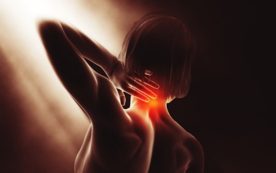
Suffering from severe cervical neck pain can drive a lot of discomfort to most of the individuals and may hamper their regular activities. However, there are various diagnosis methods through which a doctor can determine the severity of your neck pain and accordingly treatment methods. The most crucial aspect to managing your cervical pain is following a correct neck posture and giving it proper rest. This blog piece will discuss the cervical neck pain condition and the best treatment options available to ease your pain.
Understanding Cervical Neck Pain
Basically, it is the pain resulting from osteoarthritis or cervical spine spondylolysis, causing changes to the discs, bones and joints, since they connect with the neck. The Cervical Neck Pain holds various levels of intensity- from moderate to severe and each comes with specific ways of treating it. The pain in the neck can pose uncomfortableness, since the neck provides flexibility and support to our entire body posture. There are many reasons for this pain, however, considering proper exercises and some prescribed medications can help you overcome your painful condition.
Possible Cervical Neck Pain Reasons
Listed below are the most usual reasons behind the Cervical Neck Pain that you are facing:
Injury
A laceration in your neck or the spine area immensely triggers the cervical pain conditions.
Not giving proper rest to neck
Some people are stuck with jobs that require strenuous movements every day that put stress on their spines, causing wear and wear and tear conditions.
Dehydrated Spinal Discs
An individual may face dryness in the spine discs, creating pressure on them, causing chronic cervical neck pain conditions.
Herniated Discs
An individual usually suffers this when the discs of the spinal column develop cracks within, causing leakage of cushioning substance inside, putting pressure on the spinal cords and nerves. You must immediately see a medical expert in case you witness symptoms of cervical pain that may create numbness and pain to the arms.
Bone Spurs
When the bone expands in size, it presses against other spinal nerves and the spinal cord, causing severe cervical pain and this requires the person to immediately seek medical assistance.
Other Reasons- Not following the correct body posture, poor seating arrangements, incorrect mattress structure and much more.
Common Symptoms Include:
Tingling or weakness in the arms and feet as these parts directly link to cervical nerves.
Muscles spasms
Trouble walking or performing other everyday activities because of the inability to coordinate
Abnormal reflexes in legs and hands.
Neck stiffness
Aches in the lower back of the head
Diagnosis of Cervical Neck Pain
Mentioned below are some ways that a doctor may apply to diagnose the Cervical Neck Pain:
Physical test:
The medical expert may inspect the alignment of your neck and head to assess the range of motion of your neck which lets them analyze neck and supporting muscles to look for tenderness and indications of strain.
Imaging Tests:
The doctors may advise these tests to capture images of the cervical neck when they suspect you’ve suffered a serious injury or suffering from extreme pain that isn’t getting better.
Medical History:
While diagnosing your cervical pain, the specialist may ask you for details about any previous neck injuries that you suffered that might result in whiplash or herniated disks. Moreover, you must provide details regarding your work schedule and other activities causing strain to your neck. After taking all these details, the medical expert will diagnose the pain based on the pain you are experiencing and how long you’ll be facing the neck pain issue.
X-rays:
Being the most common diagnosis procedure, the doctors suggest X-rays to reveal issues in your bones or soft tissues, causing neck discomfort. The results could reveal problems with alignment of the cervical spine, fractures and slipped discs, along with the signs of arthritis.
The Magnetic Image Processing:
MRI is suggested to reveal problems in your spinal cord, bone marrow, nerves, and the soft tissue and even to diagnose any tumor or cyst in the body.
CT Scans:
The doctors usually suggest CT scans to diagnose cervical pain where MRI is not possible to reveal bone spurs or indicate bone loss.
Treatment Options
Mentioned below are some treatment options available for such conditions:
Medications: If the general painkillers don’t work, the doctors may recommend higher doses of medication to relax muscles in pain relief, while reducing inflammation.
Surgery: Usually the medical experts recommend homely remedies initially, however, in some cases, surgical procedures may prove good alternatives to treat cervical neck pain.
Steroid injections: A medical professional may inject steroid drugs near nerve roots or the muscles of the neck for a simple reason to alleviate neck discomfort.
Pain Management Home Remedies
Check out below:
Follow Exercise Routine: Practice exercises prescribed by your doctor to ease neck pain and increase mobility as avoiding these cause you to suffer a serious neck injury or a pinched nerve.
Alternate Cold and Heat: Inflammation can be reduced by using cold for example an Ice pack or placed in towel for a maximum of 15 minutes every day for in the initial 48 hours. After that, you can use the heat. Take a shower with a warm temperature or use a heating pad that is on the lowest setting.
Stress Reduction Techniques: Follow some stress management tricks, including meditation and mindfulness breath exercises to relax your tension situations that might cause neck pain.
Stop Smoke: Smoking can damage bone structure and increases the risk of the degenerative disc disease and delays healing.
Prevention Tips:
Practice good posture
Adjust your sleep position
Stay active
Don’t carry heavy weight on your shoulders
Exercise your upper back extensor muscles
Conclusion
Since we tried our best to cover every relatable aspect about Cervical Neck Pain for our readers, it is essential to note that visiting a trusted medical hospital for your treatment will maximize your process. Axon Health by Dr. Eshan offers top quality treatments for cervical pain by deeply analyzing and determining the root cause of your neck pain. Our well-trained professionals ensure that all the patients are treated appropriately in a comfortable period. Book an appointment with us today!
1 note
·
View note
Quote
What is the cause and treatment of Cervical Spondylosis?
Cervical spondylolysis treatment in Hod Hasharon includes physical therapy in which immobilization of the cervical spine is the mainstay for conservative treatment for patients who have severe cervical Spondylosis with evidence of myelopathy. Soft cervical collars are also recommended for daytime use only but they are unable to appreciably limit the motion of the cervical spine. Patient tolerance and compliance are considerations when any of the braces are used. Isometric survival exercise can also help to limit the loss of muscle tone that results from the use of more restrictive orthoses. Mechanical traction is a widely used technique and this form of treatment may be useful when it promotes immobilization of the cervical region and widens the foraminal opening. The most common type of mild to moderate neck pain usually responds within two to three weeks to self-care. There are some pain relievers and the use of heat might be helpful for neck pain.
Neck pain treatment Hod Hasharon does not include pain relievers that include nonsteroidal anti-inflammatory drugs such as ibuprofen or naproxen. The patient can take these medications only as directed by doctors, over use of these medications can also cause serious side effects if the pain relievers you can buy without the doctor's prescription that will don’t help. Therapy for neck pain treatment includes physical therapy using the 2Zone therapy technique specifically for soft tissues around the neck area. In addition, the physical therapist can teach alignment, correct posture, and neck strengthening exercises. Physical therapy might also involve the use of heat or ice and any other measures to help to ease the pain. A soft collar is the best helpful for neck pain treatment that supports the neck and may help relieve pain by taking pressure off the neck. However, if it is used for more than three years at a time or for more than one to two weeks it can do more harm than good. Other than this surgical and other procedures can also be helpful for neck pain treatment like steroid injection or surgery. If doctors prescribe steroid injections, be aware that your body will get used to the steroids and by the 3rd injection you gain no benefits. The areas where the injection will be applied are near the nerve roots into the spinal joints or the muscles in the neck. Apart from pain relievers, self-care measures can also include neck pain treatment that is alternate heat and cold like ice wrapping a towel for up to 15 minutes several times a day during the first 48 hours. Keeping the neck moving is also important for exercise. Several alternative treatments might ease back pain of which the 2Zone Therapy treatment is chosen but many as their preferred alternative to the medical route. Muscle pain treatment Hod Hasharon depends upon the cause the first these steps may help you feel better like resting and elevating the painful area, soaking in a warm bath with Epsom salts or taking a warm shower, and trying complementary therapies like 2Zonetherapy technique, massage meditation or acupuncture.
0 notes
Text
How Should I Perform Setu Bandhasana?

The three words Setu, Bandha, and Asana make up its Sanskrit-derived name. Setu, which means bridge, Bandha, which means lock in this context, refers to contracting or regulating specific body parts, and Asana, which means position. Bridge Pose got its name because when you're in it, your body looks like a bridge. This backbend position is excellent for opening the heart and strengthening the legs, hips, spine, and waist. When performed consistently and methodically, this position can not only be energetic but also revitalizing and restorative.
Sometimes it is not possible to do it on my own, look out for an online yoga class and follow the practices with professionals.
Our bodies naturally have two back-bending curves at the neck and the lumbar region, which are present from birth. To ensure that the less mobile portions of the spine—the thoracic, upper back, and sacrum—as well as the flexible elements of the spine—the lumbar and neck—bear weight equally, this position must be performed with complete awareness. While performing this pose, it is important to engage the spine's entire set of muscles.
Setu Bandhasana, also known as the Bridge Pose, is a fantastic posture to master as a beginner even though it is an inverted back bending posture. Yoga practitioners have the option to reap the rewards of an inversion without actually being upside-down by engaging in the bridge pose. For a deep frontal stretch and to get ready for more challenging backbends, the bridge position is a fantastic addition to your yoga routine.
Setu Bandhasana restrictions and contraindications
1. Sprains or injuries to the neck and shoulders
2. An ankle sprain or damage
3. Surgery for the hip, shoulder, and spine
4. A herniated or acute cervical spondylolysis disc
5. Elevated Blood Pressure
6. Patients undergoing heart surgery
7. Major Osteoporosis or Knee Replacement Surgery
8. Intestinal Hernias
Getting Ready for Setu Bandhasna Practice
It is a good idea to warm up thoroughly before performing backbends. We can perform a few Surya Namaskars before doing Setu Bandhasana (Sun Salutations) Your body will be better prepared for the asana practice if you perform some neck and shoulder stretches. Warm up the spine for this back-bending pose by performing a few rounds of Urdhvamukhasvanasana (Upward Dog Pose) and Bhujangasana (Cobra Pose).
0 notes
Text
Cervical pain is related to neck pain. It usually arouses due to wrong sitting or sleeping posture. Cervical pain is caused by abnormalities with the cervical spine’s joints and discs, which are located in the neck.
More specifically we can say that cervical pain is caused by spondylolysis or osteoarthritis in the neck. Changes in the bones, discs, or joints that are linked to the neck characterize this illness, which most commonly affects the elderly. Cervical pain can be fatal if not addressed promptly, thus people are more concerned about it. More information should be collected to control it. We are here to serve you the best cervical pain treatment in Delhi and back pain treatment in Delhi.
0 notes
Text
Ayurveda treatment for cervical spondylosis
Cervical spondylosis, also known as cervical osteoarthritis or arthritis of the neck, is a common age-related condition affecting the joints and discs in the neck. It develops from the wear and tear of cartilage and bones present in the cervical spine, located in the neck. Here will discuss how Ayurveda treatment for cervical spondylosis is helpful.
SYMPTOMS OF CERVICAL SPONDYLOSIS
The basic symptoms are neck pain and shoulder pain with stiffness. Most people with cervical spondylosis don’t have significant symptoms as well. Few more symptoms are like
Tingling, numbness and weakness in your arms, hands, legs or feet
Lack of coordination and difficulty walking
Loss of bladder or bowel control
Headaches
Dizziness
Loss of balance
Causes for cervical Spondylosis
The bones and protective cartilage in your neck are prone to wear and tear that can lead to cervical spondylosis. Some of the causes are like
Over Usage: Due to our regular activities which involve repetitive movements or heavy lifting can put extra pressure on the spine, resulting in early wear and tear. This is the most common reason found in cervical spondylosis
Stiffness in Ligaments: The tough cords which connect spinal bones to each other can become even stiffer over time of period, due to this neck movement affects and start pain.
Injury: It is also most common reason for cervical spondylosis. Any kind of neck injury faster the process of ageing.
Dehydrated Spinal Disks: Disks are like cushions between the vertebrae of the spine. Most people’s spinal disks begin drying out and shrinking, which allows more bone-on-bone contact between the vertebrae
Disk Degeneration and Bone Spurs: These overgrowths of bone are the result of the body trying to grow extra bone to make the spine stronger. However, the extra bone can press on delicate areas of the spine, such as the spinal cord and nerves, resulting in pain.
Overweight: Overweight leads to premature degeneration of the spine and other skeletomuscular structures by virtue of their metabolic effect.
Ayurveda treatment for cervical spondylolysis:
Ayurvedic treatment of Cervical spondylosis comprises herbal medicines and therapies that help to relax the muscles, as well as relieve inflammation to the nerves and surrounding tissues, healing the damage to the nerves and strengthening the nerves and muscles.
Greeva Stambha (A Vataja Disorder) simulates cervical spondylosis, which is a chronic degenerative condition of the cervical spine. In the management of cervical spondylosis Abhyangam, Swdana, Virechanam, Basti, Shalishastika pinda swdena, Greeva basti, Greeva Pichu, can improve conditions. These therapies relieve pain and inflammation, strengthen the tissues and prevent a recurrence.
Ayurveda Medicines for cervical Spondylolysis
Yogaraj Guggulu
Ashwagandha
Brahmi
Sahacharadi Kashayam
Dhanvantaram kashayam,
Dashamooladi Kashayam
Ksheerabala 101
Kaishora Guggulu
Home Tips to avoid Cervical Spondylosis
Avoid Dry, Fried, Cold, Junk, and Chilled Food.
Exposure to Cool Breezes, Air Conditioning and Fan,
Proper Posture When Sitting, Walking, and Sleeping Change your seat or bed as per your comfort.
Avoid Sleep during the day and sleep early at night.
Exercising regularly, turning the neck clockwise and counterclockwise is helpful. Bending and stretching your neck and nodding your head is also helpful.
The use of garlic, ginger, turmeric, sesame seeds in the daily diet helps in reducing Vata and as well as reduce inflammation
Applying warm sesame oil to the neck area and a hot bath also relieves pain.
Bhujangasana (Cobra Pose), Matsyasana (Fish Pose), Marjariasana (Cat Pose), as well as Ardha Matsyendrasana (Sitting Half Spine Pose), Setu Bandhasana (Bridge Pose) are some of the recommended yoga poses equally important that relieve spondylosis of the cervical spine.
Avoid lifting heavy weights and works that rather pressurize the neck.
Last but not least avoid sitting in front of a computer for a long duration, take breaks in between work for 10 minutes for every hour and that will not only reduce pain but also keeps you relaxed to work better.
1 note
·
View note
Text
Cervical Pain and Its Innovative Solution - Pillza Pillow
Most of the people from cervical pain which is usually caused by cervical spondylolysis or spondylitis or muscle strain or osteoarthritis which results in changes in the formation of the disc, bones due to tear and wear of the cartilages and bones connected to the neck. A cervical vertebra of the spine located at the base of the skull is the most delicate and thinnest bone. http://bestcomfortablepillows.over-blog.com/preview/f6220be4c0b586085669d6c1f9ae9627382afda2
#nature's best cervical support pillow#pure comfort pillow#soft bed pillows#sleep comfort pillow#natural comfort pillows
1 note
·
View note
Text
What is Minimal invasive spine surgery?
Minimally invasive spine( MIS) surgery is to stabilize the vertebral bones and spinal joints or relieve the pressure being applied to the spinal nerves — frequently a result of conditions similar as spinal instability, bone spurs, herniated discs, scoliosis, or spinal tumor.
Minimally invasive spine surgery doesn’t correspond to any particular surgery but refers to surgical procedures and the techniques developed with keyhole surgery procedures with less invasive surgery in mind.
When you think about what is minimally invasive spine surgery, it includes any surgery with the smallest amount of incision to surrounding tissues possible. Minimally invasive procedures are even as safe and effective as back surgeries performed with a traditional approach.
Advantages of Minimally invasive surgery
It reduces operative times.
Reduces soft tissue damage
Surgical incisions are less painful.
It reduces blood loss.
Recovery is faster with less post-operative pain.
The hospital stay is shortened.
Is Minimally Invasive spine surgery right for me?
The patient must be evaluated by the best spine surgeon in the field and an expert in MIS techniques to determine if their treatment requires the MIS options. Not every spinal procedure or patient may be a candidate for MIS.
Are there risks involved in minimally invasive surgery?
Some of the risks associated with MIS and spine surgery, in general, include adverse reactions to the anesthetic, blood loss, infection, blood clots, pneumonia, instrumentation tools causing damage to the spine and surrounding tissues, and even paralysis (extremely rare in major spinal surgeries).
Why and when Minimally Invasive surgery is advised?
As there are lots of patients having Back pain, not all require the MIS surgery. It is advised that pain persists after going through all other treatments like medicine or physical therapy. Then you need to take advice from the best spine surgeon to evaluate and proceed with further procedures. The conditions which require the MIS surgery are –
1- Herniated Disc
2- Spinal Stenosis
4- Scoliosis
5- Infection in the spine
6- Fracture in spine or vertebrae
7- Spondylolysis
This needs special expertise and the Best doctor for Minimally invasive spine surgery.
Dr. Puneet Girdhar
MBBS, MS, M.Ch (Ortho.)
Dr. Puneet Girdhar is presently working as Director Orthopedic Spine Surgery at BLK Centre for Orthopedics, Joint Reconstruction & Spine Surgery at BLK Super Speciality Hospital in New Delhi.
He has deep and extensive experience in the following practice areas: Microendo Discectomies, Minimally invasive spinal decompressions and fusions, Artificial disc replacements and deformity correction in Cervical, thoracic and lumbar spine in a wide array of Congenital, Degenerative,
Neoplastic and Traumatic conditions of spine. His key areas of practice are minimally invasive spine surgery and correction of spinal deformities both in the adult and pediatric age groups.
Address :- BLK Super Speciality Hospital, Pusa Road, New Delhi -110005
For more information
Call on :- +91 88002 16260
Mail id :- [email protected]
Or you can visit our website :- https://www.drpuneetgirdhar.com/
1 note
·
View note
Text
Spondylolisthesis - Causes, Symptoms, Treatments | Wascher Spine Institute
Spondylolisthesis is a cervical condition in which a vertebra in the spinal column is not properly aligned with its neighbor column.

Overview
This condition occurs when a lumbar vertebra slips out of place. It slides forward, distorting the shape of your spine. This may compress the nerves in the spinal canal. The nerves that exit the foramen (open spaces on the sides of your vertebrae) may also be compressed. These compressed nerves can cause pain and other problems for lifelong.
Common Causes
Spondylolisthesis has a variety of causes. In children, it is often due to a birth defect in that area of the spine. Some people develop this condition because of an overuse injury called "spondylolysis." This is a stress fracture of the vertebral bone. In adults, arthritis and the loss of disc elasticity that results from aging are the most common causes of spondylolisthesis.
Other Causes
Less commonly, spondylolisthesis can result from a sudden injury that leads to a broken vertebra. Diseases or tumors that weaken the spine can also result in spondylolisthesis.
Symptoms
Symptoms vary from person to person. Many people who have this condition have no symptoms at all. If you do have symptoms, you may experience pain in your lower back. You may have hamstring spasms. Pain may spread down your leg to your foot. You may also have foot numbness and tingling.
Treatment
Treatment options depend on the severity of your condition. You may benefit from rest. Medications may relieve your pain. A back brace may also help. And, you may benefit from physical therapy. If those methods are not successful, you may benefit from a Anterior & Posterior surgical procedure avialable to reduce nerve compression or to stabilize your spine.
1 note
·
View note
Text
What is Spinal Stenosis?

Spinal stenosis is defined as a narrowing of the spinal space and/or compression of the spinal cord and nerve roots as they exit each vertebra. A common cause is changes in your spine as you age. Back and/or neck pain, as well as numbness, tingling, and weakness in the arms and legs, are symptoms.
What is spinal stenosis?
Spinal stenosis refers to the narrowing of one or more sections of your spine. The amount of space available to your spinal cord and nerves spreading off it is reduced. As a result of the narrowed space, the spinal cord or nerves may become inflamed or pinched, causing back pain and claudication pain known as neurogenic claudication.
Spinal stenosis usually develops over a long period of time, especially beyond the age of 50. The most prevalent cause is osteoarthritis, or “wear and tear” changes in the spine that occur naturally as people age. As a result, if certain changes are detected on X-rays or other imaging tests done for a different reason, you may not experience any symptoms for a long time.
To get proper treatments for spinal stenosis visit Pentagon Hospital, which has the best brain and spine Surgeon in Aurangabad.
What parts of the body does spinal stenosis affect?
Spinal stenosis can affect any section of the spine, however it most commonly affects two:
Neck (cervical spinal stenosis)
Lower back (lumbar canal stenosis)
Who has spinal stenosis affected?
Although anybody can get spinal stenosis, it is most common in men and women over the age of 50. Younger people who were born with a narrow spinal canal can develop spinal stenosis. A range of illnesses that affect the spine, such as scoliosis or a spinal injury, can induce spinal stenosis.
Spinal stenosis can be caused by a variety of factors.
Spinal stenosis can occur for a variety of reasons. They all have one thing in common: they change the structure of your spine, narrowing the space around your spinal cord and the nerve roots that exit through it. Symptoms such as low back pain and sciatica are caused by compression or pinching of the spinal cord or nerve roots.
The causes are:
Bulging disks/herniated discs: the vertebral disc is a flat, circular cushioning pad that rests between each vertebra and acts as a stress absorber along the spine. Due to age-related drying out and flattening of vertebral discs, as well as cracking in the exterior border of the discs, the gel-like center of these discs breaks through a weak or damaged outer layer. The bulging disc then compresses the nerves near the disc.
Herniated discs and bone spurs usually induce spinal stenosis by narrowing the spinal and neural foramina.
Ligaments have thickened, which means the fiber bands that hold the spine together have thickened. As a result of arthritis, ligaments might expand and bulge into the spinal canal space (and can cause lateral recess syndrome)
Spinal stenosis is frequently caused by both disc prolapse and ligament and facet enlargement.
Damage to the spine can cause inflammation, which might narrow the canal space or put pressure on the nerves.
Bone overgrowth/ arthritic spurs: Osteoarthritis is a “wear and tear” condition in which cartilage in your joints, including your spine, breaks down. Cartilage is the protective layer that covers joints. When cartilage is lost, the bones begin to rub against one another. Your body responds by making more bone tissue. Bone spurs, also known as bone overgrowth, are very frequent.
Vertebral bone spurs extend into the spinal canal, constricting space and squeezing nerves.
Spinal stenosis symptoms include:
1. Lower back (lumbar) spinal stenosis:
Lower back pain is a very prevalent condition. Pain can range from a minor ache or discomfort to a burning or electrifying sensation. Pain is natural and will come and go.
Sciatica is a type of pain that begins in the buttocks and travels down the leg, possibly into the foot.
Leg cramps in one or both legs owing to a heavy sensation in the legs after walking for a short period of time in the beginning, later phases even a few steps might cause leg pain ( neurogenic claudication)
Numbness or tingling in the buttocks, leg, or foot (sometimes known as "pins and needles") is a common symptom.
2. Neck (cervical) spinal stenosis:
Neck pain
Numbness or tingling in the arm, hand, leg, or foot. (Symptoms can appear anywhere below the location of nerve compression)
Weakness or clumsiness in an arm, hand, leg, or foot.
Instability of the body's balance.
Hand dysfunction, such as difficulties writing or buttoning clothing.
Control issues with the bladder or bowels (in severe cases)
3. Abdomen (thoracic) spinal stenosis:
There is pain, numbness, tingling, or weakness at or below the level of the abdomen.
There are issues with equilibrium.
Diagnosis
Neuroimaging – MRI to see the degree of canal stenosis and disc herniation; X-rays to see the slide vertebrae and spondylolysis
Treatment
If your spine is showing, Medically, mild to severe canal stenosis and isometric spinal exercises will often suffice.
Surgery to relieve the compression is recommended in severe situations.
Our expert Brain and Spine Surgeon in Aurangabad provides consultation and treatment to all neuro problems.
#spinal stenosis exercises#spinal stenosis treatment#spinal stenosis meaning#spinal stenosis surgery#spinal stenosis#spinal stenosis progression#brain and spine Surgeon in Aurangabad#Neurosurgeon in Aurangabad
0 notes
Text
What is spinal stenosis?

Spinal stenosis is narrowing of spinal space or potentially pressure your spinal string and nerve roots as they leave every vertebra. Changes in your spine as you get old is a typical reason. Indications are – back pain and additionally neck pain, just as deadness, shivering and shortcoming in the arms and legs.
What is spinal stenosis?
The narrowing of at least one regions inside your spine is known as spinal stenosis. The measure of room accessible for your spinal line and nerves that branch off your spinal line is diminished . The spinal line or nerves may become kindled or squeezed because of a limited space, coming about in back uneasiness and causing claudication pain called neurogenic claudication.
Spinal stenosis regularly consumes a large chunk of the day to grow, particularly following 50 years . Osteoarthritis, or “wear and tear” changes in the spine that grow normally as you age, are the most widely recognized causes. Accordingly, if a few changes are seen on X-beams or other imaging tests led for another purpose, you might not have any manifestations for a significant stretch.
Where does spinal stenosis influence?
Spinal stenosis can influence any piece of the spine, anyway it typically influences two regions:
Lower back (lumbar canal stenosis)
Neck (cervical spinal stenosis)
Who gets influenced by spinal stenosis?
Spinal stenosis can influence anybody, yet it is generally successive among people more than 50 years old. Spinal stenosis can likewise influence more youthful people who were brought into the world with a limited spinal canal. Spinal stenosis can be brought about by an assortment of issues that influence the spine, like scoliosis or a spinal physical issue.
Reasons for spinal stenosis:
There are various purposes behind spinal stenosis. What they all share for all intents and purpose is that they change the state of your spine, narrowing the region around your spinal line and nerve roots that departure through it. Pressure or squeezing of the spinal rope or nerve roots causes manifestations like low back pain and sciatica.
The causes are:
Bulging discs/herniated disc: a level, round padding cushion (vertebral disc) sits between every vertebra and fills in as a pressure safeguard along the spine. The gel-like focus of these discs gets through a powerless or torn external layer because old enough related drying out and leveling of vertebral discs, just as breaking in the external line of the discs. The nerves close to the disc are then packed by the Bulging circle.
Spinal stenosis is regularly brought about by herniated discs and bone spurs causing narrowing of spinal and Neurol foramina.
Thickened ligaments: ligaments, the fiber groups that keep the spine together, have thickened. Ligaments can amplify and swell into the spinal canal space because of joint inflammation (and can cause sidelong break condition)
So often both disc prolapse and ligaments and features extend may cause spinal stenosis.
Irritation from harm along the spine, can limit the waterway space or apply tension on the nerves
Bone overgrowth/arthritic spurs: osteoarthritis is a “wear and tear” condition that makes ligament separate in your joints, including spine. The defensive covering of joints is
ligament. The bones start to rub against each other when ligament goes down. Your body responds by delivering new bone tissue. Bone spurs or bone abundance are a typical event.
Bone spurs on vertebrae jut into the spinal canal, restricting the space and crushing nerves.
Indications of spinal stenosis:
1. Lower back (lumbar) spinal stenosis:
Lower back pain is a typical infirmity. Pain can go from an unpretentious acheing or distress to an electric or burning sensation. It’s not unexpected for pain to travel every which way.
Sciatica: this aggravation is which begins in your hindquarters and spreads down your leg, perhaps into your foot.
Leg cramps in one or the two legs because of weighty inclination in the legs , subsequent to strolling for at some point before all else, later stages even couple of steps likewise pain in legs ( neurogenic claudication)
Deadness or shivering in the posterior, leg or foot (once in a while known as “pins or needles”)
2. Neck (cervical) spinal stenosis:
Neck ache
Arm, hand, leg or foot deadness or shivering. (side effects can happen anyplace underneath the nerve pressure point)
Arm, hand, leg or foot shortcoming or ungainliness.
Issues with body balance.
Loss of hand working, like trouble recorded as a hard copy or securing garments.
Bladder or entrail control issues (in serious cases)
3. Abdomen (thoracic) spinal stenosis:
At or underneath the level of the Abdomen, there is pain, deadness, shivering or shortcoming.
Issues with balance.
Diagnosis
Neuro imaging – MRI to see the disc hernation and level of canal stenosis
X Rays to see the slip vertebrae, spondylolysis
Treatment
On the off chance that your spine shows Mild to direct canal stenosis restoratively and isometric spinal excercises will be adequate ordinarily.
In serious cases a surgery is encouraged to deliver the pressure
Our master Neuro group at NeuroWellness a neuro hospital in Bangalore gives consultation and treatment to all neuro issues.
Please visit
NeuroWellness
Brain and Spine Clinic
#1224, Ground-Floor, 26th Main, Jayanagar 9th Block
BANGALORE – 560069
Phone No
+91 72596 69911
+91 73490 17701
Website www.neurowellness.in
Facebook www.facebook.com/neurowellness.in/
#What is spinal stenosis?#Neurosurgeon in Bangalore#Best Neurosurgeon in Bangalore#Neurosurgeonnearme#Famous Neurosurgeon in Bangalore#BangaloreSpineSpecialistClinic#BestspinesurgeoninBangalore
0 notes
Text
Neck Pain Treatment

Cervical pain is the pain caused due to cervical spondylolysis or osteoarthritis. Neck pain is a very common medical condition found in people these days. Neck pain can arise from a number of disorders and diseases of any tissues in the - neck pain.
The pain and other symptoms which are involved in neck problems typically cause functional difficulties which show themselves in complicated limitations of movement and activity. Psychological factors may in some cases be of crucial importance in how the patient is behaving in response to neck pain - cervical pain.
Our physiotherapist works to remove the cause of the damage to the spine and prevent further occurrence. Once the most likely cause of your problem has been determined (your diagnosis has been made), you and your healthcare professional can decide on a treatment plan. One of the keys to managing back pain or neck pain is to actively engage in rehabilitation and exercise. For more information, please visit our site https://omnipaincare.com/
0 notes
Text
300+ TOP ORAL RADIOLOGY Interview Questions and Answers
ORAL RADIOLOGY Interview Questions for freshers experienced :-
1. what is oral radiology?
A: oral radiology is a medical specialty which focuses on the creation and interpretation of diagnostic images of the mouth and surrounding area. Dentists utilize oral radiology in their practices, as do head and neck surgeons, maxillofacial surgeons, and other members of the medical profession who work around the head and neck. In some regions, oral radiology is a recognized medical subspecialty and people such as dentists and radiologists can apply for board certification in this area
2. what are the disadvantages of being a Dental Assistant?
A: getting f#cked by the dentist being elbow deep in the mouth of a crackhead whose mouth looks like he garggled acid and smells like something died. Plus you won''t make any money while the dentist hauls down six figures. People''s mouth, need I say more? It''s almost as bad as being a proctologist assistant. Well, smelling peoples breath all day and having to see what they had for lunch every day would seem to be drawbacks of the profession! Well, I couldn''t spend my day looking into other peoples mouths, but I''m glad some people can or else there would be nobody to fix our teeth when problems arise
3. oral surgery
A: Dianne - Both redoing the upper jaw surgery and moving it forward along with setting back of the lower jaw is definitely possible. Before any surgery is done, a full radiologic work up along with a dental model work up must be done to achieve a result that is not only functionally sufficient, but is also physically pleasing. The full work up is necessary and you need to see the work up after it isdone. The surgeon must go over the different options and show what your face will look like with both upper and lower at the same time, with only upper or only lower.
4. oral tingling
A: facial pain, facial tingling, peri-oral tingling, numbness, DDD, degenerative disc disease, degenerative joint disease Dear Lynn, Sorry to hear of your complaints. It sounds like you have been having a good thorough workup. Obviously a thorough examination with Xray, MRI, CT, EMG, EEG, blood tests, etc. is the first step. Facial nerve, trigeminal nerve, TMJ are all areas to be evaluate. I also agree with you, and not the doctors, that perhaps (even indirectly) the cervical spine can be contributing to this problem. I recommend a Doctor of Chiropractic with additional credentials
5. CT of abd and pelvis w/oral contrast
A: 1. Prominent = large or increased in size 2. You are probably feeling Fat accumulations. Fat is what we refer to as lucent or easily passed through by radiation so this would not be readily seen with the normal penetrating power of the X-ray beam. Endometriosis isthe thickening of the lining inside of the uterus
6. what is Potassium Iodide?
A: Potassium iodide is the active ingredient in ''fallout pills,'' pills that prevent the accumulation of radioactive iodines in the thyroid, which can cause thyroid cancer. Potassium iodide pills or potassium iodide powder should be consumed in regular doses if one might be exposed to radiation, such as during a nuclear war. The recommended oral dosage is 16 mg for infants under one month old, 32 mg for infants 1-36 months old, 65 mg for children 3-12, and 130 mg for adults. In the US, whether to stockpile potassium iodide for possible use during a nuclear war or Trans-Pacific fallout from an overseas war is a decision made at the state level by governors
7. what are the disadvantages of anyone a Dental Assistant?
A: If you are considerate and understanding, near an amazing personality and your not judgemental. Then self a Dental assistant, has giant amount of advantages. Fallow your heart, and proceed with your aspiration. It''s your life, not anyone Else''s. Unfortunately, you know individual negative family in your vivacity. So, be a leader and grasp out there and do what your heart desires! Good luck, you can do anything. But not everything will preserve you happy, so generate your own choice, most important what make you happy! getting f#cked by the dentistyou should do it if you be aware of you want to, i think putting your hand in someones mouth would be really grossA dental ass
8. what different kinds of dentistry jobs are their?
A: Denistry is a job with many opportunities, some jobs related to dentistry are: dental public health, endodontics, oral and maxillofacial pathology, oral and maxillofacial radiology, oral and maxillofacial surgery, orthodontics and dentofacial orthopedics, pediatric dentistry, periodontics(deals with the tissues in the mouth), and prosthodontics..
9. what types of doctors are there?
A: Ok, you asked for thorough! Addiction Medicine: The branch of medicine that concentrates on helping people overcome repetitive behaviors that can range from and alcohol dependency to tobacco use and eating disorders. Adolescent Medicine: The specialty of physicians with the experience and training to help young people meet the medical, psychological and social challenges that occur during the transition from childhood to adulthood. AIDS/HIV Care: A multidisciplinary effort that’s often led by primary-care physicians working in cooperation with case managers, registered nurses, nutritionists, physical and occupational therapists, and others
10. what in the ER for an asthma attack, what O2 saturation level is o.k. for discharge?
A: A normal oxygen saturation (sat) for any person is 98%. An acceptable sat for a 7 year old depends on the doctor and the presentation of the child. Recent studies show that a 90% sat is acceptable to go home. Still, our doctors usually like it to be 92% or better -- or even 94%. So you can see there is some flexability. This would be a great question to ask your doctor. Do you need a pulse oximeter for home use? I think it would be rare for a doctor to recommend this for home use, but I''m sure there are exceptions

ORAL RADIOLOGY Interview Questions
11. what Dental Work is Safe During Pregnancy?
A: An exerpt from americanpregnancy.orgPreventive dental cleanings and annual exams during pregnancy are not only safe, but are recommended. The rise in hormone levels during pregnancy causes the gums to swell, bleed, and trap food causing increased irritation to your gums. Preventive dental work is essential to avoid oral infections such as gum disease, which has been linked to preterm birth.what about other regular dental work during pregnancy?Dental work such as cavity fillings and crowns should be treated to reduce the chance of infection. If dental work is done during pregnancy, the second trimester is ideal
12. Failed Dental Implant
A: Susan - Let me first tell you that my wife is a psychotherapist and I know the responsibilities in your life. Getting away from the above, let me tell you that I think your surgeon is just plain lousy. If he was an oral surgeon, he should have been an oral and maxillary surgeon. If not, he is not really a surgeon. Of course, not seeing your surgeries or your xrays, it is a little tough for me to tell you what is going on exactly, but it does sound like the surgeon pushed the implant into the soft palate during removal or the implant tha
13. crown or extraction?
A: Corinne - The main reason that a microscopic fracture cannot be identified as opposed to evaluating bone for an implant is the size of the fractures. Most of these fractures are so small and thin that they cannot be seen on any radiologic examination. If your dentist and endodontist cannot determine if there is a fracture and you are in almost constant pain I would sort of follow your line of reasoning and get the extractions and implant insertions. The key to the success of this treatment is to be evaluated and treated only by a board certified oral and maxillofacial surgeon
14. spondylolisthesis, spondylolysis,
A: A recap for you on Adult isthmic Spondylolisthesis (Slipped Vertebra)as you know. Other than what I have established below, the only way to get satisfaction is to have surgical intervention. what is it? what treatments are available? The spine is made up of a series of connected bones called "vertebrae." In about 5 percent of the adult population, there is a developmental crack in one of the vertebrae, usually at the point at which the lower (lumbar) part of the spine joins the tailbone
15. Dental vs Chest x-ray
A: The absorbtion dose is dependent on the machine used for these procedures. Are we talking about Digital Radiograpy or the older form of diagnostic radiography. There is a huge difference and the dosages are greatly controlled with digital Vs General. The question failed to offer these options. As for the lead shields! The concentration of the xray beam to the local area of the jaw/oral, isconfined, and far away from the reproductive system. With a lead shield covering the abdomen, there is no chance of radiation exposure, and no potential risks. The thyroid collar is acceptable, but my personal opinion
16. Question for Dental Hygenist?
A: Dental hygiene is a great second career. I went to hygiene school when I was twenty eight. I have found that having more life experience has helped me a lot when it comes to hygiene. I love my job and would not trade it for anything. Pros: Great working hours Great money Being able to help people understand and take care of their teeth. esp making them understand that oral health care isan indication of their whole body health. Good benefits, sick leave, paid holidays, some medical, free dentistry minus the lab fee. The ability to make the difference in someone's life, esp older patients who look forward to their cleanings because they don't have anyone
17. Liver CT - RCC
A: Regarding the type of contrasts you were given, barium is an oral contrast. you might or might not have the oral contrast for the other studies as oral contrasts are not needed to evaluate the liver. IV iodine contrast is the essential contrast to study the liver. If possible, i would have the images of the 2 middle studies forward to the hospital or have this hospital send you the 2 scans that they did. To be able to compare all 4 studies would be very helpful in determining what"s going on. Renal cell cancer are many times not picked up on PET scan due their metabolic state not picking up
19. can you interpret my MRI in simple english please.
A: Dear Shaz, Having a leg length inequality is actually a fairly common finding in patients, however, most doctors never actually look for the relationship of a short leg to spinal or pelvic dysfunction. I am actually impressed that your Osteopath has analyzed the relationship and given you a heel lift to correct the structural differences. In regards to your previous question about the MRI, leg length inequality can lead to accelerated degeneration in the lumbar spine and sacroiliac joints of the pelvis due to long term altered weight bearing on the joints. So this can easily be a causative factor in the degeneration found, however, it is probably not the sole factor.
20. HIDA Scan Results
A: If you have several symptoms and that the gallbladder is found to be the cause it will be removed what matter the results of a statistical testing but so far the symptoms that you are giving are not much related to gallbladder but I can be mistaken without a physical exam it is hard to assess.this technique is used in acute cholecystitis and congenital biliary atresia and you do not present neither of both symptoms There are various patterns of radioactivity that can be seen following the injection of the radioactive chemical, and each has a different meaning. If the test chemical
ORAL RADIOLOGY Questions and Answers Pdf Download
Read the full article
0 notes
Text
Dr. Yogesh K. Pithwa Best Spine Surgery in India
Dr. Yogesh K. Pithwa is one of the finest Doctors in Bangalore, India. He has years of experience in Scoliosis and Best Spine Surgery in Bangalore at Sattvik Spine & Scoliosis Center. If in case you have any Scoliosis and spinal problems, feel free to consult with Dr. Yogesh K. Pithwa. At Sattvik Spine & Scoliosis Center we treated several common spinal problems including Low Back Pain, Lumbar Disk Herniation Slipped Disk Sciatica, Spondylolysis and Spondylolisthesis, Cervical Spondylosis, Tuberculosis. For further more information about us, feel free to get in touch with us.

#Top Scoliosis Surgeon in Bangalore#Best Spine Surgery in Bangalore#Best Scoliosis Surgeon in Bangalore#Best Spine Surgeons in Bangalore#Top Scoliosis Surgeon in India#Best scoliosis surgeon in India#Best spine surgeon in India#Top Spine Surgeons in India
0 notes
Text
Centaur Anatomy
I had to write this for my J.K.R. Lit. class. I’m a Bio major, not English. Sorry for any grammar errors.
The centaurs are half-human, half-horse creatures from mythology. Their body consists of that of a horse with the torso, head and arms of a man. The history of the centaur can be dated back to the thirteenth century BC, from the East (Sykes 2014). In Greek mythology, they were considered to be the children of Ixion, king of the Lapiths, and Nephele, a cloud made in the image of Hera. According to a different myth, however, they were all born from the union of a single Centaurus with the Magnesian mares.
Whatever their origin, it was during a time when the anatomy of humans and creatures weren’t as understood as today. With this little knowledge, this led the centaur to be a conceivable combination of two animals. This leads the creature to be a, though unofficial and often unthought of, scientific conundrums. To try and explain how one could exists, the centaur’s morphology will be broken into systems.
The first system discussed will be the skeletal system. Much of what you would expect out of a fusion of a human and a horse’s skeleton can be considered correct. The proportionally elongation of the arms is the only change many considered. However, this is not the only change that would have to occur. Depending on the location of the lungs, ideally in the human torso, the rib cage would have to be larger with the addition of ribs. This allows the lungs to be larger. The next change would be in the spine. Mammals almost always have seven cervical vertebrae, followed by twenty-four are articulating and separated from each other by intervertebral discs, and the lower nine, in humans, are fused in adults, five in the sacrum and four in the coccyx or tailbone. The vertebral column in a horse usually contains fifty-four bones: seven cervical vertebrae, including the atlas (C1) and axis (C2) which support and help move the skull, eighteen thoracic, five to six lumbar, five sacral (which fuse together to form the sacrum), and fifteen to twenty-five caudal vertebrae with an average of eighteen. After getting rid of the seven cervical vertebrae in the horse and nine from the human’s scrum and coccyx, this leaves a creature with seventy-one vertebrae. In many fan-made diagrams and hypotheses a centaur’s spine is curved where the lumbar spine in the human torso connects to the thoracic spine of the horse torso. This connection site can be considered dangerous. The fifth lumbar vertebra is the most common site of spondylolysis, a defect or stress fractures in the pars interarticularis of the vertebral arch, and spondylolisthesis, the forward or anterior displacement of one vertebra over another. The jerking movement of a horse could cause the formation of spondylolysis and spondylolisthesis early on. To absorb shock the spine would need to be curved. Another possible fix for this would be for the muscles in this area to be more developed.
The muscular system of the centaur is also relatively fine. The large problem comes from, once again, the connection between the human torso and the horse’s. The rectus abdominis of the human torso would have to extend. The could attach to the horse’s sternomandibularis, brachiocephalicus, and/or pectoralis descendens muscles. The external oblique could merge with the horse’s serratus ventrallis cercicis and the omotransversarius. This depends more on how you picture your centaur. The lumbodorsal fascia of a human is also where muscles that act as the trapezius, rhomboideus would be located. As the lumbodorsal fascia is where muscles come together, these replacement muscles will most likely be on top of this area. There is a chance that the fascia would not be located there, or be as large, to compensate for the muscles. The merging locations would have to have more connections. In the end, this abdominal region would have to be a glorified neck. The diaphragm would also be more developed to be able to take more air into the lungs and push out more for optimal air flow.
The lungs, as mentioned before, would be located in the human torso. Even with a torso proportional to the horse the lungs would be larger. To keep the one set of lungs, the blood would be able to absorb and transport oxygen. The creature would also have a higher tolerance for carbon dioxide for mammals. This would compensate for the small lung size to body mass. The heart would also be in the same location as it would be on a normal human. The heart would be larger than a ‘normal’ proportional heart with slightly thicker and stronger muscles. Centaurs would most likely have some form of low blood pressure or anemia. This would allow their heartbeat to be more rapid for their size and allow for better blood circulation. The digestive track would be in the horse torso. The beast’s digestive system would behave just like that as a normal horse.
A centaur’s nervous system would function relatively normal. The brain would be in the skull with a spinal cord going down the spine. Nerves would extend from the spine. At the bend of the spine, a nervous ganglion would be located to help control the digestive system. A nervous ganglion is a nerve cell cluster or a group of nerve cell bodies located in the autonomic nervous system and sensory system. The movement of the lower limbs would mostly be voluntary. However, the quicker reflexes and more focused movement would also be controlled by a ganglion.
With some changes, a centaur can hypothetically exist. This is all viewed from an underclassman pursuing a biology major. There is a high chance I missed many things and should, in no way be viewed as the accurate interpretation on how the body should function.
#centaur#anatomy#biology#had to do this for class#why did i do this#i'm so sorry#not that good#mythology
95 notes
·
View notes
Text
New Post has been published on Dr. Attaman and Dr. Cartier | Pain Management Physicians | (425) 247-3359
New Post has been published on https://bit.ly/2zhvsFj
Neck Pain / Cervical Pain Treatment
Some neck pain is caused by sprains, strains, muscle tension, or misalignment. Often those kinds of pain can be solved with massage or chiropractic therapy.
But cervical pain is a different kind of neck pain. It’s caused either by osteoarthritis or cervical spondylolysis, in which spinal discs develop cracks. The discs in your spine dry out, your bones rub together, and bone spurs appear. It can be the result of age or trauma, or both. In a case like that, a muscle rub won’t help, and so cervical pain calls for interventional pain management procedures. Dr Jason Attaman and Dr. Cameron Cartier offer several treatment options (below) for cervical pain in your neck.
Cervical medial branch blocks
This treatment requires us to inject a local anesthetic near the medial branch nerve. This will numb your pain and give you immediate relief. Unless you have other complications you can expect to enjoy relief from your pain for up to two years after this procedure. It’s an outpatient procedure, and most patients return to work the very next day.
Radiofrequency lesioning
This technique allows us to disrupt pain signals via electrical impulse. This destroys damaged nerves or tissues and stops the transmission of pain signals, putting an end to your pain. It is a more aggressive procedure than a branch block. We use an X-ray and a local anesthetic to guide the needle correctly, because we’ll be creating a small burn inside of your nerve.
CerviCool is a newer variation of this procedure and is more technologically advanced. It can safely create larger lesions, removing additional pain pathways. Water-cooled technology allows us to perform this procedure safely.
The shelf-life on pain relief varies for this procedure. It lasts six months for some patients, twelve months for other, and years for others. Sometimes the lesioned nerves regrow, and when they do the pain can return. We will help you by monitoring the situation so that you continue to receive the best solutions for your pain.
Cervical interlaminar epidural blocks
This procedure allows us to target inflamed nerve roots in your neck. As with the other procedures, we use an X-ray to guide the needle to the precise nerves. We then use an epidural steroid injection to reduce inflammation. It’s different from a lesion or a block, in that we target inflammation rather than nerve signals.
You will usually receive three injections two weeks apart, but in many cases these three injections can put an end to your pain entirely.
The right treatment at the right time
Those aren’t the only cervical pain / neck pain treatment methods in our repertoire. We stay current on new pain management technologies, and often add new treatment options. Some additional options include techniques like cervical epidural catheters, zygapophyseal blocks, and superficial cervical plexus blocks.
As our patient, you can expect a no-nonsense approach to ending your pain. A thorough diagnosis will tell us exactly which solution is right for you, and we will lay out the treatment options for you. If you’ve got neck pain, you’ve got treatment options. Call our office today to make an appointment.
0 notes
Link
Practicing Shoulderstand with a chair protects your cervical spine and allows for the security you need to turn within.
Sarvangasana (Shoulderstand) is a classic yoga asana. But it wasn’t one I enjoyed during my first two years of practice. Physically, I felt like I was choking, and mentally, I was restless whenever I tried it. Fortunately, within a few years I learned to use blankets to elevate my shoulders. That definitely improved my experience. But when I started using a chair, I was in heaven. I also was able to stay in the pose much longer—an important point: The positions our bodies take change the biochemistry and neurology of our brains. Think about sleeping; we instinctively know it’s best to lie down. And when we lie down, a cascade of events is set in motion in our bodies that facilitates the state of sleep. Inversions held several minutes or longer not only affect brain states, they change the hemodynamics (blood flow) of our bodies, stimulate our abdominal organs, help drain lymph from our legs, and calm our minds. They also shift our perspectives.
Anatomical Reality
You must understand the anatomy of your neck in order to do Shoulderstand safely. The cervical spine flexes (when your chin moves toward your chest) up to only 55 degrees. If you practice the posture directly on the floor, you force your cervical spine beyond that range of motion—and pile your body’s weight onto it. You also stress your upper thoracic spine. When you prop up your shoulders, you protect your neck, because your shoulders bear most of your weight. (This also keeps the pose true to its name.) Plus, the rest of your spine is free, and your lungs, heart, and abdominal organs aren’t compressed. Not only does this feel better, but it allows for a freer excursion of your diaphragm and therefore better breathing. When you practice Shoulderstand with a chair, things get even better for your cervical spine. Instead of your head and neck, your pelvis—which is designed to bear the weight of your head, arms, and trunk when you stand and sit—now carries your body’s load.
See also Understand Neck Safety in Supported Shoulderstand
Energetic Qualities
Yoga asana are not just physical exercises. Each posture affects your subtle energy differently. Shoulderstand represents the energies of the Mother archetype—compassionate, nurturing, benevolent—and focuses your gaze on yourself, as compared with Sirsasana (Headstand), for example, which has you face outward. Shoulderstand lets you be introspective. This is especially true for propped versions; the chair frees you from effort and striving. Accepting support allows you to give up your ambition in the pose and take deep rest physically, mentally, emotionally, and spiritually.
Watch Yoga Inversions: Shoulderstand and Plow Pose
Shoulderstand Prep
1. Enlist assistance.
Get a qualified yoga teacher to guide you at first, no matter your experience level, in order to enter the pose safely; using the chair is a little tricky. This pose is not appropriate for beginning students. Contraindications for practicing any form of Shoulderstand are issues with the cervical spine, such as a pinched nerve; nerve pain in one or both arms; diagnosed disk disease; whiplash; or chronic neck pain or dysfunction. Also, don’t practice this pose if you have hypertension; gastroesophageal reflux; a sinus infection or stuffy cold; diagnosed spondylolysis or spondylolisthesis; or are pregnant, menstruating, less than three months postpartum, or under the age of 14.
2. Assemble your props.
Backless yoga chair: It won’t have front rungs or a backrest, but its legs will create a wide base for stability.
At least three blankets: You can pad the seat with a blanket for comfort, but it can make the pose more difficult to execute; be sure it doesn’t compromise your alignment. Place enough blankets under your neck to ensure it flexes 55 degrees, no more or less, and that your weight is borne mostly by your pelvis, moderately by your shoulders, and not at all by your cervical spine.
Sticky mat: It keeps the chair in place. Also, make sure the chair has rubber footings so it doesn’t puncture or tear your mat.
See also Spine Anatomy: How to Prevent and Alleviate Back Pain
Entering the Pose
Experiencing the Pose
Concentrate on pressing your tailbone into the chair’s seat in order to arch your back. Don’t tuck your tailbone. The more you maintain an arch in your lower back, the more your upper back will lift. Remember, your pelvis, including your sacrum, should bear most of your weight. Your legs should be exactly vertical, which makes it easier to hold them up because your femurs (thighbones) are maximally congruent in your hip sockets. Also, your knee joints will be in a neutral position, and you can rest the bones of your knees against each other, requiring less effort to keep your legs straight. Drop your eyes into your lower eyelids, and soften your face.
Now that you are steady and present, take your attention to the exact center of your brain. Withdraw into this place, into the sensations of the moment. Take refuge in your own silence and stillness. Patanjali, in his Yoga Sutra (II.46), defines asana as stirham sukham asanam: abiding in ease is asana. This is truly possible in Chair Shoulderstand.
Keep your breath steady and easy. Gradually work up to staying in the pose for 5 minutes. For me, this version of Shoulderstand with variations (not shown) can be nearly a complete yoga practice.
Watch Treat Insomnia + More in Shoulderstand (Sarvangasana)
Resonance
To come out of the posture, bend your knees one by one, and place your feet on the upper back rim of the chair. Then straighten your knees over that same part of the chair. Release your grip. Slide down until your pelvis rests on the blankets; you will probably enjoy holding onto the bottom front chair legs as you do this. Drop your arms to your sides. Rest your lower legs on the seat. You now get to reap the sweetness of this version. Close your eyes and drink in the residue of the pose. Remember, the asana is not the yoga; yoga is the residue the asana leaves in your nervous system.
To finish, slide your pelvis off the blankets and onto the floor. Turn to your side for a few breaths, and then sit up slowly. A 20-minute Savasana is a perfect final pose after Chair Shoulderstand.
If this version feels awkward, don’t abandon it. Do it three times before deciding it’s not for you. The first time my students try it, they sometimes don’t like it. But the second time, they usually do, and the third time, they smile and refuse to come out of the pose. Give your body time to adjust. I believe Chair Shoulderstand can be practiced safely by experienced students, even during their senior years.
See also Iyengar 201: Build Resilience by Adding Variety to Your Yoga Practice
Learn More
Post a photo of yourself to Facebook or Instagram doing Chair Shoulderstand and tag @yogajournal and @experiential.anatomy for a chance to win free enrollment in the digital course Judith Hanson Lasater and Mary Richards teach, Experiential Anatomy: Movement Literacy for Yoga Teachers. Learn more at judithhansonlasater.com/yj.
About the author
Judith Hanson Lasater, PhD, has taught yoga since 1971 and is the author of nine books on yoga. Find her at judith.yoga.
0 notes
Last Seen Blogs

bob2muffins
Artsy AF

hellosizzlernordhessen
Unbetitelt

ehara-project2021
江原プロジェクト2021

keigochu
(๑ᵔ⤙ᵔ๑)

silverlewd
Shame