#diffuse large B-cell lymphoma
Explore tagged Tumblr posts
Visit Tumblr Blog
Explore Tumblr blogs with no restrictions, modern design and the best experience.
Last Seen Tumblr Blogs


Fun Fact
Tumblr has a low social media market share in South America.
Text
Primary dural-based parafalcine diffuse large b-cell lymphoma mimicking meningioma by Amr El Mohamad in Journal of Clinical Case Reports Medical Images and Health Sciences
Abstract
Background: Primary dural-based diffuse large B-cell lymphoma is very rare. Only few cases were reported in the literature. Case presentation: Herein, we present a case of an immunocompetent patient with primary dural-based diffuse large B-cell lymphomas mimicking meningioma associated with ghost tumor phenomenon without any evidence of a systemic lymphoma. Conclusion: Primary central nervous system lymphomas are rare. Clinicians should always consider this lesion as a differential diagnosis if radiological findings are not indicative of typical one meningiomas.
Key words: Dural-based tumor, diffuse large B-cell lymphoma, ghost tumor, MATRIX regimen, central nervous system.
Introduction
Primary central nervous system lymphomas (CNSLs) (PCNSLs) are rare and account for 2%–5% of all brain tumor cases, whereas secondary CNSLs are more common [1,2]. One study has shown that the most common intraparenchymal histological type is diffuse large B-cell lymphoma, as among 26 patients with PCNSL, 25 had diffuse large B-cell lymphoma [3]. Although primary dural-based lymphomas are rare, the most common area of involvement is the cerebral hemispheres. Most dural-based lymphomas are secondary and present as extra-nodal systemic diffuse large B-cell lymphomas. Primary dural-based lymphomas are usually histologically marginal-zone lymphomas, representing a group of lymphomas that have been historically classified together because they appear to arise from post-germinal center and marginal-zone B cells and share a similar immunophenotype, and few cases were reported to be diffuse large B lymphomas [4]. Here, we present a case of an immunocompetent patient with primary dural-based diffuse large B-cell lymphomas mimicking meningioma associated with ghost tumor phenomenon without any evidence of systemic disease.
Case Presentation
A 58-year-old male individual previously healthy and immunocompetent presented with headache, recurrent vomiting, and memory problems lasting for 3 days. No loss of consciousness, seizure, subjective weakness, or fever was observed. On physical examination, the patient’s Glasgow coma scale score was 15; his pupils were 3 mm in diameter, equal, and reactive; and the patient had nominal aphasia without motor and sensory deficit. He had normal cerebellar functions, and cranial nerve exams revealed no deficit. Head computed tomography (CT) (Fig. 1) showed a 2.2 × 3.8 cm (transverse × anteroposterior) iso-dense lesion with internal hypodensity in the left parasagittal frontal region extending to the right frontal region. Extensive perilesional edema was observed with effacement of the sulci and mass effect on bilateral frontal horns, associated with 3-mm midline shift. Head magnetic resonance imaging (MRI) showed an isointense parasagittal lesion on T1 and heterogeneous intense on T2, with redemonstration of perifocal edema (Fig. 2). Head T1-weighted imaging with contrast enhancement (Fig. 3) showed a large, left frontal, parafalcine, irregular-shaped mass located below the superior sagittal sinus level. It measured 4 × 3 × 3.3 cm in anteroposterior, mediolateral, and craniocaudal, respectively. It showed diffusion restriction (Fig. 4). There was central hyperintensity on T2-weighted imaging, without post-contrast enhancement area representing cyst formation. It exerts a mass effect characterized by effacement of the adjacent sulci, compression of the left lateral ventricle, and a 3-mm shift of the midline structures to the right side, and the impression of our neuroradiologist was atypical meningioma. Regarding extensive edema, dexamethasone was started at a dose of 4 mg, thrice a day, and the patient was planned for craniotomy and resection of the tumor. Initially, the patient was reluctant to undergo surgery; however, subsequently, the patient agreed to undergo surgery after approximately 10 days. During surgery, parasagittal craniotomy was performed; however, to our surprise, no definite mass lesion was found at the proposed site, in contrast to the findings described on imaging. The falx was thinned out and partly deficient. A biopsy sample was obtained from this abnormally appearing falx. Moreover, we obtained biopsy samples under neuronavigation guidance from abnormally appearing tissue, which was completely intra-axial, deep down in the lesion visualized on navigation. On postoperative day 1, MRI head with contrast enhancement (Fig. 5) showed that the previously seen lesion had a significant regression in size. Its right frontal extension and adjacent enhanced meningeal tail showed size reduction. Moreover, some regression in the perilesional vasogenic edema was observed. A significant regression in the previously described enhancement was noted at the left-side lentiform nucleus and external capsule. The MR spectroscopy study showed an increased choline/N-acetyl aspartate ratio and elevated lactate level within the lesion.
The histopathology results of the first brain biopsy samples (Figs.6–7) obtained from the falx cerebri showed meningothelial hyperplasia with calcification and focal perivascular lymphocytic infiltrate composed of small and large, atypical lymphocytes. Immunohistochemical staining was performed; however, the area of interest disappeared. The pathology team recommended another fresh biopsy to have the final diagnosis and flowmetry studies. So, the patient underwent redo craniotomy using the same incision, and multiple biopsy samples were taken. The second fresh brain biopsy (Figs. 8–9) showed multiple brain fragments with predominant perivascular atypical lymphoid infiltrates. Most cells were medium to large with moderate cytoplasm, atypical irregular nuclei having vesicular chromatin, variably prominent nucleoli, and several mitoses, including atypical one. Necrotic areas were also seen. Immunohistochemistry of the second biopsy (Fig.10 A-D) showed that atypical perivascular cells were positive for CD45, CD20, CD79a, BCl2, BCl6, MUM1, OCT2, and C-MYC, and negative for CD10, CD21, TDT, ALK1, EBV-LMP1, CD3, and CD5; however, few reactive/residual lymphocytes were positive for these enzymes. Moreover, 80% of lymphoid cellular nuclei were positive for Ki67. These findings were consistent with diffuse large B-cell lymphoma, not otherwise specified.
Whole-body positron emission tomography (PET) showed intense fluorodeoxyglucose (FDG) uptake higher than that in the healthy brain cortex, without evidence of coexisting systemic disease. In addition to PET scan, contrast-enhanced chest, abdomen, pelvis CT did not show any other lesions in the body; furthermore, workup for viral markers and autoimmune conditions were all unremarkable, thus confirming the diagnosis of “primary dural-based diffuse large B-cell lymphoma,” distinguishing it from secondary CNSL. The patient was transferred to the Oncology Department and started on three cycles of the methotrexate, cytarabine, thiotepa, and rituximab (MATRIX) protocol, which is the current standard treatment regimen for PCNSLs [5]. Three months after the diagnosis and after receiving two cycles of the MATRIX protocol, brain MRI with contrast enhancement (Fig. 11A, B) showed regression of the lesion, and PET scan showed complete metabolic resolution in terms of decreased FDG activity of the previously seen PCNSL without signs of lymphoma activity elsewhere. Subsequently, the patient received the third cycle of the MATRIX protocol without specific complications. Two weeks later, autologous stem cell transplantation (50 × 106/kg) was performed as part of the consolidation phase of treatment. Six weeks later, conditioning chemotherapy with carmustine–thiotepa was administered, followed by stem cell infusion (CD34 = 12 million/kg). The post-transplant course was complicated with mucositis, folliculitis, diarrhea, febrile neutropenia, and prolonged thrombocytopenia. Two months after transplantation, PET scan was repeated and showed complete metabolic resolution of initially seen PCNSL involvement. Currently, the patient is being followed by the hematology team; the patient is in good health and remission. The last outpatient follow-up was 8 months after the first surgery. The patient was seen by the vascular surgery (for permcath removal) and oncology teams. At this time, the patient was stable with complete remission; then, the patient was lost to follow-up. Another head MRI was performed and showed almost total regression of the lesion.
Discussion
Lymphomas in CNS are classified as primary, arising de novo from brain parenchyma, leptomeninges, eye, and spinal cord and as secondary to systemic lymphoma, which can be dural-based lesions. Secondary CNSLs are more common than PCNSLs. Most PCNSLs are intraparenchymal diffuse large B-cell lymphomas with a predilection to occur in the frontal lobe and then deep nucleic and periventricular locations; the infratentorial cerebellum is the most common location. However, primary dural-based lymphomas are rare, and even when found, they are histologically marginal-zone lymphomas. Few cases of primary dural-based diffuse large B-cell lymphoma have been reported in the literature [4,6]. Furthermore, PCNSLs are more common in immunocompromised patients with a mean age of 34 years, and they occur in immunocompetent individuals at an older age with a mean of 52 years [7]. The patient in this case report was 58 years old and immunocompetent without significant previous medical conditions. The latest review of the literature on primary dural-based lymphoma has been conducted by Quinn et al., who have found only 24 reported cases of primary dural-based diffuse large B-cell lymphoma, which confirms the rarity of the disease and subsequently the limited knowledge regarding this disease entity [8]. CNSLs have rapid response to steroids with shrinkage in size and initial remission [9]. Moreover, the initial response to steroids is associated with a better response to chemoradiotherapy and good prognosis [9]. In the patient in this case report, there was an unintentional delay of surgery for approximately 10 days, and the patient was on steroids (dexamethasone). In this case report, the failure to identify a discrete lesion of the size expected as perceived on initial imaging, despite proper surgical planning using neuronavigation, was probably due to the rapid regression of the tumor in response to steroids. This phenomenon agrees with the scientific literature reporting about the disappearance of lymphomas in response to steroids (ghost tumors) [10,11]. The pathogenesis of primary dural-based lymphoma remains unknown as there is no lymphoid tissue in the dura. It is hypothesized that it is related to chronic infection, autoimmune disease, or chronic inflammatory condition, which recruits polyclonal lymphocytes resulting in monoclonal lymphomas [6]. In contrast, the patient in this case report did not have any chronic conditions. All workups were negative, including the entire viral panel and autoimmune markers. Basic research is needed to determine the etiology of PCNSL, especially dural-based lymphomas. In the patient in this case report, the initial radiological findings were mimicking those of a meningioma: dural-based and uniformly enhanced. There was significant surrounding edema, significant diffusion restriction, and blooming in susceptibility-weighted image, which goes more with higher-grade meningioma or another high-grade lesion. One review has shown that primary dural-based lymphomas can display the “dural tail “sign, further confusing the preoperative diagnosis with meningioma [12], which did happen in the patient in this case report. Therefore, we suggest that in case of a dural-based lesion that has non-typical features of grade 1 meningioma, clinicians should consider lymphoma in the differential diagnosis and avoid steroids unless necessary due to edema and mass effect keeping in mind the ghost tumor phenomena of lymphoma.
The role of surgery in PCNSLs is limited mainly to histological diagnosis through biopsy or tumor debulking in case of increased intracranial pressure or impending brain herniation. Some studies have shown no benefit of complete surgical resection of PCNSLs; however, a recent systematic review of 244 articles has shown evidence in support of cytoreductive surgery [13]. Previously, whole-brain radiotherapy (WBRT) was the recommended treatment; however, this treatment modality resulted in a high rate of relapse and a decrease in performance status and cognitive impairment, and with the improvement in survival with high-dose methotrexate, WBRT is no longer recommended. Currently, newly diagnosed PCNSLs are initially treated with induction chemotherapy until complete radiological response, followed by consolidation therapy, to prolong the overall survival [14]. The International Extra Nodal Lymphoma Study Group-32 trial has shown that a methotrexate-based MATRIX regimen results in a good outcome and control rate in PCNSL [5], and it is the standard induction chemotherapy. Ferreri AJM, in his article “The role of autologous stem cell transplantation in PCNSL” has compared various consolidation phase treatment modalities, including beam radiation, carmustine–thiotepa regimens, and autologous stem cell transplantation, and the results showed that autologous stem cell transplantation resulted in good outcomes [15]. The patient in this case report showed a good response to treatment with almost total resolution of PCNSL with three cycles of MATRIX chemotherapy, followed by conditioning chemotherapy with stem cell infusion.
Conclusion
PCNSL is a rare entity. Clinicians should always consider it in differential diagnosis of meningioma if the radiological findings are not typical for meningioma. When there is a high index of suspicion of lymphoma, repeating neuroimaging, particularly MRI, before surgery, especially if the surgery is delayed while the patient is on steroids, may help develop a better management plan while dealing with this rare lesion. In case of lesion disappearance, falx biopsy can be an option. The aim of surgery in PCNSL is mainly biopsy or debulking to decrease intracranial pressure in case of significant mass effect.
List of abbreviation:
Primary central nervous system lymphomas (PCNSLs)
Central nervous system lymphomas (CNSLs)
Head computed tomography (CT)
magnetic resonance imaging (MRI)
positron emission tomography (PET)
fluorodeoxyglucose (FDG)
whole-brain radiotherapy (WBRT)
#Dural-based tumor#diffuse large B-cell lymphoma#ghost tumor#MATRIX regimen#central nervous system#jcrmhs#Clinical decision making#Is Journal of Clinical Case Reports Medical Images and Health Sciences PubMed indexed
2 notes
·
View notes
Text
youtube
#Diffuse Large B-Cell Lymphoma#DLBCL#plasma D-dimer#fibrinolysis#coagulation markers#cancer biomarkers#lymphoma prognosis#hematologic malignancies#non-Hodgkin lymphoma#chemotherapy response#immunotherapy#treatment evaluation#prognostic indicators#blood clotting factors#disease progression#oncology research#personalized medicine#survival analysis#risk stratification#predictive analytics.#Youtube
0 notes
Text
Report Overview: Summary of the clinical trial landscape for Diffuse Large B-Cell Lymphoma, including phase-wise analysis, sponsor type, regional distribution, trial status, and primary endpoints. Key Findings: Highlights of major clinical advancements, notable sponsors, promising treatment approaches, and regional activity hotspots.
0 notes
Text

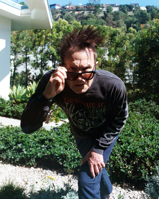
What’s His Age Again? Blink-182’s Mark Hoppus (Now 53) Looks Back.
The sometimes fraught relationship between Hoppus and DeLonge is what Ozzi described as the “real bromance” at the center of the memoir. Fellow skate punks and self-taught musicians with a penchant for phallus jokes, the pair first met in San Diego County in 1992. Hoppus — the son of a homemaker mother and an aerospace engineer father who split up when he was in the third grade — was 20 and drifting through college. DeLonge, three years his junior, was a high school miscreant.
FULL NY TIMES ARTICLE UNDER THE CUT
In early March, Mark Hoppus, the singer and bassist for the long-running pop-punk trio Blink-182, and his wife, Skye, were special guests at a Sotheby’s modern and contemporary art auction in London. The sale featured a piece from their collection, a rare Banksy titled “Crude Oil (Vettriano),” up alongside works by Yoshitomo Nara, Gerhard Richter and Vincent van Gogh.
“It was such rarefied air that we’ve never been a part of before,” Hoppus recalled at his home a week later, outfitted in chunky black glasses, a Dinosaur Jr. long-sleeve T-shirt, navy blue Dickies and Gucci Mickey Mouse sneakers. The painting sold for nearly $5.5 million, part of which will go to charity.
It would have been hard to predict such a highfalutin turn for Hoppus back in 1999, when Blink-182 released its magnum opus, “Enema of the State,” which catapulted the band to MTV “Total Request Live” stardom and sold five million copies domestically. The video for the album’s first single, the jocular “What’s My Age Again?,” famously features the band members running unclothed through the streets of Los Angeles. (“Naked dudes are so ridiculous,” Hoppus said. “It just looks comical to me.”) Blink-182 followed up that LP with its first No. 1 album, “Take Off Your Pants and Jacket,” two years later.
Despite Blink-182’s reputation for high jinks, naughty puns and charmingly adolescent hits like “All the Small Things,” Hoppus is remarkably thoughtful in person. Jim Adkins, whose group, Jimmy Eat World, supported Blink-182 and Green Day on a 2002 tour, said in an interview that Hoppus exhibited “human empathy.”
“I know ‘Mark from Blink-182 is emotionally mature’ might seem like an oxymoron if you don’t know him,” Adkins admitted, “but I would say that.”
That maturity translates to the page. In his memoir, “Fahrenheit-182,” written with the music journalist Dan Ozzi and out April 8, Hoppus details Blink-182’s turbulent history and contemplates his own mortality with grace and good humor. The band’s “Behind the Music”-worthy history includes near-death experiences, bitter splits and world-conquering tours. In 2021, Hoppus was diagnosed with Stage 4A diffuse large B-cell lymphoma and underwent an arduous course of chemotherapy. (“I was all decay and poison,” he writes. “Everyone I talked to cried. Every conversation felt like goodbye.”) He now has a clean bill of health.
Hoppus, a 53-year-old California native, was sitting cross-legged on a chair in the round sunken den at the heart of his exquisite midcentury modern house, which was designed by the architect Harold Levitt. “This room is where I’ve suffered the most,” said the musician, who wore his hair, which he had lost during chemo, in a towering front spike. “This room is where I’ve had the most difficult self-reflection and conversations of my whole life.” He compared it to Superman’s Fortress of Solitude.
“When the band broke up, I sat right here on our couch and just despaired,” he said, referring to the first of two times the singer and guitarist Tom DeLonge walked away from Blink-182, only to eventually return. “I was so filled with animosity and hatred and rage, and I just wanted to get back in our band,” he continued, dropping a number of expletives.
But “Fahrenheit-182” never turns meanspirited or dour. “The book has no demons in it,” Hoppus said. He mentioned that he’d discussed his memoir on the phone with his psychiatrist — Hoppus is treated for obsessive-compulsive disorder, intrusive thoughts, depression and anxiety — earlier that day. “I think that writing the book helped solve a lot of ongoing issues in my life, because I was trying to write it with an even hand,” he said.
The sometimes fraught relationship between Hoppus and DeLonge is what Ozzi described as the “real bromance” at the center of the memoir. Fellow skate punks and self-taught musicians with a penchant for phallus jokes, the pair first met in San Diego County in 1992. Hoppus — the son of a homemaker mother and an aerospace engineer father who split up when he was in the third grade — was 20 and drifting through college. DeLonge, three years his junior, was a high school miscreant.
“Our musical styles fit exactly, and his humor was just as abrasive and as offensive as mine was,” DeLonge, 49, recalled in an interview. “We both came from broken families and saw the world the same way.”
The fast friends formed a band, originally known simply as Blink, with an even younger drummer, Scott Raynor. Blink’s first studio album, “Cheshire Cat” from 1995, did surprisingly well for the independent Cargo Music, and the band leaped to a major label, MCA. In 1997, Blink-182 released the LP “Dude Ranch,” scoring a hit with “Dammit,” a boisterous track with an indelible refrain, delivered by Hoppus: “Well, I guess this is growing up.” Hoppus and DeLonge ended up firing a troubled Raynor and replacing him with the tattooed powerhouse Travis Barker, of another California band, the Aquabats, before recording “Enema of the State.”
It was easy to dismiss Blink-182 in the early days. “When we first came on the scene, the gatekeepers and the people in charge were so focused on Blink’s comedy side, our silliness, that it prevented them from looking deeper,” Hoppus said. “But we did it to ourselves. We played naked. We do mom jokes, Tom and I, back-and-forth onstage, nonstop.” If it weren’t for the many hits Blink-182 scored in the wake of “What’s My Age Again?,” Barker said in an interview, “We very well could have been pigeonholed as the naked band.”
Over the years, Blink-182 only grew in stature among fellow musicians, inspiring emo bands in the 2000s — Fall Out Boy, Panic! at the Disco, Paramore — and the latest generation of pop-punk acts, including MGK (formerly Machine Gun Kelly) and Meet Me @ the Altar. “Look how big Blink-182 is now,” said the Meet Me @ the Altar drummer Ada Juarez, 26, who pointed out she was born the year “Enema of the State” came out. “You can listen to it today, and it still fits. A lot of it has to do with Tom and Mark’s parts and the way that their voices just fit so well together.”
The band’s sphere of influence extends to less-expected genres. “There are emo rappers who say, ‘I grew up listening to Blink-182,’” Hoppus said. “There are dudes playing in the shreddiest heavy metal bands saying, ‘I grew up listening to Blink-182.’ The Chainsmokers are like, ‘We grew up listening to Blink-182.’ I love that celebration and that connection.”
Readers coming to “Fahrenheit-182” for gossip will be disappointed for the most part. There is, for instance, barely a mention that Barker is married to a Kardashian. “It’s not ‘The Dirt,’” Hoppus said, referring to Mötley Crüe’s debauched tell-all. “It’s a PG-13 book.” The memoir does, however, provide insight into strife within Blink. After DeLonge’s 2005 departure broke up the group for the first time, he and Hoppus didn’t talk for several years. “It was awful,” Hoppus said. “I felt like my world had been rugged.”
DeLonge called that rupture “a tale as old as time.”
“When you start a band, it’s just you guys,” he said. “You all have the same dream, same aspirations, same work schedule, same passion, same drive. Then each person finds a spouse, might have kids, might start extracurricular activities.” (DeLonge, who formed the alternative rock band Angels & Airwaves in 2005, is a well-known U.F.O. researcher.) “That just creates issues amongst the band members that I wasn’t even emotionally intelligent enough to communicate or understand or be able to remedy.”
Barker’s near death in 2008 — he survived a plane crash that killed four of the six people onboard — precipitated a Blink reunion, but after five years and just one album, DeLonge bailed again. “I don’t think we were all healed, and we didn’t fully trust each other,” he said. This time, Hoppus and Barker replaced him with Matt Skiba, the singer and guitarist for the Chicago punk band the Alkaline Trio.
“I remember that first Roxy show being mildly terrified and looking over at Mark cracking jokes,” said Skiba, who went on to record two albums with Blink. “His joy would just bring me back into the moment.”
Just as the world was beginning to emerge from the pandemic, Hoppus learned he had cancer. “It got really dark,” he said, recounting a conversation with Skye, with whom he has a 22-year-old son, Jack, a video game designer. “We were sitting in our kitchen and I was dying — the medication, the chemo, was just so gnarly,” he recalled. “Felt like I was being crushed between two trucks. I was like, ‘I don’t know if I can do this.’
“My wife goes, ‘What are you saying? Are you going to kill yourself?’” he continued. “And that moment really crystallized the fight for me. That was when I was like, ‘This is a losing battle, but I have to fight the fight. I can’t just give up in front of my wife and son.’”
When DeLonge found out about Hoppus’s illness, the resentments dropped away. “I was very quick to say, ‘I’m all in, Mark,’” DeLonge said. “‘When — not if — you’re done with these treatments, the north star is we’re going to play again. Let’s do that for each other.’”
Hoppus was cleared of cancer in September 2021, and a year later, he, DeLonge and Barker announced that they were reuniting again. In 2023, Blink released its ninth studio album, “One More Time…,” which became the group’s third record to hit No. 1 on the Billboard 200.
“I don’t think the band’s relationship has ever been as healthy or as strong as it is now,” Barker said. “We love this version of our band.” DeLonge marveled at Blink’s longevity: “Plane crash, cancer, top of the charts, breakups. It’s absolutely bananas that we’re still here.”
Blink-182 will continue so long as it’s still fun, Hoppus said. “The one thing that we have all agreed on, and promised one another, is we have no desire to become a legacy band,” he said. “We don’t want to ride off into the sunset playing ‘All the Small Things’ at casinos ad nauseam. I want to play ‘All the Small Things’ forever, but I also want to keep creating new music that connects with people.”
47 notes
·
View notes
Text
Fuck Cancer, especially Diffuse Large B-Cell Lymphoma. But thank the universe for modern medicine that will give a teenager a fighting chance. They could still use prayers/vibes.
2 notes
·
View notes
Text
I've gotten several asks from you fine folks asking where I've been the last couple years.
Shortly after my last post, I had several family medical emergencies.
My grandfather (89 at the time) was in and out of the hospital.
Then when he got better, my dad was diagnosed with Diffuse Large B-Cell Lymphoma that started in his brain. I spent 10 months taking him back and forth to the hospital, taking my mom back and forth when my dad was admitted, or caring for him at home while he was on hospice. He spent 2 months at home on hospice when he passed in April 2024 at the age of 70.
The literal day after my dad died, my grandfather was diagnosed with another form of lymphoma, aged 90 at that point. Thankfully, his is slow moving and we're in an observe and monitor plan. I've been slowly caring for him more and more over the last 10 months and hopefully he'll get to see his 92nd birthday this July.
I've continued caring for my mom over all of this. I've had my very supportive fiance by my side. My son has grown so much and is becoming a fantastic person as he learns to navigate this world.
It's been a strange 2 years. Death and cancer are bitches. I'm still here. Doing the best I can.

#dead dad club#cancer#tw death#caregiving#autistic adult#tw hospice#lymphoma#last year sucked#checking in#family caregivers#anticipatory grief
7 notes
·
View notes
Text
Seven years ago on this day I was waiting to be wheeled into surgery for doctors to remove a growth and send it to pathohistology. A month later I got the results and a diagnosis of stage three diffuse large B-cell Non-Hodgkin lymphoma. Soon after they started me on chemotherapy.
It was a long and difficult fight, but I managed to pull through. They say passing a five-year mark is a good sign; I am now two years beyond that point and I intend to continue moving forward, one year after another.

8 notes
·
View notes
Text

The decision to undergo bone marrow transplantation (BMT) in a lymphoma patient is highly personalized and depends on several key factors:
• Histological Subtype: The specific form of lymphoma greatly influences treatment choices. For example, aggressive diffuse large B-cell lymphoma (DLBCL) or refractory Hodgkin lymphoma may require BMT.
• Disease Stage: The stage at the time of diagnosis plays a crucial role. BMT is often considered for advanced-stage lymphomas or those that have recurred after initial treatments.
• Response to Previous Therapies: Patients who do not achieve remission or experience a recurrence after traditional therapies may explore BMT as a treatment option.
• Age and Overall Health: The patient's age and general health significantly impact their ability to withstand the demanding BMT process.
• Donor Availability: In the case of allogeneic stem cell transplantation (allo-SCT), locating a compatible donor, typically a sibling or a well-matched unrelated individual, can be a complex undertaking.
A bone marrow transplant can be recommended in other medical conditions besides lymphoma, like leukemia, aplastic anemia, thalassemia, myeloma, and sickle cell disease. You can choose to undergo a bone marrow transplant from some of the best hospitals in Delhi. The cost of bone marrow transplant in Delhi will vary depending on the type and severity of condition being treated and the type of bone marrow transplant being performed.
#bone marrow transplant#cost of bone marrow transplant#bone marrow transplantation#lymphoma#leukemia#myeloma#aplastic anemia#sickle cell disease#thalassemia
6 notes
·
View notes
Text
Reliable PCR Solutions for Detecting ROS1 and MYD88 Mutations in Oncology Labs

Molecular diagnostics has moved beyond research settings and is now a routine part of clinical practice. Today, routine cancer diagnosis often depends on identifying genetic mutations that help classify the disease and guide treatment decisions. Two critical targets in this field are ROS1 gene fusions and MYD88 mutations. These are linked to cancers such as non-small cell lung cancer and B-cell lymphomas, and detecting them early can have a direct impact on patient outcomes.
3B BlackBio Biotech India Ltd provides dedicated real-time PCR kits for both targets. Their ROS1 PCR Kit and MYD88 PCR Kit are designed for diagnostic laboratories that need fast, reliable, and clinically relevant mutation detection. These kits are optimized for compatibility with most thermal cyclers used in India and meet the technical needs of oncology labs without unnecessary complexity.
ROS1 rearrangements are found in a subset of non-small cell lung cancers. Although the prevalence is low—approximately 1 to 2 percent—the clinical significance is high. ROS1-positive tumors often respond to tyrosine kinase inhibitors. The challenge is timely identification. The ROS1 PCR Kit from 3B BlackBio enables labs to detect ROS1 gene fusions using real-time PCR with fluorescent probes. This approach offers high specificity and sensitivity while remaining simple enough for routine use.
What makes this ROS1 mutations kit valuable is its straightforward protocol. Labs don’t need high-throughput sequencing systems to get actionable results. The kit works with extracted RNA from FFPE samples, which are common in hospital pathology departments. The total run time is short, and results can be interpreted without additional post-PCR analysis. That makes it suitable for both high-volume diagnostic labs and smaller centers with limited infrastructure.
The MYD88 mutation, particularly the L265P substitution, is frequently detected in hematological cancers such as Waldenström’s macroglobulinemia and certain forms of diffuse large B-cell lymphoma. It serves not only as a diagnostic marker but also influences how treatment is selected. 3B BlackBio’s MYD88 Mutation Detection Kit uses real-time PCR to enable rapid identification of the mutation from DNA extracted from FFPE tissue samples.
The advantage of this MYD88 PCR Kit lies in its accuracy and ease of integration into standard lab procedures. It uses a TaqMan-based probe system, which avoids cross-contamination risks associated with gel-based or nested PCR. The workflow is clean, and results are ready within hours. For clinicians managing B-cell malignancies, having MYD88 status early in the diagnostic process helps confirm the disease subtype and plan targeted therapies or clinical management accordingly.
In both cases, the kits are built for use in real-world conditions. They are tested for lot-to-lot consistency, include positive and negative controls, and are validated on commonly used thermal cyclers like ABI 7500, Bio-Rad CFX96, and Roche LightCycler. That ensures labs don’t have to modify equipment or protocols to adopt these assays.
From a clinical standpoint, time is critical in cancer diagnosis. A lung cancer case with a ROS1 fusion can shift quickly from chemotherapy to targeted therapy if the mutation is identified early. Similarly, confirming MYD88 L265P in a lymphoma case can help exclude other subtypes, making treatment selection more precise. These are not just technical results—they directly affect what happens next for the patient.
The broader benefit is accessibility. While large hospitals may use sequencing panels, most diagnostic centers and pathology labs still rely on PCR due to its cost-effectiveness and speed. 3B BlackBio’s ROS1 and MYD88 mutation kits provide a reliable solution that fits into this setting. There’s no dependency on high-throughput platforms, which reduces cost per test and makes it feasible to run these assays routinely.
These kits are also useful in labs where sample volume is limited. Many cases rely on small tissue biopsies or archival FFPE blocks. The kits are optimized to perform with low input, which improves reliability when working with compromised or minimal material.
The ROS1 PCR Kit and the MYD88 Mutation Detection Kit by 3B BlackBio are essential tools for molecular diagnostics. They serve real clinical needs with accurate, reproducible results, and offer labs a practical way to keep up with modern oncology demands. These are not research-use-only tools. They are designed to fit seamlessly into the workflow of any molecular lab that needs dependable mutation detection without complexity or delays.
For those in diagnostic settings looking to improve testing accuracy and reduce turnaround time, these PCR kits are proven options. More importantly, they contribute to faster clinical decisions and better patient outcomes—something every diagnostic lab ultimately strives for.
0 notes
Text
CAR-T Cell Therapy: A Breakthrough in Cancer Treatment
CAR-T cell therapy is revolutionizing cancer treatment by harnessing the body’s immune system to fight cancer in an extremely targeted way. Unlike chemotherapy or radiation, CAR-T therapy uses genetically engineered T-cells (a type of white blood cell) to seek out and destroy cancer cells. This advanced treatment is showing promising results, especially in patients with relapsed or refractory blood cancers like acute lymphoblastic leukemia (ALL), non-Hodgkin lymphoma (NHL), and multiple myeloma.
India is rapidly emerging as a global destination for affordable CAR-T therapy, thanks to indigenous innovations and the expansion of its healthcare infrastructure. In this article, we explain what CAR-T therapy Cost in India is, how it works, its benefits, risks, costs in India and abroad, and why India is a preferred destination for international patients seeking advanced cancer care.
What is CAR-T Cell Therapy?
CAR-T (Chimeric Antigen Receptor T-cell) therapy is a type of immunotherapy in which a patient’s own T-cells are collected, genetically modified in a lab to express special receptors (CARs), and then re-infused into the body to identify and kill cancer cells.
CARs enable the T-cells to recognize specific proteins on cancer cells, making them highly effective in targeting malignancies that evade traditional treatments.
How Does CAR-T Therapy Work?
The CAR-T cell therapy process involves the following steps:
Cell Collection (Apheresis): Blood is drawn from the patient, and T-cells are separated.
Genetic Modification: In the laboratory, T-cells are modified using viral vectors to express chimeric antigen receptors (CARs) that can recognize cancer cells.
Cell Expansion: The modified cells are multiplied to reach billions in number.
Conditioning Therapy: Before re-infusion, the patient may receive mild chemotherapy to reduce other immune cells and make space for CAR-T cells.
Infusion: The modified CAR-T cells are infused back into the patient.
Monitoring: Patients are closely monitored for side effects and response.
Types of Cancers Treated with CAR-T Therapy
CAR-T therapy is approved or under investigation for:
Acute Lymphoblastic Leukemia (ALL)
Diffuse Large B-cell Lymphoma (DLBCL)
Follicular Lymphoma
Mantle Cell Lymphoma
Multiple Myeloma
Chronic Lymphocytic Leukemia (CLL)
Clinical trials are underway for solid tumors (e.g., glioblastoma, pancreatic, lung cancers)
Benefits of CAR-T Cell Therapy
High Success Rates: Especially in blood cancers resistant to other treatments.
Single-dose Treatment: Often requires only one infusion session.
Personalized: Tailored to each patient’s immune cells.
Potential for Long-Term Remission: Many patients achieve lasting results.
Risks and Side Effects
While CAR-T therapy is highly effective, it may cause side effects:
Cytokine Release Syndrome (CRS) – fever, low blood pressure, and breathing difficulty due to immune overactivation.
Neurotoxicity – confusion, tremors, or seizures.
Infections – due to weakened immune response.
Tumor Lysis Syndrome – rapid breakdown of cancer cells leading to metabolic imbalance.
Most side effects can be managed with medications and close hospital monitoring.
CAR-T Therapy Cost in India vs. Other Countries
Country Estimated Cost (USD)
India $45,000 – $65,000
USA $400,000 – $600,000
UK $ 350,000 – $500,000
Thailand $ 100,000 – $200,000
Turkey $ 80,000 – $150,000
India offers a major cost advantage without compromising on treatment quality or safety. Indigenous CAR-T products like NexCAR19 and Qartemi have brought down the cost significantly.
Top Hospitals in India Offering CAR-T Cell Therapy
Several premier cancer hospitals and research centers are offering CAR-T therapy:
Tata Memorial Hospital, Mumbai
Apollo Cancer Centres (Pan-India)
HCG Cancer Hospitals, Bangalore
Max Super Speciality Hospital, Delhi
Medanta – The Medicity, Gurugram
Amrita Hospital, Kochi
Fortis Memorial Research Institute, Gurugram
Why Choose India for CAR-T Cell Therapy?
Cost-Effective Treatment
CAR-T therapy in India costs 5–10 times less than in Western countries due to local manufacturing and affordable healthcare systems.
Skilled Oncologists & Researchers
India is home to globally trained hematologists and oncologists. Many doctors are part of CAR-T trials and collaborative research projects.
Indigenous CAR-T Products
India has approved its own CAR-T therapies like:
NexCAR19 by ImmunoACT
Qartemi (varnimcabtagene autoleucel) by Immuneel Therapeutics
Global-Standard Infrastructure
State-of-the-art labs, ICUs, transplant units, and sterile environments support safe and effective delivery of this therapy.
Medical Tourism Support
Hospitals in India offer:
Visa assistance
Translators
International patient coordinators
Affordable accommodation for patient and caregivers
Treatment Timeline for CAR-T Therapy
StepDurationInitial evaluation2–3 daysT-cell collection (apheresis)1 dayCell engineering & expansion2–3 weeksConditioning therapy2–3 daysCAR-T cell infusion1 dayIn-patient observation7–14 daysFollow-up & monitoring1–3 months
CAR-T Therapy Success Rates
Clinical trials and real-world results show impressive outcomes:
Acute Lymphoblastic Leukemia (ALL) – ~85–90% remission in children
DLBCL & NHL – 50–60% long-term remission
Multiple Myeloma – 70–75% response rate
These results vary by disease type, patient age, and prior treatments.
Is CAR-T Therapy Safe for Everyone?
CAR-T therapy is usually recommended for patients who:
Have relapsed after multiple lines of chemotherapy
Have aggressive or treatment-resistant cancers
Are medically fit for immune-based therapies
Not all patients qualify. Thorough pre-treatment evaluation is required including:
Blood tests
Imaging
Bone marrow biopsy
Cardiac and liver function tests
Final Thoughts
CAR-T cell therapy has transformed the landscape of cancer care—offering hope where conventional treatments fail. India’s rising role in CAR-T innovation, manufacturing, and delivery has made it a global hub for affordable and cutting-edge cancer treatment.
If you or a loved one is battling a relapsed or refractory blood cancer, CAR-T therapy in India may offer a lifeline. Speak to a certified oncologist, check eligibility, and explore this breakthrough treatment that could change your future.
0 notes
Text
CAR T-Cell Therapy in India: A New Hope for Blood Cancer Patients
When it comes to advanced cancer treatment, few therapies have created as much buzz in recent years as CAR T-Cell Therapy. This groundbreaking form of immunotherapy has redefined the possibilities in treating blood cancers like leukemia, lymphoma, and multiple myeloma. And now, CAR T-Cell Therapy in India is becoming a global beacon of hope—thanks to cutting-edge research, clinical innovation, and cost-effective care.

🔬 What Is CAR T-Cell Therapy?
CAR T-Cell Therapy (Chimeric Antigen Receptor T-cell therapy) is a highly advanced form of immunotherapy that involves genetically modifying a patient’s T-cells to recognize and destroy cancer cells. This personalized approach trains your immune system to be a precision-guided missile aimed at your cancer.
The procedure begins with a leukapheresis, where T-cells are collected from the patient’s blood. These cells are then re-engineered in a lab to produce special receptors (CARs) that target cancer cells. After being multiplied, they are infused back into the patient, where they go to work fighting the cancer from within.
🩸 Who Needs CAR T-Cell Therapy?
CAR T-cell therapy is primarily used in patients with:
Acute Lymphoblastic Leukemia (ALL) – especially in children and young adults with relapsed or refractory cases.
Non-Hodgkin Lymphoma (NHL) – including Diffuse Large B-cell Lymphoma after two or more lines of failed treatment.
Multiple Myeloma – for patients who have tried and not responded to other standard treatments.
💉 Types of CAR T-Cell Therapy Available in India
India offers multiple FDA-approved and emerging CAR T-cell therapies, including:
ABECMA
BREYANZI
CARVYKTI
KYMRIAH
NexCAR19 – Developed by IIT Bombay & Tata Memorial Centre.
Varnimcabtagene Autocel – Co-developed by Immuneel Therapeutics and Hospital Clinic de Barcelona.
Each therapy is designed to target specific types of blood cancers and is offered under strict medical guidance at certified treatment centers.
🧪 Pre-Therapy Evaluation
Before undergoing therapy, a thorough pre-treatment diagnostic is conducted to determine eligibility and optimize results. This includes:
Cancer staging and treatment history
Organ function testing (heart, liver, kidney)
Tumor burden assessment (MRI, PET-CT)
Bone marrow biopsy
Neurological and pulmonary evaluations
💲 Cost of CAR T-Cell Therapy in India
One of the most compelling reasons patients choose India is affordability. The Cost of CAR T-Cell Therapy in India ranges between $100,000 to $110,000, significantly lower than in the US or Europe, where costs can easily exceed $400,000.
Despite the lower cost, the treatment quality remains world-class, making India a leading medical tourism destination for cancer care.
⚕️ Success Rate of CAR T-Cell Therapy in India
Clinical trials in India have demonstrated remarkable outcomes:
NexCAR19 showed a 73% success rate in patients with relapsed/refractory leukemia and lymphoma.
Varnimcabtagene Autocel recorded an 83.3% response rate in lymphoma cases.
These numbers highlight India’s potential to become a global hub for advanced immunotherapies.
🌿 Recovery & Long-Term Care
Post-therapy recovery involves:
Intensive monitoring for 2–4 weeks post-infusion
Regular follow-ups for infections, neurotoxicity, or cytokine release syndrome
Long-term tracking for relapse prevention
Mental health support and immune system rebuilding
With the right care team and patient commitment, long-term remission is increasingly achievable.
🇮🇳 Why Choose India for CAR T-Cell Therapy?
Advanced Research Facilities (e.g., Tata Memorial Centre, IIT Bombay)
High Success Rates with innovative therapies
Affordable Pricing
Medical Tourism Support including visa assistance, accommodation, and interpreter services
Access to internationally trained oncologists and hematologists
🔚 Conclusion
CAR T-Cell Therapy in India is more than just a medical procedure—it’s a lifeline for patients who have exhausted traditional treatments. With a combination of world-class expertise, innovative therapies, and affordable care, India is quickly emerging as a global leader in precision oncology.
Whether you're a local patient or an international traveler seeking hope, the breakthroughs in CAR T-cell therapy could be the transformative answer you’ve been waiting for.
#CARTCellTherapy#CARTinIndia#CancerTreatmentIndia#BloodCancerCare#ImmunotherapyIndia#MedicalTourismIndia#AffordableCancerCare#LeukemiaTreatmentIndia#LymphomaTherapy#IndiaFightsCancer
1 note
·
View note
Link
Updated data from the pivotal phase III STARGLO study continue to demonstrate a clinically meaningful improvement in overall survival with a 40% survival benefit for people with R/R DLBCL who are not candidates for transplant189% of patients whose c #BioTech #science
0 notes
Text

CAR T-Cell Therapy in Non-Hodgkin’s Lymphoma (NHL)
CAR T-cell therapy has become a groundbreaking treatment option for Non-Hodgkin’s Lymphoma, particularly in cases that are relapsed or resistant to standard therapies. This cutting-edge therapy involves genetically engineering a patient’s T cells to express chimeric antigen receptors (CARs) that specifically target and destroy cancer cells. It has demonstrated strong efficacy, especially in aggressive forms like Diffuse Large B-Cell Lymphoma (DLBCL), and has received regulatory approvals, including from the FDA. However, the therapy still faces challenges, such as potentially serious side effects (e.g., cytokine release syndrome and neurological toxicity), as well as its high cost. Continued advancements in CAR T-cell research and ongoing clinical trials are expected to broaden its application across NHL subtypes and improve treatment effectiveness and safety.
Want more data like this? Get the infographic now
Epidemiological Segmentation
CAR T-cell therapy eligibility for NHL is segmented by:
Incident cases by indication (e.g., Mantle Cell Lymphoma, DLBCL, Follicular Lymphoma, Marginal Zone Lymphoma, PMBCL, CLL/SLL)
Indication-specific eligible patient populations for CAR T-cell therapy
Epidemiology Highlights (2023)
An estimated 126,000 patients in the 7MM were eligible for CAR T-cell therapy in 2023.
DLBCL was the most common NHL subtype in the EU4 and the UK, accounting for around 32,000 cases.
Japan reported approximately 23,700 incident cases of NHL across key CAR T-suitable subtypes in 2023.
Visualize the trends—then explore the data behind them in our latest report : Click Here
Market Overview
The US market for CAR T-cell therapy in NHL was valued at approximately USD 1.2 billion in 2023.
Market Drivers
Strong clinical success in hard-to-treat NHL cases.
Rapid technological progress in CAR T-cell development.
A growing number of FDA-approved CAR T therapies.
Rising demand for individualized and immune-based treatments.
Market Barriers
High production and administration costs of CAR T-cell therapies.
Limited availability of treatment centers with necessary infrastructure.
Risks of serious adverse events, including CRS and neurotoxicity.
Regulatory hurdles and insurance coverage issues impacting accessibility.
Download the full infographic to uncover detailed insights : Click Here
Pipeline Therapies
Emerging CAR T-cell products include:
Cemacabtagene ansegedleucel
Rapcabtagene autoleucel
Zamtocabtagene autoleucel
AUTO4
CD30.CAR-T
PMB-CT01
And others
Key Industry Players
Allogene Therapeutics
Novartis
Miltenyi Biomedicine
Mustang Bio
Crispr Therapeutics
2seventy Bio
Imugene
Among others
Make informed decisions—start with the full report
0 notes
Text
0 notes
Text
Diffuse Large B Cell Lymphoma: Symptoms, Diagnosis, and Treatment
Introduction to Diffuse Large B Cell Lymphoma
Diffuse Large B Cell Lymphoma (DLBCL) is the most common type of non-Hodgkin lymphoma (NHL), accounting for about 30% of all cases. This aggressive cancer originates in the B cells, a type of white blood cell responsible for producing antibodies. DLBCL can develop in lymph nodes or other organs and spreads rapidly if left untreated.
Understanding the symptoms, diagnostic procedures, and treatment options for Diffuse Large B Cell Lymphoma is crucial for early detection and effective management.
Symptoms of Diffuse Large B Cell Lymphoma
The symptoms of DLBCL can vary depending on where the cancer is located. Some common signs include:
Swollen Lymph Nodes – Painless swelling in the neck, armpits, or groin.
B Symptoms – Fever, night sweats, and unexplained weight loss (more than 10% of body weight in six months).
Fatigue – Persistent tiredness due to the body’s immune response.
Abdominal Pain or Swelling – If the lymphoma affects the abdomen or digestive tract.
Shortness of Breath – If the tumor presses against the lungs or airways.
Neurological Symptoms – Confusion, weakness, or seizures if the central nervous system is involved.
Because these symptoms can mimic other conditions, proper medical evaluation is essential.
Causes and Risk Factors of DLBCL
The exact cause of Diffuse Large B Cell Lymphoma is unknown, but several risk factors have been identified:
Age – Most common in people over 60, though it can occur at any age.
Weakened Immune System – Individuals with HIV/AIDS, autoimmune diseases, or those taking immunosuppressive drugs are at higher risk.
Infections – Certain viruses, such as Epstein-Barr virus (EBV) and hepatitis C, have been linked to lymphoma development.
Genetic Mutations – Abnormalities in genes like MYC, BCL2, and BCL6 can contribute to aggressive lymphoma growth.
Environmental Factors – Exposure to pesticides, herbicides, or certain chemicals may increase risk.
Diagnosis of Diffuse Large B Cell Lymphoma
Accurate diagnosis of DLBCL involves multiple tests:
1. Physical Examination
Doctors check for swollen lymph nodes, liver, or spleen enlargement.
2. Blood Tests
Complete blood count (CBC) to detect abnormalities.
Lactate dehydrogenase (LDH) levels, which are often elevated in aggressive lymphomas.
3. Imaging Tests
CT Scan – Identifies enlarged lymph nodes or tumors.
PET Scan – Helps determine the cancer’s stage and spread.
MRI – Used if central nervous system involvement is suspected.
4. Biopsy
A lymph node or tissue sample is examined under a microscope to confirm DLBCL and classify its subtype.
5. Bone Marrow Biopsy
Determines if lymphoma has spread to the bone marrow.
6. Molecular and Genetic Testing
Tests like fluorescence in situ hybridization (FISH) detect genetic abnormalities that influence treatment choices.
Staging Diffuse Large B Cell Lymphoma
The Ann Arbor staging system is used to classify DLBCL:
Stage I – Cancer is in one lymph node region or a single organ.
Stage II – Two or more lymph node regions on the same side of the diaphragm are affected.
Stage III – Lymph nodes on both sides of the diaphragm are involved.
Stage IV – Cancer has spread to organs like the liver, lungs, or bone marrow.
The presence of B symptoms (fever, night sweats, weight loss) is also noted as "B" designation, indicating a more aggressive disease.
Treatment Options for DLBCL
Treatment for Diffuse Large B Cell Lymphoma depends on the stage, patient’s age, and overall health. Common approaches include:
1. Chemotherapy
The R-CHOP regimen (rituximab, cyclophosphamide, doxorubicin, vincristine, and prednisone) is the standard first-line treatment.
2. Immunotherapy
Rituximab (anti-CD20 antibody) targets B cells.
CAR T-cell therapy (e.g., axicabtagene ciloleucel) is used for relapsed/refractory cases.
3. Radiation Therapy
Used for localized disease or to relieve symptoms in advanced stages.
4. Stem Cell Transplant
High-dose chemotherapy followed by autologous stem cell transplant may be recommended for relapsed patients.
5. Targeted Therapy
Drugs like ibrutinib (BTK inhibitor) or lenalidomide are options for specific subtypes.
6. Clinical Trials
New treatments, such as bispecific antibodies and checkpoint inhibitors, are being studied.
Prognosis and Survival Rates
The prognosis for DLBCL varies based on factors like:
International Prognostic Index (IPI) score (age, stage, LDH levels, performance status).
Cell of origin (germinal center vs. activated B-cell subtype).
With treatment, the 5-year survival rate is approximately:
60-70% for early-stage DLBCL.
30-40% for advanced or high-risk cases.
Relapse can occur, but newer therapies like CAR T-cell therapy offer hope for refractory patients.
Living with Diffuse Large B Cell Lymphoma
Managing DLBCL involves:
Regular follow-ups to monitor remission.
Managing side effects (fatigue, infections, neuropathy).
Nutritional support and physical activity to improve recovery.
Emotional and psychological support through counseling or support groups.
Conclusion
Diffuse Large B Cell Lymphoma is a fast-growing but treatable cancer. Early diagnosis and advances in immunotherapy have improved outcomes significantly. Patients should work closely with their healthcare team to determine the best treatment plan.
Ongoing research continues to explore novel therapies, offering hope for better survival and quality of life for those affected by DLBCL.
This article provides a comprehensive overview of Diffuse Large B Cell Lymphoma, covering symptoms, diagnosis, treatment, and prognosis. If you or a loved one are experiencing concerning symptoms, consult a hematologist or oncologist for evaluation.
1 note
·
View note