#fluorescence microscopy
Text
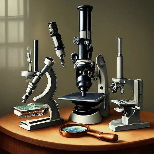
A Journey into the World of Microscopy: From Humble Beginnings to High-Tech Magnification
The science of looking into the hidden invisible Microscopy has transformed our understanding of the world around us. It can explore the universe beyond the reach of our naked eyes, with complex cellular structures, red blood cells, viruses and other viruses and microorganisms taking on amazing perspectives
The history of the microscope is a fascinating story of human curiosity, scientific genius, and relentless exploration. From the humble beginnings of simple magnifying glasses to the sophistication of modern electronic microscopes, the invention of microscopes has shaped our understanding of the microscopic world
In the 1600s, Dutch opticians such as Hans and Zachary Janssen are credited with inventing the first microscope. Known for this hybrid microscope, many lenses were used to magnify objects up to 30 times.At the end of the 17th century, Antony van Leeuwenhoek, Dutch draper some changed our perception of thumbnails. Armed with a well-made single-lens microscope, and explored the hidden reaches of nature. In 1674, Leeuwenhoek discovered microorganisms in lake water, which he aptly named “animalcules”. His discovery laid the foundations of biology and inspired generations of scientists. This incredible feat allowed him to uncover a hidden universe – the first sightings of bacteria, red blood cells, and other microorganisms.
Formation of the scientific environment (17th-19th centuries): Leeuwenhoek’s discoveries boosted scientific research. Robert Hooke, an English scientist, established these developments. In 1665, his book "Micrographia" recorded his observations with a compound microscope. Notably, the term "cell" was coined by Hooke when he examined cork tissue, laying the foundation for cell biology.Microscope systems flourished throughout the 18th and 19th centuries Joseph Lister and other scientists addressed the limitations of the early lenses, introducing improvements that reduced image distortion.
Beyond the Limits of Light: The Beginning of the New Age (19th-20th century): As the 19th century progressed, the limitations of optical microscopy became apparent and scientists yearned for a tool which can go deeper into cells. This research culminated in the development of the electron microscope in the 1930s. The 20th century was revolutionary with the invention of the electron microscope. Unlike light microscopes, which use visible light, electron microscopes use electron beams to achieve much higher magnification.Formation of the scientific environment (17th-19th centuries): Leeuwenhoek’s discoveries boosted scientific research. Robert Hooke, an English scientist, established these developments. In 1665, his book "Micrographia" recorded his observations with a compound microscope. Notably, the term "cell" was coined by Hooke when he examined cork tissue, laying the foundation for cell biology.Microscope systems flourished throughout the 18th and 19th centuries Joseph Lister and other scientists addressed the limitations of the early lenses, introducing improvements that reduced image distortion.
Beyond the Limits of Light: The Beginning of the New Age (19th-20th century): As the 19th century progressed, the limitations of optical microscopy became apparent and scientists yearned for a tool which can go deeper into cells. This research culminated in the development of the electron microscope in the 1930s. The 20th century was revolutionary with the invention of the electron microscope. Unlike light microscopes, which use visible light, electron microscopes use electron beams to achieve much higher magnification.
In the 1930s, German experts Max Knoll and Ernst Ruska made the first electron microscope. This tool let us see tiny things like cells and even atoms by using electron beams, not light, getting images many times bigger. This cool invention showed us the tiny parts inside cells, viruses, and stuff too small to see before. The 1900s brought even more cool microscopes. New kinds like phase-contrast and confocal microscopy let scientists look at live cells without using stuff that could hurt them. Now, the world of looking at tiny things is getting even better. Today, we have high-tech microscopes that use computers and lasers. These let us see and even change tiny things in ways we never could before.
Modern Microscopy's Diverse Arsenal - Today, the field of microscopy boasts a diverse range of specialized instruments, each tailored to address specific scientific needs. Here's a glimpse into some remarkable examples:
Scanning Electron Microscope (SEM): Imagine a high-tech camera that captures images using a beam of electrons instead of light. That's the essence of a SEM. By scanning the surface of a sample with a focused electron beam, SEMs generate detailed information about its topography and composition. This makes them ideal for studying the intricate structures of materials like insect wings, microchips, and even pollen grains.
Transmission Electron Microscope (TEM): While SEMs provide exceptional surface detail, TEMs take us a step further. They function by transmitting a beam of electrons through a very thin sample, allowing us to observe its internal structure. TEMs are the go-to instruments for visualizing the intricate world of viruses, organelles within cells, and macromolecules like proteins.
Confocal Microscopy: Ever wished to focus on a specific layer within a thick biological sample and blur out the rest? Confocal microscopy makes this possible. It utilizes a laser beam to precisely illuminate a chosen plane within the sample, effectively eliminating information from out-of-focus regions. This allows researchers to create sharp, three-dimensional images of cells, tissues, and even small organisms.
Atomic Force Microscopy (AFM): This technique takes a completely different approach, venturing into the realm of physical interaction. AFM employs a tiny cantilever, akin to a microscopic feeler, to physically scan the surface of a sample. By measuring the minute forces between the cantilever and the sample's surface, AFM can map its topography at an atomic level. This provides invaluable insights into the properties of materials at an unimaginable scale, making it crucial for research in fields like nanotechnology and surface science.
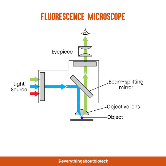
Fluorescence Microscopy: Imagine illuminating a sample with specific wavelengths of light and observing it glowing in response. That's the essence of fluorescence microscopy. This technique utilizes fluorescent molecules or tags that bind to specific structures within a cell or tissue. When excited by light, these tags emit their own light, highlighting the target structures with remarkable clarity. This allows researchers to visualize specific proteins, DNA, or even pathogens within biological samples.
Super-resolution Microscopy (SRM): Overcoming the limitations imposed by the wavelength of light, SRM techniques like STED (Stimulated Emission Depletion) and PALM (Photoactivated Localization Microscopy) achieve resolutions surpassing the diffraction limit. This allows researchers to visualize structures as small as 20 nanometers, enabling the observation of intricate cellular machinery and the dynamics of individual molecules within living cells.
Cryo-Electron Microscopy (Cryo-EM): This powerful technique takes a snapshot of biological samples in their near-life state. Samples are rapidly frozen at ultra-low temperatures, preserving their native structure and minimizing damage caused by traditional fixation methods. Cryo-EM has been instrumental in determining the three-dimensional structures of complex molecules like proteins and viruses, providing crucial insights into their function and potential drug targets.
Correlative Microscopy: Combining the strengths of multiple microscopy techniques, correlative microscopy offers a comprehensive view of biological samples. For instance, researchers can utilize fluorescence microscopy to identify specific structures within a cell and then switch to electron microscopy to examine those structures in high detail. This integrated approach provides a deeper understanding of cellular processes and their underlying mechanisms.
Light Sheet Microscopy (LSM): Imagine illuminating a thin slice of a sample within a living organism. LSM achieves this feat by focusing a laser beam into a thin sheet of light, minimizing photobleaching and phototoxicity – damaging effects caused by prolonged exposure to light. This allows researchers to observe dynamic processes within living organisms over extended periods, providing valuable insights into cellular behavior and development.
Expansion Microscopy (ExM): This innovative technique physically expands biological samples by several folds while preserving their structural integrity. This expansion allows for better resolution and visualization of intricate cellular structures that would otherwise be difficult to distinguish using traditional microscopy methods. ExM holds immense potential for studying the organization and function of organelles within cells.
Scanning Near-Field Optical Microscopy (SNOM): This innovative technique pushes the boundaries of resolution by utilizing a tiny probe that interacts with the sample at an extremely close range. SNOM can not only image the surface features of a sample with exceptional detail but also probe its optical properties at the nanoscale. This opens doors for research in areas like material science and photonics, allowing scientists to study the behavior of light at the interface between materials.
X-ray Microscopy: Stepping outside the realm of light and electrons, X-ray microscopy offers unique capabilities. By utilizing high-energy X-rays, this technique can penetrate deep into samples, making it ideal for studying the internal structure of dense materials like bones and minerals. Additionally, it allows for the visualization of elements within a sample, providing valuable information about their distribution and composition.
From revealing the building blocks of life to aiding in the development of new medicines, the microscope has played an undeniable role in shaping our scientific understanding. As technology continues to evolve, one can only imagine the future breakthroughs this remarkable invention holds in unveiling the secrets of our universe, both seen and unseen. These advancements hold the potential to revolutionize our understanding of biological processes, develop new materials with extraordinary properties, and ultimately pave the way for breakthroughs in medicine, nanotechnology, and countless other fields. As we continue to refine and develop novel microscopy techniques and the future holds immense promise for further groundbreaking discoveries that will undoubtedly revolutionize our perception of the world around us.
#science sculpt#life science#science#molecular biology#biology#biotechnology#artists on tumblr#microscopy#microscope#Scanning Electron Microscope#Transmission Electron Microscope#Confocal Microscopy#Atomic Force Microscopy#Fluorescence Microscopy#Expansion Microscopy#X-ray Microscopy#Super-resolution Microscopy#Light Sheet Microscopy#illustration#illustrator#illustrative art#education#educate yourself#techniques in biotechnology#scientific research#the glass scientists#scientific illustration#scientific advancements
6 notes
·
View notes
Video
Traffic and Travel
Carrying messages to and from our moving limbs, neurons rely on signalling endosomes to ferry supportive chemicals along their branches or axons. Although ten million times smaller than postable parcels, here researchers use intravital imaging to capture a bustling traffic of endosomes (highlighted in blue) shuttling through a mouse’s sciatic nerve. The technique involves anaesthetising the mouse and exposing its nerve under a microscope – it reveals endosomes carrying BDNF, a type of neurotrophin which helps to keep neurons healthy. Certain genetic mutations can block BDNF traffic at junctions where nerve meets muscle, leading to weakness or muscle wasting. The team hopes designing drugs to boost BDNF may restore endosome transport and bring relief to human patients with disorders like Charcot-Marie-Tooth disease.
Written by John Ankers
Video from work by James N. Sleigh and colleagues
Department of Neuromuscular Diseases, Queen Square Institute of Neurology, University College London, London, UK
Video copyright held by James N. Sleigh
Research published in JCI Insight, March 2023
You can also follow BPoD on Instagram, Twitter and Facebook
#science#biomedicine#neuroscience#axons#nerves#neurons#charcot-marie-tooth#cmt#neurotrophin#endosomes#fluorescence microscopy
19 notes
·
View notes
Text
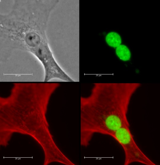
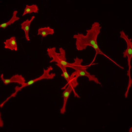
look at my cells boy

#i took these pics how sick is that#the thing speaks#should i put this in the study tags#yeah fuck it lets go#biology#cell biology#studyblr#microscopy#fluorescence microscopy#microscope#cells
30 notes
·
View notes
Text

So I'm working with Daphnia and their uptake of microplastic under different environmental influences at the lab. Took some fluorescence pictures and really liked how they turned out...
#daphnia#Daphnia magna#fluorescence#microscopy#fluorescence microscopy#lab#laboratory#biology#science#microplastic
7 notes
·
View notes
Text

Plant Microverse 🦠
Autofluorescence of plant tissues can reveal amazing colors. Autofluorescence in UV light.
1 note
·
View note
Text
What is Fluorescence Microscopy?
What is Fluorescence Microscopy? #FluorescenceMicroscopy
Fluorescence microscopy is a technique used to study the structure and function of molecules in living cells. This type of microscopy uses light to excite fluorescent molecules, which then emit light of a different color that can be used to track the movement of these molecules or to visualize specific structures within cells.
Photo by Artem Podrez on Pexels.com
What is Fluorescence…

View On WordPress
0 notes
Text
Pistils of dandelion were collected and made into slide samples. Different fluorescence patterns of its parts were observed using confocal microscopy.
By Liu Ruming (China)
Light Microscopy Awards
#liu ruming#photographer#china#pistils of dandelion#fluorescence patterns#nature#light microscopy awards#micro photography
23 notes
·
View notes
Text
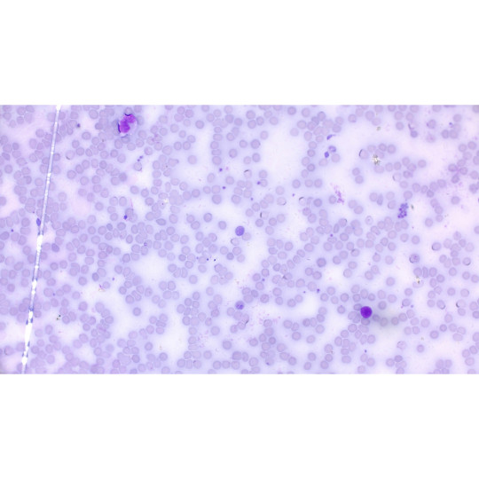
This is malaria captured under a basic fluorescence microscope.
15 notes
·
View notes
Text
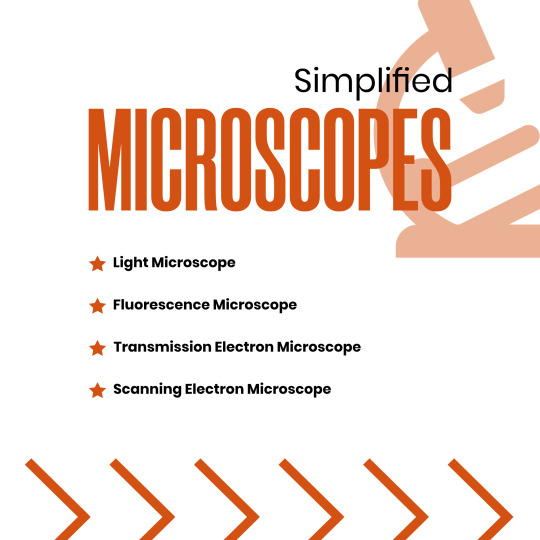


Microscopes Simplified
Light Microscope
Fluorescence Microscope
Transmission Electron Microscope
Scanning Electron Microscope
#microscope#light microscope#Fluorescence Microscope#Transmission Electron Microscope#Scanning Electron Microscope#microscopic#science#microscopy#microscopic life#biotechnology#biotech#biology#biochemistry#studyblr#molecularbiology#notes#class notes#students#science illustration#illustration#illustragram#scientific illustration#graphic design#graphicwork#study notes#study#study motivation#study tips#studygram
82 notes
·
View notes
Text
Uses of Different Types of Microscopes in Forensics
Uses of Different Types of Microscopes in Forensics
The forensic and microbiological labs include a variety of microscopes that may be used. The value of microscopes is increased by how widely they may be used and modified.
#microscope #forensicscience
(more…)

View On WordPress
#comparison microscope#comparison microscope in Forensic Science#dark-field microscope in forensic science#Fluorescent microscope#Forensic science#forensic science notes#microscopy notes#polarising microscope#Scanning Electron Microscope#stereoscopic microscope#stereoscopic microscope uses in forensic science#Transmission Electron Microscope#use of compound microscope in forensic science#use of digital microscope in forensic science#use of fluorescent microscope in forensic science#use of inverted microscope in forensic science#use of phase contrast microscopy in forensic science#use of polarising microscope in forensic science#use of scanning electron microscope#use of scanning electron microscope in forensic science#Uses of Different types of Microscope In Forensics
7 notes
·
View notes
Text
youtube
The confocal microscope at Imperial College's Sir Alexander Fleming Building lab is used for imaging the interior of living plant and animal cells.
During my PhD project, I used the confocal microscope to view the interior of Nicotiana benthamiana plant cells which were expressing Green Fluorescent Protein (GFP) tagged genes of interest. I aimed to find out where the proteins encoded by the genes of interest were localised in the plant cell, which turned out to be in the cytoplasm.
From Wikipedia's entry on Confocal Microscopy: "Confocal microscopy, most frequently confocal laser scanning microscopy (CLSM) or laser scanning confocal microscopy (LSCM), is an optical imaging technique for increasing optical resolution and contrast of a micrograph by means of using a spatial pinhole to block out-of-focus light in image formation. Capturing multiple two-dimensional images at different depths in a sample enables the reconstruction of three-dimensional structures (a process known as optical sectioning) within an object. This technique is used extensively in the scientific and industrial communities and typical applications are in life sciences, semiconductor inspection and materials science. Light travels through the sample under a conventional microscope as far into the specimen as it can penetrate, while a confocal microscope only focuses a smaller beam of light at one narrow depth level at a time. The CLSM achieves a controlled and highly limited depth of field."
Music by the Fiechter Brothers
Images by Katia Hougaard & the Facility for Imaging by Light Microscopy at Imperial College London
#katia plant scientist#botany#plant biology#plants#plant science#biology#science#science and technology#confocal microscopy#microscopy#microscope#scientific instruments#laboratory#research#phdblr#phd#phd life#molecular biology#cell biology#green fluorescent protein#plant scientist#Youtube
1 note
·
View note
Photo

On Your Nerves
Sensory neurons in our skin are the first to warn us about threats in the outside world. They relay temperature, pain and stress as electrical impulses fired towards the central nervous system, but these messages start at the fringe where skin meets nerve. To investigate further, scientists develop living models inside microfluidic devices. Green fluorescence highlights these rat nerve cells (with their DNA in blue) reaching out in three dimensions into a nurturing environment of chemicals, mimicking their natural extracellular matrix (ECM). Each column tests a different type of ECM, finding more developed growth on the right over 4 days (top to bottom). Later, the researchers grew skin cells down into the ECM to mingle with the nerve cells – similar to innervated skin. Such models could be used to test treatments for pain-related disorders, or mimic disorders like diabetic neuropathy.
Written by John Ankers
Image from work by Jinchul Ahn and Kyungeun Ohk, and colleagues
School of Mechanical Engineering, Korea University, Seoul, South Korea
Image originally published with a Creative Commons Attribution 4.0 International (CC BY 4.0)
Published in Nature Communications, March 2023
You can also follow BPoD on Instagram, Twitter and Facebook
#science#biomedicine#neuroscience#microfluidics#lab-on-a-chip#skin cells#skin#skin pain#pain#pain treatment#immunofluorescence#microscopy#fluorescence microscopy
11 notes
·
View notes
Text
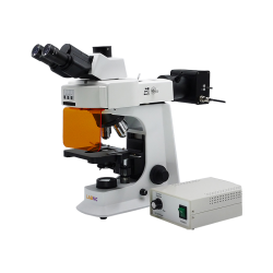
Fluorescence Microscope
Fluorescent Microscope incorporates a Siedentopf trinocular head with 30° inclination and 360° rotation. The WF10x/20 mm high-eyepoint eyepiece and a quadruple inwardly rotating nosepiece make up the microscope. It concentrates on fine and coarse specimens and has an Abbe condenser with an iris diaphragm. A modernized halogen lamp with Kohler lighting as the light source is also featured.
0 notes
Text
Why should Science and Art be antithetical?!
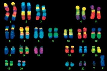
Chromosome painting -> Fluorescent in situ hybridization (FISH)
#science#art#stem#stemgirls#stemwomen#biology#biologystudent#biologycell#gene#genome#cariotype#microscopy#microscopyart#microscopyphotography#chromosome#chromosomepainting#fluorescent#fluorescentart#fluorescentinsituhybridization
1 note
·
View note
Text
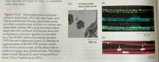
When young Arabidopsis plants are rapidly frozen by plunging them in slush nitrogen, then freeze-substituted and fixed, sieve plate pores are often unobstructed in the tissue (Figure 11.12A). (...) While a meshwork of protein filaments was often shown to extend throughout the lumen (Figure 11.12B), masses or agglomerates of protein were frequently observed to fill large portions of the sieve tube lumen at or close to the sieve plate. The structure of these masses was highly variable, but multiple large masses sometimes filled the entire lumen of the sieve tube (Figure 11.12C).
"Plant Physiology and Development" int'l 6e - Taiz, L., Zeiger, E., Møller, I.M., Murphy, A.
#book quotes#plant physiology and development#nonfiction#textbook#arabidopsis#snap frozen#nitrogen#sieve plate#meshwork#protein filaments#lumen#mass#agglomerate#sieve tube#plant cells#microscopy#fluorescence
0 notes
Text
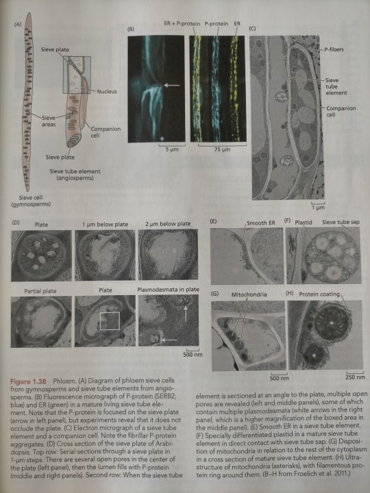
Specialized proteins are made in the phloem cells, such as the P-protein (see Figure 1.38B).
"Plant Physiology and Development" int'l 6e - Taiz, L., Zeiger, E., Møller, I.M., Murphy, A.
#book quote#plant physiology and development#nonfiction#textbook#plant cells#cell differentiation#cell development#proteins#fluorescence#microscope#microscopy#micrograph#endoplasmic reticulum#sieve tube
1 note
·
View note
Last Seen Blogs

dearskyzinha
writer in the dark.

just-anoth3r
Asteroid Fields

hackerpenguin
Absolute Fool

benjamin-13
Untitled

rvnarzkyr
hai rav!