#oncogene expression
Explore tagged Tumblr posts
Visit Tumblr Blog
Explore Tumblr blogs with no restrictions, modern design and the best experience.
Last Seen Tumblr Blogs


Fun Fact
The average Tumblr user visits about 67 pages every month.
Text
youtube
#m6A modification#28S rRNA#oncogenic mRNA translation#tyrosine catabolism#RNA epigenetics#ribosomal biology#cancer metabolism#tumor progression#mRNA regulation#epitranscriptomics#oncogene expression#translational control#metabolic pathways#RNA modification#ribosome recruitment#cancer research#mRNA stability#oncogenic signaling#molecular oncology#targeted cancer therapy.#Youtube
0 notes
Text
Drug resistance in multiple myeloma: When cancer cells say "NO" to treatment
Drug resistance is like a game of cat and mouse. Cancer cells are the cat, and researchers are the mouse. The cat is always trying to find new ways to catch the mouse, but the mouse is always trying to find new ways to avoid getting caught.
Read More
#multiple myeloma#molecular mechanisms#signaling pathways#Health#stem cell transplantation#oncogenes#Lifestyle#cancer cells#gene mutations#drug resistance#cellular environment#gene expression
0 notes
Text

[ UV Albert Wesker Uroboros & PG67 HCs ]
These take after the knowledge that PG67 contains hastily-added leech genes. This is full of medical jargon related to virology and genetics. It's hard sci-fi styled!
tags: medical:genetic;neurology (hardly);virology;needles (PG67-A/W), cannibalism mention, childhood trauma, body horror, mental decay

Such a far cry from his beginnings... he's truly taken after the man he hates the most.
Unfortunately for Wesker, adding Uroboros in intensely massive quantities to an existing Progenitor-67 infection is a poor choice that makes him spend much of his fight with Chris alternating between pouncing him like a rabid animal and curling like the death throes of a dying spider.
And unfortunately for Chris, Wesker doesn't die when he's left at the volcano to stew in a puddle of himself. Even with the awakened lack of thermophilic qualities as a result of the genetic influence of G, there is enough local flora and fauna to keep him alive... albeit in a subdued state.
Physiological:
Uroboros, combined on the initial foundation of Progenitor 67, takes after Progenitor 67's metabolic inefficiency and drives that far higher than it was ever meant to go in an attempt to sow compatibility.
Due to this, Wesker needs a truly ridiculous amount of calories (~6,000 - ~9,000 kcal/d) to remain fully intelligent.
A lesser amount will generally retain him, though he will experience a plethora of negative symptoms related to starvation (~3,500 - ~5,500 kcal/d).
The very brink of his sanity and humanity will find him lacking if he consumes the average human male's daily caloric intake (~1,800 - ~3,000 kcal/d).
Wesker cannot digest iron bound to salt or any organic chelates except heme (or hemeprotein) as a result of initial Progenitor 67 infection.
Progenitor 67 epigenetically alters his enterocytes. The genes involved with this are the upregulation of HCP1 and the downregulation of DMT1 and, less intensely, Dcytb, and is a result of both Progenitors' own natural leaning and, primarily, the hasty introduction of leech genes when microinjection became available.
This ultimately means that, as time drags on, he begins to find his palate shifts from a lot of power smoothies to meat.
But there is something crucial here: 67% or more of heme iron is lost when you cook meat. That means...
↑ He slowly slips into preferring his meat blue or raw, otherwise he won't be able to properly supplement his need for heme iron.
↑ With the addition of Uroboros, which dethrones and replaces Progenitor, this change persists and couples roughly with his modified caloric needs.
Usually present in brown adipocytes, UCP1 and UCP2 are both overactive as a result of the improper inclusion of Uroboros' gene signalling.
As a result of this, Wesker's natural temperature is hotter than a normal human being's.
He's probably uncomfortably warm to others - almost naturally feverish.
UCP1 and UCP2 cause proton leakage across the mitochondrial membrane - UCP1 being the primary uncoupler regarded in thermogenesis.
↑ As a result of this leak, ATP use is less efficient. This exacerbates his metabolic disorder.
↑ The inclusion of UCP2 upregulation alongside UCP1 was done out of an abundance of caution.
The gluconeogenesis enzyme PEPCK-C, involved in the burning of fat to produce glucose, is overexpressed in dual infection.
While this gives Wesker higher stamina and physical endurance, it comes at the expense of contributing to insulin resistance and is a key player in his bodies' teetering metabolic imbalance.
↑ This is because Uroboros doesn't upregulate and express PEPCK-C in just skeletal muscular cells - it does so where it is already present, too; this is a personal oversight.
↑ PEPCK-M was personally avoided in its' development because it is a popular oncogene.
Uroboros; a monster that eats itself continually without end. It physically presents a sign of Wesker's malingering, endless need to consume across his skin.
Patches of necrotizing, hardening skin and flesh form across his arms and legs in uneven patterns.
Scaly, itchy lesions creep along his cheekbones and the sides of his neck like a corrupted skin flush.
These are a weaker, useless vestigial presentation of the hardening of skin he undergoes when he's being flash-fried by high temperatures.
They're positive for many of the proto-oncogenes related to melanomas and are undoubtedly at least oncogenetic, but aren't strictly cancerous; instead, they're a misguided attempt at defense that vacillates 'on' without facing heat danger.
One of the scars of Wesker's infection and subsequent mutative transformation are hypermelanotic deposits that coat previously-mutated skin in a dark fade.
They start strongest at his fingertips and weaken at the base of his shoulders.
They are also strongest in the center of his chest where his macronuclei is present and taper off approaching his shoulders and abdomen.
His face has some of the same tinting in uneven splotches descending strongest from where he was burned.
Uroboros enhances the strength of many of his bones - more potent the closer to the surface of his body - by fortifying them with calcium and double-thickening them.
As a feature of the beginning stages of Uroboros' starvation, it will begin to leach calcium out of bones and the deposits it creates.
↑ These deposits are not found in the brain or the skull.
When resources are threateningly low, the bones will be stripped for the calcium they uniformly contain, causing him to experience an effect similar to calcium gout / pseudoarthritis, malaise, and brittling.
This leaching undoes itself and becomes re-fortifying under the duress of stress hormones like cholecystokinin and epinephrine. Less specific but still counted in re-fortification is cortisol and norepinephrine.
↑ Re-fortifying can be triggered accidentally through rigorous & sustained physical exercise or copulation.
Epigenetic changes brought on by Progenitor physiologically are stacked upon inappropriately and pseudorandomly by Uroboros like re-patchwork, which leads to a general instability and accidental incorporation of Progenitor's physical defense mechanisms.
This is what ultimately enables him to survive the events of RE5.
Neurological:
Progenitor infection spiraling into his temporal lobes, brain stem, and, minorly, his frontal lobes as years progress are the cause of his irritation and aggression in RE5.
It does this because it has no antimutagenic properties unlike the more developed t-Virus, slowly mutating out of control into a dysfunctional beast as Wesker experiences more and more stress and trauma.
A bit like G in this regard...
His SERT is disrupted by its' presence in the midbrain raphe nuclei along the brain stem.
This causes elevated extracellular serotonin levels initially, followed by receptor downregulation and eventual serotonin deficiency.
The lack of available serotonin leads him to experience, at first, heightened periods of aggression. Later, this becomes mania and even delirium if he forfeits sleep or a PG/67 shot, because...
As time ticks on, his dopamine begins to become dysregulated as well.
This imbalance contributes to mood swings, aggression, and impaired decision-making.
It also leads to a loss of the ability to feel pleasure or joy in things, making him manic and paranoid, taking risks uncharacteristically to feel something, anything.
As Progenitor 67 degrades brain function, particularly areas involved in impulse control and aggression, Wesker experiences the uncontrollable urge to consume.
This leads him to cannibalize interns and people he calculates won't be missed, as well as people who piss him off enough. During this time it does change - and become true - that a misplaced boop! or headpat! might get you killed.
His ego begins to vacillate and strengthen itself in an effort to maintain itself despite his decline. If he can just finish Uroboros, everything will be okay again, he can fix himself...
His brain practically relies on his PG67-A/W dose before he gains Uroboros.
A rapid decline in cognitive and emotional stability when he’s off the dose is present.
It's not unlike sundowning...
You would not want to see his MRIs during this time. They look awful - his brain has lesions and there are parts that are grossly overgrown; there are tiny black specks where Progenitor has gnawed holes in the fabric of him. If he hadn't roped it in, he would probably end up eventually going completely feral from his brain swelling up against the sides of his skull; he'd just become another victim of Cannibal Disease and turn, simply painfully slowly.
Uroboros is not supposed to crawl into the brain. It's supposed to leave it unaffected - but Progenitor having already curdled and spread unchecked into it gives it an opening for opportunistic infection.
It attempts to knit the holes and tamp down overgrown regions, but in doing this, it feeds from a sort of 'genetic template' that restores Wesker's ability to feel and feel greatly. This includes the reactivation (and reanimation) of the greatly disordered regions of his brain responsible for empathy, sleep regulation, attachment, and guilt.
It also cannot perfectly recreate lost tissue or resurrect memories from dead, liquefied tissue, so he retains some of his symptoms, like mood swings.
Rage becomes consuming, hunger becomes voracious, and guilt becomes haunting; he is plagued with an unfortunate (to him) tendency to consider others.
He finds it a prying weakness, but he can no longer tamp it down into nonexistence as he did; he is, essentially, thermally reduced to his Arklay days in behavior. He’s still intelligent and cunning, but he must now wrestle with intense emotional responses that don’t align with his goals at all...
Guilt and empathy are so foreign to him they are nearly out-of-body; he views them clinically and tries to rationalize them away, but his inability becomes the stake that impales him and imparts a malingering sense of conflict.
...and he must take a good, hard, long look at his goals and how far they've diverted from their initial precedence.
... what is he now? What use does he serve in this world - to what master does he heel, clearly dethroned from his position? These questions and more are things he asks himself now.
Is he doomed to roam aimlessly without purpose until he perishes?

#albert wesker headcanons#albert wesker#resident evil#resident evil rp#hard sci fi as in I tried to make it come off as highly realistic but its probably still running along the edges of sci-fant.#this guy is more aimed towards my roleplaying BUT I CAN WRITE FOR HIM TOO!!#the irritation and aggression paper is literally 107 people that's... not nearly high enough#we copped 1 wikipedia article for raphe nuclei#i couldnt support his cannibalism with the movies but in the movies just trust me bro he ate someone once#ego vacillation source: ME and my DARK MIND /j#/dev/writing/
78 notes
·
View notes
Text
Folks, the mRNA vaccine scam is unraveling, and a top HHS insider is spilling the truth. Dr. Steven Hatfill, senior medical advisor at the U.S. Department of Health and Human Services (HHS), just gave an explosive interview with Dana Parish. He’s a virologist and biodefense expert, serving as a special advisor in the Administration for Strategic Preparedness and Response (ASPR) under HHS Secretary Robert F. Kennedy Jr. since early this month. He also worked in the first Trump administration. Hatfill’s seen the cover-ups firsthand, and he’s calling out the mRNA vaccines for what they are: a dangerous experiment.
"We’re not seeing a dramatic cancer association with the virus, but we are with the vaccinated people!" Dr. Steven Hatfill, HHS Special Medical Advisor
Dr. Hatfill is crystal clear about one of the mRNA vaccine’s dirtiest secrets: the SV40 enhancer. He knows this genetic “afterburner” supercharges gene expression, like slamming the gas on a car with no brakes. It’s a disaster waiting to happen. The vaccines also contain plasmid DNA, and your cells’ LINE-1 mechanism grabs this foreign DNA, shoving it into your chromosomes. These “jumping genes” can land next to cancer-related genes, triggering chaos. Dr. Hatfill’s aware that the SV40 enhancer makes this worse, amplifying genes that can turn your body into a tumor factory. Random insertions disrupt pre-oncogenes, leading to cancer. Studies show spike protein from the VACCINE—not the virus—in tumors, months after the jab. “We’re not seeing a dramatic cancer association with just the virus,” Hatfill says. “But we are with the vaccinated people.” Autopsy data and published papers confirm it. This is real, and it’s terrifying.
10 notes
·
View notes
Text
White spotting II.
Genetics
Right now we have one identified gene (tyrosine kinase receptor - KIT), with several identified alleles. Let's look over them, in order of dominance.
*Usual eye color means here blue for pointed patterns and yellow, green and similar shades (so all non-blue cat eye colors) for everything else.
Full white. The cat is completely white. Eye color is blue, usual or odd. Mutation type: partial retrovirus insertion.

Traditional white spotting. The coat is partly white, partly colored. Eye color is usual, rarely blue or odd. Mutation type: full-length retrovirus insertion.


No white - No white spotting, although the cat may seem white (eg: pointed, silver shaded), or can have white as part of the pattern (tabbies, smokes, points ect). Eye color is usual. Mutation type: no mutation, wild type.

White gloving - White paws, otherwise no white spotting. Eye color: na, since this allele, while it can be found in other breeds and randombred cats, was mostly studied in the birman breed which only comes in pointed colors. (But probably usual.) Mutation type: missense mutation.


Further more-or-less suspected white spotting alleles (maybe KIT, maybe different genes):
Dominant blue eyes. Often goes with other usually small white patches. Eye color is blue, odd, or rarely usual (DBE present but not expressed).

Low-level white spotting. Lockets, belly spots, tailtip ect. Unclear if it has any independent genetic component. I heard about two non-white cats giving birth to kittens with white tailtip. Eye color is usual.

Sources:
David VA, Menotti-Raymond M, Wallace AC, et al. Endogenous retrovirus insertion in the KIT oncogene determines white and white spotting in domestic cats. G3 (Bethesda). 2014;4(10):1881-1891. Published 2014 Aug 1. doi:10.1534/g3.114.013425
Montague MJ, Li G, Gandolfi B, et al. Comparative analysis of the domestic cat genome reveals genetic signatures underlying feline biology and domestication. Proc Natl Acad Sci U S A. 2014;111(48):17230-17235. doi:10.1073/pnas.141008311
26 notes
·
View notes
Text

Propaganda!
A dendritic cell (DC) is an antigen-presenting cell (also known as an accessory cell) of the mammalian immune system. A DC's main function is to process antigen material and present it on the cell surface to the T cells of the immune system. They act as messengers between the innate and adaptive immune systems.
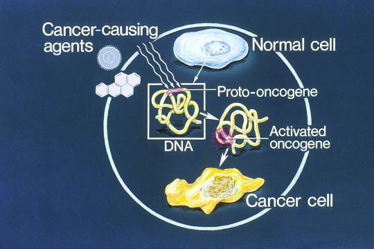
An oncogene is a gene that has the potential to cause cancer. In tumor cells, these genes are often mutated, or expressed at high levels. Most normal cells will undergo a programmed form of rapid cell death (apoptosis) when critical functions are altered and malfunctioning. Activated oncogenes can cause those cells designated for apoptosis to survive and proliferate instead.
#tournament poll#polls#wikipedia#cells of the human body#science tournament#dendritic cell#oncogene#image descriptions in alt text
12 notes
·
View notes
Text
c-Fos exhibits a dual role in memory formation and Alzheimer's disease
Immediate-early genes (IEGs), including c-Fos, are integral to the brain’s response to stimuli. Initially identified as a proto-oncogene, c-Fos is essential for neural activity, synaptic plasticity, and stress responses. While its transient expression supports memory formation in healthy brains, chronic overexpression in AD exacerbates neurotoxicity and cognitive decline. This review synthesizes…

View On WordPress
0 notes
Text
“Multiple Myeloma Oncogene”, Victor McKusick, “Mendelian Inheritance in Man”, 1966. (IRF4) 多發性骨髓瘤。idc10=C90.0
Here I present: “Multiple Myeloma Oncogene”, Victor McKusick, Mendelian Inheritance in Man’, 1966. (IRF4) 多發性骨髓瘤。idc10=C90.0 INTRODUCTION. MUM1 (multiple myeloma oncogene-1)/IRF4 (interferon regulatory factor-4) is a transcription regulatory factor that is activated as a result of t(6;14)(p25;q32) in multiple myeloma. MUM1 expression is seen in various B-cell lymphomas/leukemias and has been…
0 notes
Text
Pathophysiology of Cervical Cancer Every two minutes, somewhere in the world, a woman dies from cervical cancer (GlaxoSmithKline 2007). Caused by persistent or continuous infection by human papillomavirus (HPV), cervical cancer progresses slowly over a number of years (GlaxoSmithKline 2007) and is the second most common cancer for women around the world (Ivansson E.L. et al. 2008) . Due to its slow progression, observable symptomology usually translates into late stage illness, and indeed, within the 1940's invasive cervical cancer was the major cause of death for child bearing women in the United States (National Cancer Institute 2010). As of 2009 the number of cervical cancer diagnoses stood at 11,270 with 4,070 resultant deaths (National Cancer Institute 2010). Prior to HPV being targeted as the main culprit of cervical cancer, mitotic abnormalities and chromosome rearrangements showed a stepwise progression of tumorigenesis (Therman, E. et al. 1983) and later statistical analysis showed that the chromosome abnormalities were nonrandom, implicating viral sites and proto-oncogene locations within the chromosomes (Sreekantaiah, C. et al. 1991). Now it is known that HPV is a necessary step in the acquisition of cervical cancer and is present in 99.7% of all invasive cervical carcinomas (Gius, D. et al. 2007). Up to 80% of women will acquire HPV infection by age 50 (GlaxoSmithKline 2007) and for women between the ages of 20-24 years old, the infection rate is 44.8% (Steben 2007). Over 120 types of HPV exist and over 40 of those infect the epithelial lining of the anogenital tract along with other mucosal areas of the cervix (Steben 2007). Eighteen types of HPV are known to be oncogenic: 16, 18, 45, 31, 33, 52, and 58 with types 16 and 18 accounting for 70% of all cervical cancer cases (GlaxoSmithKline). The virus itself is a non-enveloped double stranded DNA virus (Steben 2007) containing 8,000 base pairs and encoding for two separate protein classes (Ellenson, L.H. & Wu, T.C. 2004). Because most women infected with HPV do not actually develop invasive cervical carcinoma, additional exogenous or genetic factors may be required to keep the infection persistent enough to progress into cancer (Gius, D. et al. 2007). Risk factors include, "Individual susceptibility, immune status, exogenous hormones, tobacco smoking, parity, co-infection with other sexually transmitted agents such as HIV, herpes simplex virus type 2, and Chlamydia trachomatis," (Steben 2007). The histological changes occurring within cervical tissue as a result of HPV infection and other factors are separated into three stages of cervical intraepithelial neoplasias (CINs). Following the invasion of the basement membrane by HPV-containing epithelial cells, lesions become malignant and are thus classified as squamous cell carcinoma (Gius, D. et al. 2007). Proteomic analysis using mass spectroscopy of this squamous cell carcinoma has shown 55 significantly changed protein spots, with 24 upregulated and 31 downregulated proteins, suggesting that using direct tissue samples may be the most persuasive way to find biomarkers and therapeutic targets (Xueqiong, Z. et al. 2009). Preceded by precursor legions, invasive cervical cancer is characterized by disturbances of the "cellular maturation, stratification, and nuclear atypia…" (Boulet, G.A.V. et al. 2008). Using genomic analysis, a model of cervical cancer carcinogenesis as been illuminated across all three CIN stages with early viral infection leading to CIN-1 including the altered expression of genes associated with cellular proliferation and suppression of the immune system which may allow the HPV virus to replicate without detection or destruction (Gius D. et al. 2007). Genetic risk factors for cervical cancer reflect the genetic variation influencing immune response toward HPV infection with a number of genes involved in inflammation and immunity contributing to susceptibility (Ivansson, E.L. et al. 2008). CIN-2 favors the growth of new blood vessels in both the epithelial and stromal cells suggesting communication, while CIN3 has the most pro-invasive genomic signature with changes in cell adhesion proteins and enzymes involved in the extracellular matrix remodeling of both epithelial and stromal cells (Gius, D. et al. 2007). Figure 1. Specific alterations of host tissue during CIN stages 1-3 (Gius, D. et al. 2007) Specific protein classes affected by HPV are the early and late proteins. Early proteins encode for viral DNA replication (E1, E1), RNA transcription (E2), and cell transformation (E5, E6, E7) while late proteins (L1, L2) are the structural components of the viral capsid (Ellenson, L.H. & Wu, T.C. 2004). While most viral DNA replicates extrachromosomally, HPV integrates into the cellular DNA and results in the deletion of large areas of the viral genome including L1 and L2 along with E2 and E5 which leaves E6 and E7 as the primary remaining open reading frames (Zur Hausen, et al. 2002). E2 is a functional elimination as it acts as a viral repressor, while E6 and E7 act as promoters (Yu, T. et al. 2005). Figure 2. Loss of the E2 gene during viral integration (Boulet, G.A.V. et al. 2008) The left over oncogenic proteins, E6 and E7 are implicated in proliferation with E6 expression leading to unregulated cell growth through the blocking of p53, a tumor suppressing protein, and proapoptotic BAK (Ellenson, L.H. & Wu, T.C. 2004). Indeed, HPV types and 16 and 18 have been found to complex with p53, resulting in its ubiquitin-dependent degradation, taking away its control of the cell cycle -- a hallmark of most cancers (Hu, X. et al. 2010). With these checkpoint proteins blocked, E6 activates telomerase and inhibits SRC family kinase degradation, leading to growth stimulation (Ellenson, L.H. & Wu, T.C. 2004). E7 activates genes: cyclin a and E. which stimulate S. phase while simultaneously blocking the cyclin-dependent kinase inhibitors KIP1 and WAF1 breaking down cell growth regulation. It is the co expression of both E6 and E7 that increases transforming activity (Zur Hausen, et al. 2002) as their overexpression contributes to malignant phenotype (Yu, T. et al. 2005). Additionally, alterations such as the activation of ras mutations or the deletion of DCC are required. All of this remodeling is common to viral integration of not only the HPV genome, but the surrounding integration site (Yu, T. et al. 2005). In terms of genetic heritability and susceptibility, studies have investigated TP53, MDM2, NQO1, SNPS, and ICC in conjunction with CIN-3 finding that Caucasians had a significant association between TP53 codon 72 and cervical cancer, with that association stronger in subjects infected with HPV types 16 and 18 pointing to molecular level evidence for the heritability of cancer risk (Hu, et al. 2010). Genetic predisposition had been seen prior in the observation that biological first-degree relatives of women who had been previously diagnosed with cervical cancer had double the risk of contracting it themselves (Ivansson, E.L. et al. 2008). TP53 produces the tumor suppressing protein p53 which regulates DNA repair, cell cycle arrest and apoptosis and has been observed to be blocked by E6, the oncogenic protein of HPV. Ubiquitin degradation of p53 is controlled by both MDM2 and NQO1 with the latter acting as a back up for the former since it does not need ubiquitin and is instead regulated by H. quinone oxidoreductase 1 (Ivansson E.L. et al. 2008). MDM2 traditionally works by binding directly to p53 with its promoter SNP309 T/G rs2279744 increasing its affinity (Ivansson E.L. et al. 2008). Weakening of the immune system by viral integration, in conjunction with a patients internal immunity are also risk factors in the acquisition of cervical cancer. Immune response is inherently regulated by the genes that code for proteins involved with the major histocompatibility complex (MHC), which in turn encodes for leukocytic antigens (HLA) I and II with cervical cancer being linked to the HLA II region (Ivansson, E.L. et al. 2008). When tumors lose the expression of certain MHC class I molecules, as seen in cervical cancers, the reason may be because of an immunoselection by T cells specific for he peptides presented by MHC I. The loss of the MHC I expression invariably results in non-recognition by cytotoxic T cells (Janeway, C.A. et al. 2005). The reality of cervical cancer acquisition points at HPV being a main player; however, while it is a necessary step, it is not the only sufficient risk factor since disease progression is variable among those infected with the virus. Host genetics along with environmental, hormonal, and even nutritional factors may have an influence in creating a situation wherein HPV is allowed to become a continuous infection. To date, the only way to routinely test for cervical cancer is by using HPV as a biomarker through the Pap smear procedure which involves scraping of the transformation zone of the cervix and transferring those cells to a glass slide (Boulet, G.A.V. et al. 2008). Within the past fifty years Pap smears have played a vital role in the decrease of cervical cancer incidence and mortality and its increased use worldwide is helping a number of nations, like Mexico decrease mortality to the disease (Lazcano, P.E. 2008). The test itself, however, is high subjective with a limited sensitivity of 50% along with a number of inherent individual variability (Boulet, G.A.V. et al. 2008). Indeed better screening is necessary due to the number of false-negatives from women with precancerous lesions among the most frequent reasons of medical malpractice in the United States (Steben, M. et al. 2007). In the case of having a tissue sample to be tested, early stage cervical cancer can be differentiated from healthy cervical tissue by gene expression profile due to comparisons done with healthy and lymph node metastatic tissues which found certain genes upregulated and down regulated (Biewenga, P. et al. 2008). Early stage cervical cancers can be cured by radical surgery or radiotherapy with similar effectiveness (Biewenga, P. et al. 2008). Overall the treatment options for cervical cancer are based on whatever the outcome of clinical staging is and include surgery, radiotherapy, chemotherapy, and/or chemoradiotherapy (Ellenson, L.H. & Wu, T.C. 2004). More sophisticated methods such as the use of MR imaging before, during and after radiation therapy is providing accuracy in the main evaluation of prognostic factors and staging (Engin, G. 2006). Currently there is an HPV vaccine for women that have been shown to be 100% effective against HPV types 16 and 18, preventing subsequent development of CIN (Steller, M.A. 2003). This vaccine is the direct result of over two decades of research and ongoing trials to find a male equivalent vaccine are underway utilizing a number of species. Papillomavirus infections, however, are species restricted and do not infect of induce changes in the morphology of animal tissues, thus requiring the use of species specific HPV which is hard to equate to humans due to no cervico-vaginal challenge or natural sexual transmission (Schiller, J.T., & Lowy, D.R. 2006). Because of these issues it is difficult to evaluate potential benefits or efficacy. Ultimately, cervical cancer requires HPV infection but once infection occurs the virus requires other factors such as its host's genetics, immunity, as well as environmental factors such as nutrition, hormonal supplementation like birth control, or cigarette smoking which causes vasoconstriction and overall inflammation. If given the right external factors to its own viral integration into the host's DNA than continuous HPV infection may lead to precancerous lesions and cervical cancer itself. With a number of genetic, molecular, and protein markers being elucidated, tailoring both the treatment and screening processes can bring down overall cervical cancer mortality worldwide. References Biewenga, P., et al. (2008). Gene Expression in Early Stage Cervical Cancer. Gynecologic Oncology, 108, 520-526. Boulet, G.A.V., et al. (2008). Human Papillomavirus in Cervical Cancer Screening: Important Role as Biomarker. Cancer Epidemiology, Biomarkers, and Prevention, 17(4), 810-817. Ellenson, L.H., & Wu, T.C., (2004). Focus on Endometrial and Cervical Cancer. Cancer Cell, 5, 533-538. Engin, G. (2006). Cervical Cancer: MR imaging findings before, during, and after radiation therapy. Urogenital, 16. 313-324. Gius, D. et al. (2007). Profiling microdissected epithelium and stroma to model genomic signatures for cervical carcinogenesis accommodating for covariates. Cancer Research 67, 7113-7123. GlaxoSmithKline. (2007). Cervical Cancer Fact Sheet. www.glaxosmithkline.com Hu, X., et al. (2010). TP53, MDM2, NQO1, and susceptibility to cervical cancer. Cancer Epidemiology, Biomarkers, and Prevention, 19(3), 755-761. Ivansson, E.L., et al. (2008). MHC loci affecting cervical cancer risk: distinguishing the effects of HLA-DQB1 and non-HLA genes TNF, LTA, TAP1, and TAP2. Genes and Immunity 9, 613-623. Janeway, C.A. et al. (2005). Manipulation of the Immune Response. Immunobiology: 6th Edition. 636-637. Lazcano-Ponce, E. et al. (2008). Decreasing Cervical Cancer Mortality in Mexico: Effect of Papanicolaou Coverage, Birthrate, and the Importance of Diagnostic Validity of Cytology. Cancer Epidemiology, Biomarkers, and Prevention, 17(10), 2808-2817. National Cancer Institute (2010). Cancer Advances in Focus: Cervical Cancer, www.cancer.gov Schiller, J.T., & Lowy, D.R. (2006). Prospects for Cervical Cancer Prevention by Human Papillomavirus Vaccination. Cancer Research, 66(21), 10229-10232. Sreekantaiah, C., De Braekeleer, M., & Haas, O., (1991). Cytogenic Findings in Cervical Carcinoma: A Statistical Approach. Cancer Genetics and Cytogenetics, 53, 75-81. Steben, M., & Duarte-Franco, E. (2007). Human papillomavirus infection: Epidemiology and pathophysiology. Gynecologic Oncology, 107(2), S2-S5. Steller, M.A. (2003). Human Papillomavirus, it's genes…and cancer vaccines. Cancer Cell, January, 7-8. Therman, E., Buchler, D.A., Nieminen, U., & Timonen, S. (1983). Mitotic Modifications and Aberrations in Human Cervical Cancer. Cancer Research 30(4), 185-197. Walboomers, J.M.M. et al. (1999). Human papillomavirus is a necessary cause of invasive cervical cancer worldwide. The Journal of Pathology, 189(1), 12-19. Xiaoxia, H. et al. (2010). TP53, MDM2, NQO1, and Susceptibility to Cervical Cancer. Cancer Epidemiology, Biomarkers, and Prevention, 19(3), 755-761. Yu, T., et al. (2005). The Role of Viral Integration in the Development of Cervical Cancer. Cancer Genetics and Cytogenetics, 158, 27-34. Zhu, X., et al. (2009). Proteomic Identification of differentially-expressed proteins in squamous cervical cancer. Gynecologic Oncology, 112, 248-256. Zur Hausen, H. (2002). Papillomaviruses and cancer: From basic study to clinical application. National Review of Cancer, 2, 342-350. https://www.paperdue.com/customer/paper/pathophysiology-of-cervical-cancer-every-12622#:~:text=Logout-,PathophysiologyofCervicalCancerEvery,-Length6pages Read the full article
0 notes
Text
youtube
#Cancer genomics#tumor mutational burden#genomic instability#precision oncology#somatic mutations#germline variants#next-generation sequencing (NGS)#oncogenic drivers#tumor suppressor genes#DNA methylation#transcriptomic profiling#epigenetic alterations#gene expression signatures#liquid biopsy#multi-omics analysis#genomic biomarkers#clonal evolution#chromosomal aberrations#pharmacogenomics#personalized cancer therapy.#Youtube
1 note
·
View note
Text
Perhaps the Journal editors should consult a few AI platforms before they gobble down Gates funded fact checker pie. DeepSeek from China doesn’t seem to have drank the mRNA KoolAid. The country largely avoided the scam.
The very first thing it highlights is Oncogenic potential!
Prompt- What are the hazards associated with injection 50-500B SV40 promoters wrapped in LNPs?
DeepSeek-
Injecting SV40 promoters wrapped in lipid nanoparticles (LNPs) into a biological system, such as the human body, carries several potential hazards and risks. Here are some key concerns:
### 1. **Oncogenic Potential:** - **SV40 Promoter:** The SV40 (Simian Virus 40) promoter is a strong viral promoter often used in molecular biology to drive high levels of gene expression. However, SV40 has been associated with oncogenic (cancer-causing) properties in certain contexts. The promoter itself can potentially integrate into the host genome and disrupt normal gene regulation, leading to uncontrolled cell proliferation and cancer.
### 2. **Immune Response:** - **Immune Activation:** The introduction of foreign genetic material, especially viral promoters like SV40, can trigger an immune response. This could lead to inflammation, autoimmune reactions, or other adverse immune responses. - **LNP Immunogenicity:** Lipid nanoparticles (LNPs) themselves can be immunogenic, potentially causing an inflammatory response or other immune-related side effects.
### 3. **Genomic Integration and Mutagenesis:** - **Insertional Mutagenesis:** If the SV40 promoter integrates into the host genome, it could disrupt essential genes or regulatory elements, leading to mutations that might cause disease, including cancer. - **Off-Target Effects:** The SV40 promoter might drive expression of genes in unintended tissues or cell types, leading to off-target effects and potential toxicity.
### 4. **Toxicity and Cellular Stress:** - **Cellular Stress:** High levels of gene expression driven by the SV40 promoter could overwhelm cellular machinery, leading to cellular stress, apoptosis (programmed cell death), or necrosis. - **LNP Toxicity:** While LNPs are generally considered safe, high doses or repeated administration could lead to toxicity, including liver damage or other organ-specific toxicity.
### 5. **Biodistribution and Off-Target Effects:** - **Biodistribution:** LNPs can distribute to various tissues beyond the intended target, potentially leading to expression of the SV40 promoter in unintended locations, which could have unpredictable effects. - **Off-Target Gene Expression:** The SV40 promoter might drive expression of genes in non-target tissues, leading to unintended biological effects or toxicity.
### 6. **Long-Term Effects:** - **Persistence:** If the SV40 promoter persists in the body, it could lead to long-term expression of genes, which might be undesirable or harmful over time. - **Latent Effects:** There could be latent effects that only become apparent after a long period, such as delayed immune responses or late-onset oncogenesis.
### 7. **Ethical and Regulatory Concerns:** - **Ethical Considerations:** The use of viral promoters in humans raises ethical concerns, especially given the potential for long-term and unpredictable effects. - **Regulatory Hurdles:** Regulatory agencies may have stringent requirements for the use of viral promoters in therapeutic contexts, given the associated risks.
### 8. **Environmental and Horizontal Gene Transfer Risks:** - **Environmental Release:** If the LNPs containing SV40 promoters are released into the environment, there is a risk of horizontal gene transfer to other organisms, potentially leading to unintended ecological consequences.
### Conclusion: The use of SV40 promoters wrapped in LNPs carries significant risks, including potential oncogenicity, immune activation, genomic instability, and off-target effects. These risks must be carefully weighed against the potential benefits, and thorough preclinical and clinical testing would be required to ensure safety. Regulatory oversight and ethical considerations are also critical in the development and application of such technologies.
0 notes
Text
Understanding the Importance of Primary and Secondary Cell Lines in Modern Research: A Look into Medi Ray's Contributions
Cell lines play a pivotal role in advancing biomedical and pharmacological research. By providing consistent, reproducible models for studying various cellular behaviors and responses to different stimuli, cell lines are indispensable in the development of therapeutic treatments, drug discovery, and disease understanding. Among the various types of cell lines, primary and secondary cell lines hold particular significance. In this article, we will explore the differences between primary and secondary cell lines, the role of primary cell culture in research, and how companies like Medi Ray are contributing to the evolution of cell line technology.
What are Primary and Secondary Cell Lines?
At the heart of cell culture technology lies the distinction between primary and secondary (or immortalized) cell lines. Understanding these two types of cell lines is crucial for researchers selecting the right model for their experiments.
Primary Cell Line
A primary cell line and secondary cell line is derived directly from living tissue, often through the enzymatic digestion of tissue samples to isolate individual cells. These cells retain many of the characteristics of their tissue of origin, which makes them valuable for studying normal cellular functions and behaviors. Primary cell cultures typically have a limited lifespan because the cells naturally undergo senescence (aging) after a certain number of divisions.
Despite their finite nature, primary cell cultures are highly prized in research because they offer an accurate representation of the in vivo (natural) cellular environment. Researchers can use primary cell lines to study processes like cellular differentiation, gene expression, and responses to treatments in a more physiologically relevant context than secondary cell lines.
Examples of primary cell cultures include human fibroblasts, hepatocytes (liver cells), and neurons. Each of these types of cells provides insights into how diseases manifest in different organs and tissues, offering a window into various therapeutic approaches.
Secondary (Immortalized) Cell Line
Unlike primary cell lines, secondary cell lines (also known as immortalized cell lines) are derived from primary cell cultures that have been altered to overcome senescence. This is often achieved by introducing certain genetic modifications, such as the activation of oncogenes or the inhibition of tumor suppressor genes, which allow the cells to proliferate indefinitely.
These immortalized cell lines are more commonly used than primary cell lines because of their long-term growth potential, which provides a stable, continuous supply of cells for experimentation. They are particularly useful in drug screening, gene expression studies, and cancer research. Secondary cell lines are easier to maintain than primary cell cultures and can be subcultured (replanted) indefinitely without the need for fresh tissue samples.
Popular examples of secondary cell lines include HeLa cells (derived from cervical cancer), Chinese Hamster Ovary (CHO) cells, and HEK293 cells (human embryonic kidney cells). These cell lines have become essential tools for researchers working in various fields of biotechnology, particularly in the development of therapeutic proteins and vaccines.
The Significance of Primary Cell Culture and Cell Lines in Research
Both primary and secondary cell lines are used for different research purposes, and understanding when to use each is crucial for obtaining accurate results. The choice between primary cell culture and cell line depends on the goals of the experiment and the specific cellular properties that need to be studied.
Primary Cell Culture: A Closer Look
Primary cell cultures are invaluable when a study requires an in vivo-like environment. For instance, when investigating the effects of a drug on a specific tissue, using a primary cell culture derived from that tissue provides a model that closely mimics the behavior of cells within the organism. Primary cell cultures are preferred when researchers are interested in studying cell signaling, protein interactions, or other aspects of cellular metabolism that are specific to a particular tissue type.
Because primary cell lines are more representative of the biological conditions in the human body, they are essential in areas like cancer research, immunology, and regenerative medicine. In cancer research, primary tumor cells allow scientists to explore how cancerous cells behave differently from normal cells and how they respond to various treatments.
Similarly, in drug discovery, primary cell cultures are used to test the efficacy and safety of new medications. Using primary cells from the target tissue (such as liver, lung, or heart) provides a better understanding of how a drug might behave within the body, helping researchers develop more effective and targeted therapies.
Secondary Cell Lines: Powering Biotechnology and Drug Development
While primary cell cultures are essential for many aspects of biomedical research, secondary cell lines are indispensable in large-scale production and biotechnology applications. The ability to grow indefinitely without the limitations of senescence makes immortalized cell lines ideal for the mass production of proteins, vaccines, and antibodies.
For example, secondary cell lines are widely used in the pharmaceutical industry to produce therapeutic proteins. These cell lines are engineered to express proteins of interest, which are then harvested and purified for medical use. This application is crucial in the development of biologic drugs, such as monoclonal antibodies, which are used to treat diseases like cancer and autoimmune disorders.
Furthermore, secondary cell lines are valuable for genetic studies. Researchers can manipulate these cell lines to study the effects of gene expression, mutations, and other genetic alterations. For example, in cancer research, secondary cell lines derived from tumors can be genetically modified to explore how specific mutations contribute to cancer progression and how they can be targeted by new therapies.
Medi Ray's Contribution to Cell Culture Technology
Medi Ray, a leader in the field of biotechnology and life sciences, has been making significant strides in advancing the use of both primary and secondary cell lines in research. The company provides high-quality cell culture products, tools, and services that facilitate the cultivation and maintenance of both primary and secondary cell lines.
Through Medi Ray's cutting-edge technology and expertise, researchers are able to access a broad range of primary cell cultures and secondary cell lines, allowing them to conduct experiments with confidence and accuracy. The company ensures that its cell culture systems are optimized for reproducibility, minimizing variations between experiments and enabling the generation of reliable data.
In addition to offering specialized cell lines, Medi Ray also focuses on providing the necessary tools for maintaining cell cultures, such as culture media, growth factors, and cryopreservation services. These resources make it easier for researchers to establish, grow, and maintain both primary and secondary cell cultures over extended periods of time.
Medi Ray also emphasizes education and support for the scientific community, offering detailed protocols and guidance on how to handle various cell types. By providing not only the cells but also the knowledge necessary for their proper use, Medi Ray empowers researchers to achieve their goals with precision and efficiency.
The Future of Cell Lines in Research
The future of primary cell lines and secondary cell lines in research looks promising. As biotechnology continues to advance, researchers are finding new ways to engineer both types of cell lines to meet the ever-growing demands of science and medicine. Innovations in gene editing technologies, such as CRISPR, allow for the creation of more specific and refined cell models, enabling researchers to study diseases at the genetic level and develop targeted treatments.
Furthermore, secondary cell lines are being engineered to more closely resemble the characteristics of primary cells, overcoming some of the limitations of immortalized cell lines. This convergence of primary and secondary cell lines will provide more versatile and accurate models for studying human diseases and drug responses.
At Medi Ray, the company is committed to staying at the forefront of these advancements, offering researchers access to the latest tools and technologies that will drive the next generation of scientific discoveries.
Conclusion In conclusion, the distinction between primary cell lines and secondary cell lines is fundamental in biomedical research. While primary cell cultures offer a more authentic representation of in vivo conditions, secondary cell lines provide the longevity and reproducibility required for large-scale experiments and drug development. Companies like Medi Ray play a crucial role in advancing cell culture technology, ensuring that researchers have access to high-quality, reliable cell lines and the necessary tools for their work. As science continues to progress, the role of cell lines in understanding human health and developing new therapies will only grow more critical.
0 notes
Text
MicroRNA Market Key Insights and Growth Opportunities, Forecast - 2031

The global microRNA (miRNA) market has gained substantial attention in recent years, driven by its expanding applications in disease diagnosis, therapeutics, and biomedical research. As per the latest insights from SkyQuest Technology, the global microRNA market is projected to reach a value of USD 3.58 billion by 2031, growing at a CAGR of 12.9% from 2024 to 2031. This growth is fueled by advancements in RNA-based research, rising incidences of chronic diseases, and the increasing adoption of miRNA-based diagnostics and therapeutics.
What is Driving the Growth of the MicroRNA Market?
Advancements in Biotechnology The continuous evolution of next-generation sequencing (NGS) and molecular biology technologies has significantly enhanced the detection, profiling, and understanding of miRNA. This progress has opened doors for new therapeutic and diagnostic applications.
Rising Incidence of Chronic and Genetic Diseases The increasing prevalence of chronic diseases such as cancer, cardiovascular diseases, and neurological disorders has led to a growing demand for miRNA-based diagnostics and therapeutics, as these molecules hold the potential to modulate gene expression effectively.
Emerging Role in Precision Medicine MicroRNA is becoming a vital component in precision medicine, offering solutions tailored to an individual’s genetic profile. This trend has driven research funding and investment in miRNA technologies.
Growing Focus on Early Diagnosis Early detection of diseases is crucial for effective treatment, and miRNA-based diagnostic tools are gaining popularity for their accuracy, reliability, and ability to identify diseases in their initial stages.
Request a Sample Report - https://www.skyquestt.com/sample-request/microrna-market
Market Segmentation: A Closer Look at Key Areas
By Application
Cancer Diagnostics & Therapeutics: The largest segment due to the rising prevalence of cancer and miRNA’s role in tumor suppression and oncogene regulation.
Neurological Disorders: Increasing use in diagnosing and treating neurodegenerative diseases like Alzheimer’s and Parkinson’s.
Cardiovascular Diseases: miRNA’s ability to regulate gene expression has made it essential in addressing heart diseases.
Others: Includes applications in metabolic disorders, infectious diseases, and stem cell research.
By Technology
Next-Generation Sequencing (NGS): Widely adopted for profiling and discovering novel miRNA biomarkers.
qRT-PCR: A cost-effective and reliable technology for miRNA quantification.
Microarray: Primarily used for miRNA expression profiling.
By End-User
Research Institutes: Significant demand for miRNA in academic and industrial research.
Pharmaceutical and Biotech Companies: Focused on developing miRNA-based drugs and therapies.
Clinical Diagnostics Laboratories: Increasing use of miRNA as diagnostic markers.
Speak to an Analyst for Customization - https://www.skyquestt.com/speak-with-analyst/microrna-market
Regional Insights: The Global Reach of the MicroRNA Market
North America North America dominates the market, driven by robust R&D funding, advanced healthcare infrastructure, and the growing adoption of miRNA technologies in clinical diagnostics and therapeutics.
Europe Europe represents a significant share of the market due to the rising prevalence of chronic diseases, government support for RNA research, and the strong presence of biotechnology companies in countries like Germany and the UK.
Asia-Pacific Asia-Pacific is the fastest-growing region, with countries like China, Japan, and India witnessing increasing investments in biotechnology and a growing focus on personalized medicine.
Latin America & the Middle East These regions are gradually emerging as potential markets, driven by increasing awareness of miRNA-based applications and improving healthcare infrastructure.
Make a Purchase Inquiry - https://www.skyquestt.com/buy-now/microrna-market
Key Market Players in the MicroRNA Landscape
The microRNA market is highly competitive, with numerous companies investing in R&D to develop innovative diagnostics and therapeutics. Major players include:
Thermo Fisher Scientific Inc.
Qiagen N.V.
Merck KGaA
Illumina, Inc.
Horizon Discovery Group plc
GeneCopoeia, Inc.
NanoString Technologies, Inc.
Abcam plc
New England Biolabs
Rosetta Genomics Ltd.
These companies are driving the market through strategic collaborations, partnerships, and the development of novel miRNA-based products.
Emerging Trends in the MicroRNA Market
Integration of Artificial Intelligence (AI) in miRNA Research AI and machine learning are being employed to analyze complex miRNA datasets, accelerating the discovery of novel biomarkers and therapeutic targets.
miRNA-Based Therapeutics The focus on RNA interference (RNAi) technologies is driving the development of miRNA-based drugs for treating diseases like cancer and genetic disorders.
Liquid Biopsies and miRNA Liquid biopsy technologies, which use miRNA as biomarkers, are revolutionizing non-invasive disease diagnostics, particularly in oncology.
Rising Collaborations Between Academia and Industry Academic institutions and biotech companies are forming partnerships to advance miRNA research and bring innovative products to market.
The Future of the MicroRNA Market
The global microRNA market holds immense potential, driven by its expanding applications across diagnostics and therapeutics. With increasing investments in research and the growing adoption of RNA-based technologies, the market is set to achieve remarkable growth in the coming years.Emerging regions, coupled with advancements in precision medicine and sustainable healthcare practices, will further drive innovation in this sector. Companies that prioritize R&D and focus on developing personalized solutions are well-positioned to lead the market in the future.
#MicroRNA Market#MicroRNA Market Size#MicroRNA Market Share#MicroRNA Market Trends#MicroRNA Market Growth#MicroRNA Market Outlook#MicroRNA Market Key Players#MicroRNA Market Overview#MicroRNA Market Competitor#MicroRNA Market Insights#MicroRNA Market Forecast#MicroRNA Market Analysis#MicroRNA Market Statistics#MicroRNA Market Data#MicroRNA Market PDF#MicroRNA Market Excel#MicroRNA Market Strategy#MicroRNA Market Innovations
0 notes
Text
Exploring the Antisense Therapies Market: Size, Key Players, and Future Forecast to 2034
The Antisense Therapies market is expected to grow significantly by 2034, driven by the development of novel treatments targeting various diseases, especially genetic disorders, cancers, and neurological conditions. This class of therapies works by targeting and modulating the expression of specific genes, offering new hope for conditions that are difficult to treat with traditional methods.
Antisense Therapies Market Size and Forecast
The global market for antisense therapies is experiencing robust growth, with projections suggesting that it will continue to expand significantly by 2034. This growth is largely due to advancements in biotechnology and the increasing adoption of these therapies across a range of diseases. Key drivers include the rising prevalence of genetic disorders, the increasing number of cancer cases, and the growing research into neurological conditions, such as Duchenne muscular dystrophy (DMD) and spinal muscular atrophy (SMA).
By 2034, the market size for antisense therapies is expected to be significantly larger, with increasing investments in research and development, which will drive the availability of new drugs and treatments. The global spread of these therapies, particularly in developed and emerging markets, will also contribute to market growth.
Download sample pages @ https://www.delveinsight.com/sample-request/antisense-therapies-market-forecast
Antisense Therapies Target Population
Antisense therapies target a wide range of diseases. The patient populations for these therapies are diverse, spanning both rare genetic diseases and more common conditions such as various cancers. One of the most notable examples of antisense therapy success is in genetic disorders like SMA and DMD, where the therapies target the root cause of the disease at the genetic level.
In the oncology sector, antisense therapies are being investigated for their ability to modulate cancer-related genes, offering hope for cancers that are resistant to conventional treatments. This includes the targeting of oncogenes like BCL-2 and KRAS. The growing use of antisense oligonucleotides (ASOs) in targeting specific mutations also contributes to the expanding target population.
For neurological conditions, antisense therapies are seeing potential success in managing diseases with limited treatment options. Notably, SMA treatments have already demonstrated efficacy in early clinical trials, setting a precedent for similar approaches in other disorders.
Download sample pages @ https://www.delveinsight.com/sample-request/antisense-therapies-market-forecast
Antisense Therapies Competitive Landscape
The competitive landscape for antisense therapies is dynamic and rapidly evolving. Several pharmaceutical companies and biotech firms are developing antisense drugs, with significant players such as Ionis Pharmaceuticals, Sarepta Therapeutics, and BioMarin leading the way.
Ionis Pharmaceuticals, for example, is at the forefront of antisense oligonucleotide (ASO) therapies, with a strong pipeline of products aimed at treating a variety of genetic conditions, including SMA and familial hypercholesterolemia. Sarepta Therapeutics has made significant progress in antisense therapies targeting genetic diseases like DMD, while BioMarin's efforts are focused on rare genetic disorders. These companies are collaborating with others in the industry, as well as with academic institutions, to advance antisense therapies and expand their reach.
With advancements in gene editing and CRISPR technology, antisense therapies are increasingly being combined with other modalities to improve their efficacy. Companies are also focused on overcoming challenges related to delivery mechanisms, as successfully delivering antisense oligonucleotides to the target tissues remains one of the biggest hurdles in this field.
Antisense Therapies Challenges and Opportunities
While antisense therapies show immense potential, they face challenges related to regulatory approval, delivery methods, and high treatment costs. The complexity of genetic diseases, coupled with the need for precise targeting and efficient delivery, requires significant investment in R&D and advanced technologies. However, there are numerous opportunities for growth, particularly as the understanding of molecular biology advances and more diseases are identified as suitable for treatment with antisense therapies.
The increasing focus on rare and orphan diseases presents a unique opportunity, as these conditions often have limited treatment options, and antisense therapies could provide a breakthrough. Furthermore, ongoing improvements in delivery methods, such as lipid nanoparticles and other nanocarriers, are expected to improve the uptake and effectiveness of antisense oligonucleotides.
Request for a sample report @ https://www.delveinsight.com/sample-request/antisense-therapies-market-forecast
Antisense Therapies Regional Insights
The antisense therapies market is expanding globally, with North America and Europe being the leading markets. However, emerging markets in Asia-Pacific are also expected to see significant growth, driven by increasing healthcare investments, rising awareness of advanced therapies, and growing patient populations. The development of healthcare infrastructure in these regions, coupled with government support for biotechnology innovations, will likely accelerate the uptake of antisense therapies.
In regions like Japan, a strong focus on genetic and rare disease treatments, coupled with advanced healthcare technologies, positions the market to grow rapidly. Meanwhile, in the United States and European Union, market growth is supported by a high level of investment in research and a growing number of orphan drug approvals.
The antisense therapies market is poised for significant growth in the coming years, driven by scientific breakthroughs, expanded indications, and a growing understanding of molecular genetics. Companies and research institutions continue to innovate, with the potential to address unmet medical needs in both rare and common diseases. While challenges remain, the opportunities for market expansion are vast, and the competitive landscape will continue to evolve with advancements in technology and the increasing global demand for advanced therapeutics.
For more detailed insights and projections, you can access the full Antisense Therapies Market report on DelveInsight's website.
0 notes
Text
In January 2024, a preprint was uploaded to the bioRxiv preprint server entitled: “The SV40 virus enhancer functions as a somatic hypermutation targeting element with potential oncogenic activity”.
In a Venice.ai-summarized nutshell, the paper established the following:
The SV40 enhancer has been found to function as a somatic hypermutation-targeting element with potential oncogenic activity. This was discovered through research on the Merkel cell polyomavirus (MCPyV), which causes Merkel cell carcinoma in humans by expressing truncated large tumor antigen (LT) due to APOBEC cytidine deaminase family enzymes induced mutations. Activation-induced cytidine deaminase (AID), a member of the APOBEC family, initiates the antibody diversification process known as somatic hypermutation (SHM). The SV40 enhancer's ability to target AID and induce SHM has implications for the development of cancer and the immune system's response to pathogens.
Kevin McKernan has written an article outlining the link to cancer due to the presence of SV40 (in droves) in the modified RNA COVID-19 injectable products (Pfizer). You can read that here.
Nepetalactone Newsletter
60 Billion Oops
An X follower pointed me to this PrePrint…
Read more
2 days ago · 244 likes · 60 comments · Anandamide
Here is the phrase in the abstract of the preprint that I would like to focus on.
We demonstrate that the SV40 enhancer has strong [somatic hypermutation] (SHM) targeting activity in several cell types.
Now this is no easy subject matter to grasp - even for an immunologist - but I will do my best to describe it so that you all can understand.
One of the silver linings of the COVID scam, is that most of you probably know what antibodies are because of it. For those who do not, antibodies are secreted immunoglobulin molecules derived from the membrane-bound immunoglobulin molecules on a B cell. The process by which these antigen-specific secreted proteins are produced is quite complex. The process starts in the bone marrow, and ends up in the blood (plasma), tissue fluid, tears (mainly IgA), saliva, breast milk (colostrum, specifically) and/or lymph nodes.
0 notes