#plexus axilla
Explore tagged Tumblr posts
Visit Tumblr Blog
Explore Tumblr blogs with no restrictions, modern design and the best experience.
Last Seen Tumblr Blogs


Fun Fact
The “We are the 99%” Tumblr blog became the slogan for the Occupy Wall Street movement.
Text

do you see the vision...
#the binding of isaack#plexus axilla#coincidentalanxitie#homestuck#the binding of isaac#isaack's sign is the Sign of the Penitent Sagimino Prospit Doom#sketch in a bottle
22 notes
·
View notes
Text
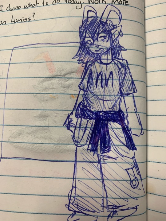


Various Lumi drawings ft his moirail @coincidentalanxitie and his Lusus (Praying Mantis)
8 notes
·
View notes
Text

so Ike sent me this one animation of vampire yuri and i had to draw it as them.
Plexus literally only live-feeds from Lumiss. if its not from her its from a bag, and its probably still some kind of high blood because if she drinks warm hued blood the Symbolism of a highblood literally drinking the blood of the warmer hues will Get To Her. Even though caste doesnt really work like that at all on continua. So she usually just drinks it out of opaque vessels so she isnt seen picking favorites for the religious implications that could have in any direction.
this of course is during early days continua, when the vast majority of the population were either lusii and wrigglers, or carapacians, so thats all yet to happen.
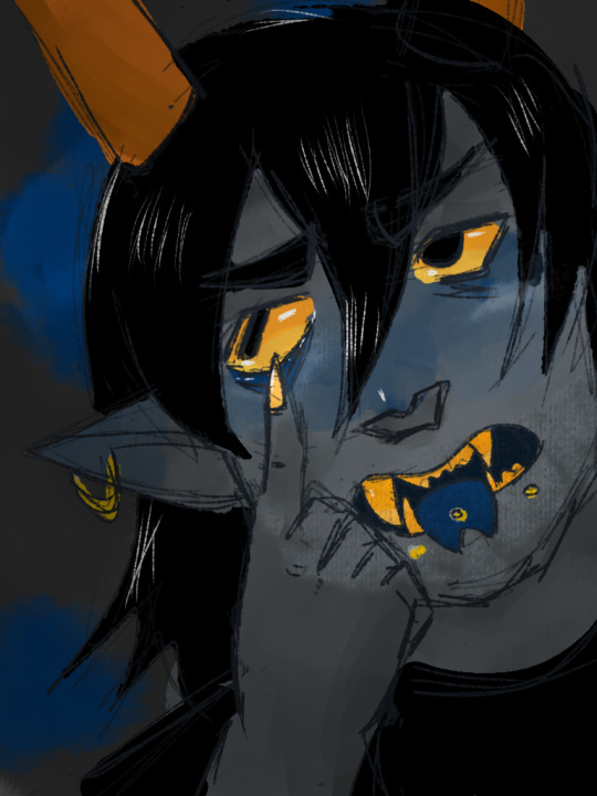
Lumiss from way later in the timeline though, as a treat

shes so pretty
0 notes
Text
November 14, 2024
Read namaz
Exercised
Classes
Physiology Action potential - stimulus, definition, graphs, ionic basis, sodium voltage gated channels, hodgkin's cycle, why does the membrane potential not reach +60 mV due to sodium influx
Anatomy - axilla - axillary artery, axillary vein, brachial plexus, lymphatics
Dissection - axilla
Histology lab - connective tissues
Attended my probably first ever Christmas party
Watched 2 episodes of house MD and learnt about Churg-Strauss syndrome (eosinophilia, granulomatus vasculitis and asthma), mixed connective tissue disease, paraneoplastic syndrome (immune system mistakes neurons for tumor cells and attacks them), tularemia (rabbit fever), melarsoprol (arsenic meds for human african trypanosomiasis)
Watched one episode each of cherry magic and from me to you


2 notes
·
View notes
Text


Brachial Plexus Anatomy! 👇🏾 Important Details Below!👇🏾
The brachial plexus is a network of nerves originating from the C5-T1 spinal nerve roots in the neck and extending into the axilla.
It comprises roots, trunks, divisions, cords, and branches, giving rise to various nerves that innervate the upper limb 💪🏾
Providers need to understand the anatomy of the brachial plexus, especially when treating their patients.
Brachial Plexus Anatomy: ⏩ Roots to Trunks: C5 and C6 roots merge to form the upper trunk, C7 continues as the middle trunk, and C8-T1 combine to form the lower trunk.
⏩ Divisions and Cords: Each trunk splits into anterior and posterior divisions, which recombine to form lateral, posterior, and medial cords.
Branches: ⚡ Musculocutaneous Nerve: Arises from the lateral cord, innervates the muscles in the anterior compartment of the arm and provides sensory fibres to the lateral forearm.
⚡ Median Nerve: Derived from lateral and medial cords, it supplies muscles in the anterior forearm and hand, including the thenar muscles and provides sensation to parts of the hand.
⚡ Ulnar Nerve: Originates from the medial cord, innervates muscles in the forearm and hand and provides sensation to the medial part of the hand.
⚡ Axillary Nerve: Comes from the posterior cord, innervates the deltoid and teres minor muscles and supplies sensation to a small area over the shoulder.
⚡ Radial Nerve: Derived from the posterior cord, it innervates the posterior compartment muscles of the arm and forearm and provides sensory input to the posterior arm and hand.
⏩ Position-Related Injuries during Anesthesia: Improper positioning during surgeries, particularly in the neck or shoulder region, can lead to brachial plexus injuries due to compression or stretching.
⏩ Stretch Injuries - Erb's Palsy: Stretching of upper trunk (C5-C6) during childbirth or trauma can cause Erb's palsy, leading to weakness in shoulder abduction, elbow flexion, and supination.
#anf therapy#anf academy#radial nerve#nerve damage#inflammation#Brachial Plexus#injury#chronic injury#muscle pain#muscle cramps#Axillary Nerve#median nerve#back pain
0 notes
Text
Back Of The Neck Short Question And Answers
0 notes
Photo

THE BRACHIAL PLEXUS ⠀ [NEUROANATOMY] ⠀ The brachial plexus is a network of nerve fibres that supplies the skin and musculature of the upper limb. It begins in the root of the neck, passes through the axilla and runs through the entire upper extremity. ⠀ As you know from my previous posts about Thoracic Outlet Syndrome, compression of the medial, lateral and posterior cords of the brachial plexus can occur between the anterior and middle scalenes, the first rib and clavicle and below the pectoralis minor muscle. ⠀ The brachial plexus originates from five nerve ROOTS: C5, C6, C7, C8, and T1. ⠀ These nerve roots coalesce to form three TRUNKS: Superior, Middle and Inferior. ⠀ The trunks divide to form six DIVISIONS: An anterior and posterior division for each of the three trunks. ⠀ The divisions coalesce to form three CORDS: Medial, lateral and posterior. ⠀ Then the cords form five terminal BRANCHES: median, radial, ulnar, axillary and musculocutaneous. ⠀ Pic 5 is a great illustration which will help you to learn the brachial plexus easily! This piece of artwork by @DrJoeMuscolino is drawn like a tree. The names of the components of the brachial plexus (roots, trunks, branches) are all parts of a tree. Even the term cord can be related to a tree, as in a cord of wood. ⠀ Video 1 explains the components of the brachial plexus. ⠀ Video 2 explains the innervation of the radial nerve which is a terminal branch nerve of the brachial plexus so it is shown as a branch of the tree. Each muscle innervated by the radial nerve is indicated by a leaf with enough letters written on the leaf (Pic 4) to indicate the name of that muscle. ⠀ Video 3 shows some of the “preterminal” branch nerves of the brachial plexus and the muscles they innervate. The brachial plexus has five “terminal” branches and eleven “preterminal” branches. ⠀ Videos 4/5 demonstrate the medial and lateral cords and the three terminal branches to which they give rise (median, ulnar and musculocutaneous). ⠀ #anatomy #fascia #chiropractic #physicaltherapy #dr #physiotherapy #osteopathy #orthopedics #shoulder #nerves #medicine #student #medstudent #education #doctor #neuroanatomy #cadaveranatomy #brachialplexus https://www.instagram.com/p/CL_h17UgPZA/?igshid=i23pdufv4uhs
#anatomy#fascia#chiropractic#physicaltherapy#dr#physiotherapy#osteopathy#orthopedics#shoulder#nerves#medicine#student#medstudent#education#doctor#neuroanatomy#cadaveranatomy#brachialplexus
2 notes
·
View notes
Text


Day 18
8th November , 2019
18/100 days of productivity
Completed some of the topics I found difficult in superior extremity.
So here , I’m doing Axilla and Brachial plexus.
I’m so stressed out for Monday. We just got to know that another topic has been added for the Anatomy practical exam. And I’m not even done with the ones we already have done classes on.
IG: Medstudentstudycorner
#firstyearmedstudent#medschool#studyblr#studyspiration#workhard#studying#study blog#studygram#studyinspo#studywithme#100daysofproductivity#late night studying#studygirl#studygrind#study space#workload#human anatomy#exam week#stressed#medblr
19 notes
·
View notes
Photo

MUSCULOCUTANEOUS NERVE
Origin
continuation of lateral cord of brachial plexus
formed from anterior divisions of superior and middle trunks
Course
it leaves the axilla by piercing coracobrachialis muscle
it then passes down the arm beneath biceps muscle
it ends as the lateral cutaneous nerve of forearm
Sensory supply
skin of lateral forearm
Motor supply
anterior compartment of arm (BBC)
biceps – flexes elbow, supinates forearm
brachialis – flexes elbow
coracobrachialis – flexes and adducts the arm at the glenohumeral joint
CLINICAL FEATURES OF MUSCULOCUTANEOUS NERVE PALSY
SENSORY LOSS
numbness over lateral forearm
MOTOR DEFICIT
paralysis of anterior compartment of arm – very weak elbow flexion and weak forearm supination
absent biceps reflex
DEFORMITY
wasting of anterior compartment of arm
elbow usually held in extension with forearm pronated
A stab wound in the axilla with loss of sensation in lateral forearm indicates injury to musculoskeletal nerve (BBC muscles)
2 notes
·
View notes
Text

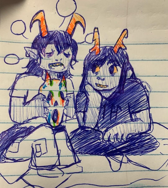
have these tiny thangs too!


wrigglers
8 notes
·
View notes
Text
Saturday Night Palsy Meaning, Definition, Symptoms, Recovery, Treatment
Saturday night palsy is a type of neuropathy that affects the radial nerve. It happens when an object or surface presses directly on the upper arm or axilla for a long time. Roots from the cervical spine (C5) to the first thoracic (T1) spinal nerve (R1) originate in the posterior branch of the brachial plexus and form the radial nerve. It first travels deep to the axillary artery, then descends…

View On WordPress
0 notes
Text
Radia nerve

Surgical procedures such as stabilization of an acute humeral fracture with humeral nailing can also cause radial neuropathies. The severity of the neuropathy depends on the level of the injury. Wrist drop is the most common presentation. Radial neuropathies occur from injury to the radial nerve due to compression, ischemia, fractures to the arm, or penetrating wounds. Muscle overuse may cause compression of the radial nerve anywhere along its path, but most commonly occurs over the elbow as it passes through the radial tunnel. Treatment for radial tunnel syndrome can be conservative or surgical if non-operative therapy fails. Radial Tunnel Syndrome typically occurs secondary to overuse or repetitive movements from pushing, pulling, gripping, pinching, or bending at the wrist typically from a job or playing sports. Less commonly these symptoms can occur at the dorsal aspect of the wrist or hand. Radial Tunnel Syndrome presents with symptoms including fatigue or dull, aching pain at the proximal portion of the forearm during use. The following is a list of the motor and cutaneous sensory functions of the radial nerve. The superficial branch then courses dorsally over the distal radius over the anatomical snuffbox to innervate the posterior lateral three and a half digits (the thumb, index, middle, and lateral half of the ring fingers) and the associated hand area. The superficial branch follows the radial artery inferiorly to the anterolateral portion of the radius, deep to the brachioradialis muscle. The deep branch is a motor branch that passes between the heads of the supinator muscle and becomes the posterior interosseous nerve to innervate the muscles of the posterior compartment of the forearm. Here, the radial nerve separates into the deep and superficial branches. The radial nerve then passes over the lateral epicondyle into the cubital fossa and forearm. After giving these two sensory branches, the radial artery passes through the lateral intermuscular septum to infiltrate the anterior compartment of the forearm between the brachialis and brachioradialis muscles. The posterior cutaneous nerve of the antebrachium also perforates through the lateral head of the triceps but continues to innervate a posterior strip of the forearm. At this point, the radial nerve gives a motor branch to the lateral head of the triceps brachii followed by two sensory branches: the inferior lateral cutaneous nerve of the arm which perforates through the lateral head of the triceps and the posterior cutaneous nerve of the forearm. After passing through the triangular interval, the radial nerve descends the radial groove before laterally wrapping around the humerus. This sensory branch is called the posterior cutaneous nerve of the arm which supplies cutaneous sensory innervation to a portion of the distal posterior arm. These motor branches innervate the medial and long heads of the triceps. It passes with the deep brachial artery and gives two motor branches and one sensory branch before traversing the triangular interval. The radial nerve derives from the posterior cord of the brachial plexus and exits the axilla posteriorly the brachial artery. It courses from the axilla to the posterior compartment of the arm, then into the anterior compartment of the arm, and continues into the posterior compartment of the forearm. The radial nerve is formed as a continuation of the posterior cord of the brachial plexus and arises from the C5-T1 nerve fibers.
0 notes
Text
November 12, 2024
Read namaz
Meditated
Exercised
Classes :
Physiology - resting membrane potential- genesis, factors affecting, Gibbs donnan equation, nernst potential, Goldman-hodgkin-katz equation, contribution of potassium and sodium diffusion potentials and chloride movement.
Anatomy - pectoral region - cutaneous and blood supply, mammary glands, pectoralis major and minor, subclavius, clavipectoral fascia, serratus anterior. Axilla - location, shape, boundaries, contents, brachial plexus, erb's palsy, klumpke's palsy, horner's syndrome
Pectoral region dissection!!!!!
Biochemistry lab - normal urine analysis - urinometer, test for calcium, phosphate, ammonia, urea, uric acid, creatinine
Helped seniors to decorate
Watched fourth episode of house MD and learnt about MRSA - methicillin resistant staphylococcus aureus
2 notes
·
View notes
Text
Radia nerve

#Radia nerve skin#
It gives sensory supply to dorsal aspect of hand, dorsal aspect of thumb, index finger, middle finger and lateral side of ring finger except the nail beds, which are supplied by proper digital branches of median nerve. It crosses brachioradialis to enter posterior of forearm near the back of the wrist and supply dorsum of hand.
The superficial branch of the radial nerve is widely separated from the radial artery in the upper one third of the forearm, closely related to radial artery in the middle third of the forearm, and in the lower third, it descends in the forearm under the tendon of brachioradialis.
In the forearm, it is divided into a superficial branch (primarily sensory) and a deep branch (primarily motor). The radial nerve also gives articular branches to supply the elbow joint. In the radial sulcus, it gives off lower lateral cutaneous nerve of the arm and posterior cutaneous nerve of the forearm.
#Radia nerve skin#
Ībove the radial sulcus, the radial nerve gives off posterior cutaneous nerve of the arm which supplies the skin at the back of the arm. After it emerges out from the radial sulcus, it supplies the brachialis, brachioradialis and extensor carpi radialis longus. Radial nerve gives out muscular branches to supply the long head, medial head, and lateral head of triceps brachii muscles before and during its course in the radial sulcus. Then, it descends down to cross the lateral epicondyle of the humerus where the nerve terminates by branching itself into superficial and deep branch which continues into cubital fossa and then into the forearm. It travel downwards together with profunda brachii artery, between the lateral and medial heads of triceps brachii until it reaches the lateral side the arm at 5 cm below the deltoid tuberosity where it pierces the lateral intermuscular septum to reach the anterior compartment of the arm. In the arm, it runs behind the brachial artery and then enters the lower triangular space to reach the radial sulcus of back of the humerus. From the brachial plexus, it travels behind the third part of the axillary artery (part of the axillary artery distal to the pectoralis minor). The radial nerve originates from the posterior cord of the brachial plexus with root values of C5 to C8 and T1. It goes through the arm, first in the posterior compartment of the arm, and later in the anterior compartment of the arm, and continues in the posterior compartment of the forearm. The radial nerve originates as a terminal branch of the posterior cord of the brachial plexus. Radial nerve of the right axilla, posterior view This nerve was historically referred to as the musculospiral nerve. The radial nerve divides into a deep branch, which becomes the posterior interosseous nerve, and a superficial branch, which goes on to innervate the dorsum (back) of the hand. The radial nerve and its branches provide motor innervation to the dorsal arm muscles (the triceps brachii and the anconeus) and the extrinsic extensors of the wrists and hands it also provides cutaneous sensory innervation to most of the back of the hand, except for the back of the little finger and adjacent half of the ring finger (which are innervated by the ulnar nerve). It originates from the brachial plexus, carrying fibers from the ventral roots of spinal nerves C5, C6, C7, C8 & T1. It innervates the medial and lateral heads of the triceps brachii muscle of the arm, as well as all 12 muscles in the posterior osteofascial compartment of the forearm and the associated joints and overlying skin. The radial nerve is a nerve in the human body that supplies the posterior portion of the upper limb.
0 notes
Photo

THE BRACHIAL PLEXUS ⠀ [NEUROANATOMY] ⠀ @StefanDuell x @DrJoeMuscolino ⠀ The brachial plexus is a network of nerve fibres that supplies the skin and musculature of the upper limb. It begins in the root of the neck, passes through the axilla and runs through the entire upper extremity. ⠀ As you know from my previous posts about Thoracic Outlet Syndrome, compression of the medial, lateral and posterior cords of the brachial plexus can occur between the anterior and middle scalenes, the first rib and clavicle and below the pectoralis minor muscle. ⠀ The brachial plexus originates from five nerve ROOTS: C5, C6, C7, C8, and T1. ⠀ These nerve roots coalesce to form three TRUNKS: Superior, Middle and Inferior. ⠀ The trunks divide to form six DIVISIONS: An anterior and posterior division for each of the three trunks. ⠀ The divisions coalesce to form three CORDS: Medial, lateral and posterior. ⠀ Then the cords form five terminal BRANCHES: median, radial, ulnar, axillary and musculocutaneous. ⠀ Pic 5 is a great illustration which will help you to learn the brachial plexus easily! This piece of artwork by @DrJoeMuscolino is drawn like a tree. The names of the components of the brachial plexus (roots, trunks, branches) are all parts of a tree. Even the term cord can be related to a tree, as in a cord of wood. ⠀ Video 1 explains the components of the brachial plexus. ⠀ Video 2 explains the innervation of the radial nerve which is a terminal branch nerve of the brachial plexus so it is shown as a branch of the tree. Each muscle innervated by the radial nerve is indicated by a leaf with enough letters written on the leaf (Pic 4) to indicate the name of that muscle. ⠀ Video 3 shows some of the “preterminal” branch nerves of the brachial plexus and the muscles they innervate. The brachial plexus has five “terminal” branches and eleven “preterminal” branches. ⠀ Videos 4/5 demonstrate the medial and lateral cords and the three terminal branches to which they give rise (median, ulnar and musculocutaneous). ⠀ #anatomy #fascia #chiropractic #physicaltherapy #dr #physiotherapy #osteopathy #orthopedics #shoulder #nerves #medicine #student #medstudent #education #doctor #neuroanatomy (hier: Frankfurt, Germany) https://www.instagram.com/p/B2gpbyxBHog/?igshid=5xlk2y1wkbn5
#anatomy#fascia#chiropractic#physicaltherapy#dr#physiotherapy#osteopathy#orthopedics#shoulder#nerves#medicine#student#medstudent#education#doctor#neuroanatomy
1 note
·
View note
Text
The median nerve, colloquially known as the "eye of the hand," is one of the three major nerves of the forearm and hand. It courses from the brachial plexus in the axilla to innervate the intrinsic muscles of the hand.


The median artery arises posterior to the pronator teres muscle, whereupon it turns medially to accompany the median nerve along its course in the distal two-thirds of the forearm, where it travels between flexor digitorum superficialis and flexor digitorum profundus.
Arteries (in red) are the blood vessels that deliver blood to the body. Veins (in blue) are the blood vessels that return blood to the heart.
0 notes