
Don't wanna be here? Send us removal request.
Statistics
We looked inside some of the posts by stefanduell and here's what we found interesting.
Average Info
Notes Per Post
26
Likes Per Post
26
Reblog Per Post
0
Reply Per Post
0
Time Between Posts
12 days
Number of Posts By Type
Text
5
Photo
12
Last Seen Tumblr Blogs


Fun Fact
The Tumblr app for Google Glass was released on May 16, 2013.
Text
6 notes
·
View notes
Text
2 notes
·
View notes
Text
instagram
2 notes
·
View notes
Photo

Nice seeing you guys again, this time at @MiamiOpen to discuss new future projects. 💪🤝🙏 Thank you @LarsLienhard from @NeuroAthleticTraining & @Ale_Xander_Will from @goimprovr for the introduction in your newest developments to improve athletes with brain based training! ➡️ Next level training methods! 👀🧠👓👌 📍Miami, Florida,🇺🇸 #TrainTheBrain #BrainBasedTraining #VirtualReality #ImprovingAthletes #NeuroAthleticTraining #Physiotherapy #Osteopathy #Sportsphysiotherapy #AppliedNeuroScience #AppliedMovementNeurology #BrainbasedFitness (hier: Miami, Florida) https://www.instagram.com/p/CqPhBqBARK0/?igshid=NGJjMDIxMWI=
#trainthebrain#brainbasedtraining#virtualreality#improvingathletes#neuroathletictraining#physiotherapy#osteopathy#sportsphysiotherapy#appliedneuroscience#appliedmovementneurology#brainbasedfitness
0 notes
Photo

First @WTA 1000 title at @BNPParibasOpen in #TennisParadise! 🎾🌵🌴🏆 Bravo Elena! 💪🙏 📍Indian Wells, California,🇺🇸 📷 credit: @WTA #Discipline #Focus #Details #TeamWork 🙌 (hier: Indian Wells, California) https://www.instagram.com/p/Cp_25cTuIOf/?igshid=NGJjMDIxMWI=
0 notes
Photo
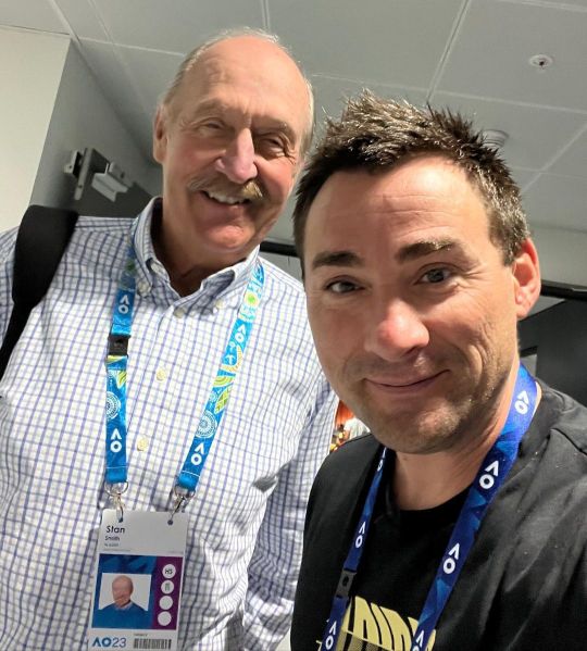
The man behind the 👟 @StanSmithOnline 🙌💪🙏👌 #StanSmith #adidas (hier: Melbourne, Victoria, Australia) https://www.instagram.com/p/CojlCx3A3gW/?igshid=NGJjMDIxMWI=
0 notes
Photo

What are you doing @_Dario_Novak_? 😎 Thank you @goimprovr for the introduction in your newest developments to improve athletes with brain based training! ➡️ Next level training methods! 🧠💪🙏👌 #TrainTheBrain #BrainBasedTraining #VirtualReality #ImprovingAthletes #NeuroAthleticTraining #Physiotherapy #Osteopathy #Sportsphysiotherapy #AppliedNeuroScience #AppliedMovementNeurology #BrainbasedFitness (hier: Germany) https://www.instagram.com/p/CoO1BffN29l/?igshid=NGJjMDIxMWI=
#trainthebrain#brainbasedtraining#virtualreality#improvingathletes#neuroathletictraining#physiotherapy#osteopathy#sportsphysiotherapy#appliedneuroscience#appliedmovementneurology#brainbasedfitness
0 notes
Photo

Bravo Elena! Amazing @AustralianOpen tournament and great start of the year! 💪🙏 📍Rod Laver Arena, Melbourne, Australia #Discipline #Focus #Details #AusOpen #AO2023 (hier: Melbourne, Victoria, Australia) https://www.instagram.com/p/Cn9uUhSP8yg/?igshid=NGJjMDIxMWI=
0 notes
Photo

At @AustralianOpen Player Restaurant… #ThomasTuchel: „I m trying to be a tennis coach from now on!“ ☝️💪🤣⚽️🎾 Thanks for the support coach! 💪💪💪 📍Rod Laver Arena, Melbourne, Australia (hier: Australian Open) https://www.instagram.com/p/Cn4ETrpPvvU/?igshid=NGJjMDIxMWI=
0 notes
Photo
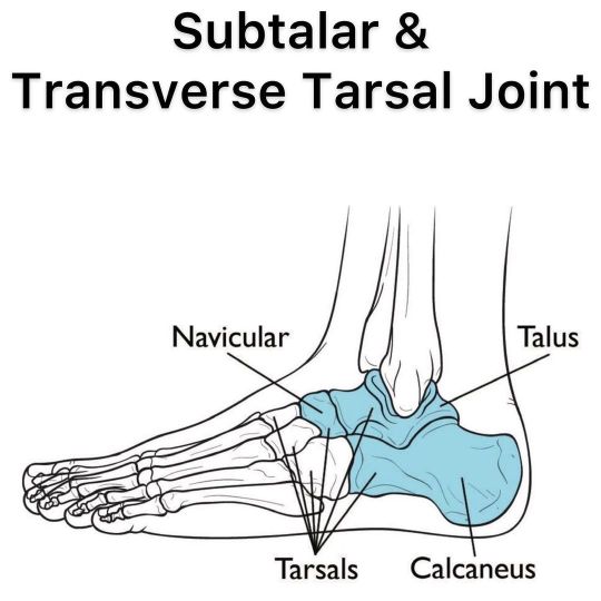
SUBTALAR & TRANSVERSE TARSAL JOINT ⠀ [CADAVER DISSECTION ANATOMY] ⠀ Swipe left! ⠀ Amazing follow up to my previous post about the subtalar joint and the importance to assess it especially after an inversion trauma (ankle sprain injury). ⠀ Video 1 shows an „open book view“ cadaver dissection of the subtalar joint, its surfaces and movements. (For more information about the subtalar joint check my previous posts.) ⠀ Video 2 shows an „open book view“ cadaver dissection of the transverse tarsal joint (also called midtarsal joint or Chopart's joint), which is formed by the articulation of the calcaneus with the cuboid (the calcaneocuboid joint), and the articulation of the talus with the navicular (the talocalcaneonavicular joint). In addition to the subtalar joint, the foot also pronates and supinates in the transverse tarsal joint. ⠀ Thank you @DrJoeMuscolino for providing me always with amazing input which is very important for us health practitioners to get a better understanding of the anatomical structures not only in 2-D (anatomy book) but also in 3-D. ⠀ Check my YouTube channel for more information about treatment and exercises: www.youtube.com/stefanduell ⠀ #physiotherapy #osteopathy #ankle #foot #rehab #dr #anatomy #movement #mobility #education #yoga #pilates #crossfit #medicine #barefoot #fitness #running #walking #exercise #therapy #ironman #sportstherapy #physicaltherapy #manualtherapy #supinationtrauma #inversiontrauma #anklesprain #medicine #doctor #runner #gym https://www.instagram.com/p/CmOcNjJgDFU/?igshid=NGJjMDIxMWI=
#physiotherapy#osteopathy#ankle#foot#rehab#dr#anatomy#movement#mobility#education#yoga#pilates#crossfit#medicine#barefoot#fitness#running#walking#exercise#therapy#ironman#sportstherapy#physicaltherapy#manualtherapy#supinationtrauma#inversiontrauma#anklesprain#doctor#runner#gym
0 notes
Photo
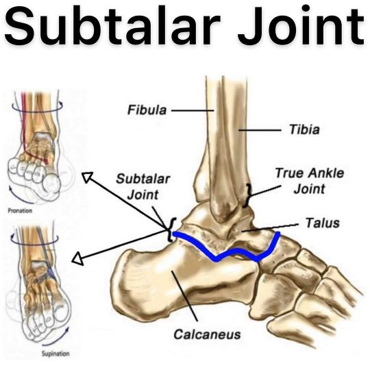
SUBTALAR JOINT ⠀ [ANATOMY, FUNCTION & BIOMECHANICS] ⠀ Regarding my previous post about myofascial treatment of the „INVERSION TRAUMA“ it is very important to mobilise the subtalar joint if it is restricted. If your patient suffers from an inversion trauma, you will most likely find a restriction in the subtalar joint. ⠀ Don’t look at the ankle only, examine the whole body! ⠀ The subtalar joint isn’t discussed as much when it comes to the ankle. More commonly, the talocrural joint is discussed, as it is involved in plantarflexion and dorsiflexion. ⠀ The subtalar joint is where the talus meets the calcaneus (heel bone) at the hindfoot. The subtalar joint is considered a ‘gliding’ joint, as it displays a combination of translational and rotational movement. The joint allows for inversion and eversion of the foot/ankle complex. This motion can be seen in the pictures 7-9. ⠀ Adequate mobility and control at this joint is important both statically and dynamically: ▪️Statically, it is important to be able to find a ‘neutral’ position. This allows for ideal foot and ankle placement, and a stacking effect through the entire lower body. Efficient use of the foot/arch depends on a well controlled and positioned subtalar joint. ▪️Dynamically, this joint gives the ankle more degrees of freedom. It acts as a buffer to allow for rotational/frontal plane movement when walking over uneven terrain for example. ▪️The combination of movement from the talocrural joint and subtalar joint turn the ankle into an important shock absorbing and movement buffering complex (when they are moving well). This helps disperse movement through the lower chain, making it easier on the knee and foot (upstream and downstream). ⠀ After an inversion trauma (ankle sprain injury) it is very important to assess this joint as well and if required to mobilise it as it can force knee problems or even hip or lower back problems through a so-called ascending kinematic chain! ⠀ #phiotherapy #osteopathy #ankle #foot #rehab #anatomy #movement #mobility #education #yoga #pilates #crossfit #medicine #pain #therapy #sportstherapy #physicaltherapy #manualtherapy #supinationtrauma #inversiontrauma #anklesprain https://www.instagram.com/p/Cl-19nPASmB/?igshid=NGJjMDIxMWI=
#phiotherapy#osteopathy#ankle#foot#rehab#anatomy#movement#mobility#education#yoga#pilates#crossfit#medicine#pain#therapy#sportstherapy#physicaltherapy#manualtherapy#supinationtrauma#inversiontrauma#anklesprain
4 notes
·
View notes
Photo
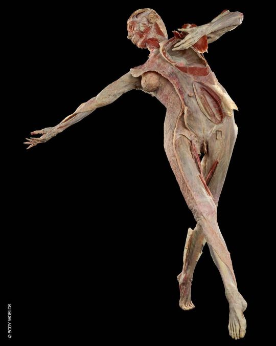
WORLD’S FIRST FULL BODY 3-D HUMAN FASCIA PLASTINATE The field of fascia research has undergone remarkable expansion and growth in the last decade, which is shaping the progression of many related fields. In order to further support fascia anatomy education, the imaging of fascia has also been expanding, but until now a true visualization has been a challenge. Based on this, the teams behind the BODY WORLDS Exhibitions and the Fascia Research Society joined forces to create an interdisciplinary team with the lofty goal to produce a full body plastinate, revealing the entire human fascial net, three-dimensionally. The result of this ambitious three-year project is the successful creation of the world’s first 3-D human fascia plastinate titled FR:EIA (Fascia Revealed: Educating Interconnected Anatomy). FR:EIA will become the new attraction and an integral part of the permanent collection at the BODY WORLDS Museum in Berlin. Congrats to everyone involved on that amazing milestone project! Watch the video: https://youtu.be/vCzX0c_D6kY Photo Credit: ©BODY WORLDS @KoerperWelten, @FasciaPlastination, @FasciaResearchSociety #fasciaresearchsociety #FRS #fasciaresearchcongress #FRC #fasciaplastination #fascia #dissection #human #plastination #plastinarium #bodyworlds #anatomy #guben #germany #fasciaresearch #faszientraining #FasciaInANewLight #UNVEILINGFREIA #pilates #physiotherapy #osteopathy #anatomy #yoga #crossfit #humanbody #cadaver #faszien #faszienyoga #faszientherapie #faszienrolle #koerperwelten (hier: Berlin, Germany) https://www.instagram.com/p/Ck-YyaOg6oD/?igshid=NGJjMDIxMWI=
#fasciaresearchsociety#frs#fasciaresearchcongress#frc#fasciaplastination#fascia#dissection#human#plastination#plastinarium#bodyworlds#anatomy#guben#germany#fasciaresearch#faszientraining#fasciainanewlight#unveilingfreia#pilates#physiotherapy#osteopathy#yoga#crossfit#humanbody#cadaver#faszien#faszienyoga#faszientherapie#faszienrolle#koerperwelten
8 notes
·
View notes
Photo

FUNCTION OF THE LUMBODORSAL FASCIA ⠀ In the pictures the lumbodorsal fascia (LDF) also known as thoracolumbar fascia is beautifully illustrated, showing the interlocking fiber arrangement of the tissue. ⠀ For more information about the anatomy of this important structure check my previous post. ⠀ The LDF is effectively the continued link of the latissimus dorsi fascia with the contralateral glute max fascia (Pic 4-9). This collagen tissue is extremely strong and due to its visco-elastic properties, it contributes to trunk extension at any angle, assuming it has been properly "loaded" or stretched. ⠀ If the fascia is stretched fairly quickly (1/3 sec) it responds with an elastic recoil, amplifying the power of the erector muscles. ⠀ If we stretch slowly, we miss the elastic window for power. This would explain why a failed deadlift always takes longer to complete than a successful one. ⠀ It has been proven that the erector spinae muscles themselves can only handle approximately 50kg of load themselves before failing. Of course the world record deadlift is half a ton, so the only physical explanation is the fascia supplies the power along with intra-abdominal pressure. ⠀ From Gracovetsky, JBMT 2008: The importance of the LDF can also be demonstrated by the well-known ‘‘muscle relaxation’’ phenomenon in which the entire erectors spinae muscle shuts down when the trunk is sufficiently flexed, even while holding significant loads. Relaxation is possible because the tightened fascia takes on the lion share of the forces transmitted from the legs to the upper extremities. It is only when the trunk returning to the erect stance crosses a threshold of about 45 degrees of flexion that the fascia begins to slacken forcing the erectors spinae to contribute to the lift. When this switchover happens, the most difficult part of the lift has already been done and the erectors have enough residual power to take control of the posture in the near erect stance. ⠀ Simply put, the LDF is a hydraulic amplifier for lifting loads where the spine goes from flexion to extension as in deadlifting. One cannot lift the massive loads we see in powerlifting without this tissue. ⠀ #Fascia https://www.instagram.com/p/CjvL8PLgHVv/?igshid=NGJjMDIxMWI=
0 notes
Photo

THORACOLUMBAR FASCIA ⠀ [FUNCTIONAL ANATOMY] ⠀ Although the connective tissue network is continuous and difficult to separate, there is one structure that is directly or indirectly linked to many tissues in the body. ⠀ The thoracolumbar fascia (TLF) is the strong connective tissue bridge that covers the dorsal spine region. ⠀ The TLF (light blue in Pic 2) is a 3D structural composite built out of aponeurotic and fascial planes that unite together to surround the paraspinal muscles and stabilize the lumbosacral spine while having fascial connections superiorly to the cranium and into all extremities. ⠀ To understand interconnected function of the body, one must understand the TLF structure! ⠀ The TLF is composed of 3 layers (Pic 3-6): 1. Deep/Anterior layer: the anterior fascia of the QL and the posterior visceral wall 2. Middle layer: composed of the TVA, oblique and QL muscles which anchor to the transverse processes of the lumbar vertebrae 3. Posterior layer: which has two lamina, superficial, which is a continuation of the Latissimus dorsi and the deep, which is the compartment that houses the paraspinal muscles ⠀ The links from lower extremity superiorly: biceps fem - sacrotuberous ligament - inferior TLF posterior layer - lumbar erectors/deep posterior layer - serratus post inferior - thoracic paraspinals - rhomboids - splenius cervicus - occiput (Pic 7,8). ⠀ The other links are the ipsilateral Latissimus dorsi - contra Glute max sling which is continuous with the posterior superficial lamina (Pic 9,10). ⠀ The obliques and TVA blend and connect with the middle and posterior layers as seen on the left (darker blue fibers in Pic 2). This functionally links the vertical spinal muscles to the transverse abdominal muscles. ⠀ The TLF serves as a functional centerpiece for the trunk and is the common linking tissue between the lower extremity, trunk and upper extremity. ⠀ This allows us to move like a connected, functional system and explains why fascial dysfunction can affect areas far from the local problem. ⠀ Credit: @AnatomyLinks ⠀ #anatomy #fascia #yoga #pilates #physiotherapy #osteopathy #fitness #medicine #yogainspiration #yogalove #yogapractice #fitness https://www.instagram.com/p/Cjp2G22rWvt/?igshid=NGJjMDIxMWI=
#anatomy#fascia#yoga#pilates#physiotherapy#osteopathy#fitness#medicine#yogainspiration#yogalove#yogapractice
0 notes
Photo

POSTERIOR PELVIS LINKS ⠀ [FASCIAL ANATOMY & OSTEOPATHIC LINKS] ⠀ The posterior pelvis is a unique area in that it shows the direct relationships between ligaments and muscles in function. ⠀ Fascia connects everything in the body. Specifically, the collagen tubules that make up one structure are often the same tubules that make up another. ⠀ Even though man has given names to "separate" or organize ligaments from muscles, they are virtually the same continuous tissues on a micro level. ⠀ In the first picture, the relationships are indicated by numbers (blue colored): 1 Biceps Femoris links to the 2 Sacrotuberous Ligament (Pic 1/2/3/4) 3 Gluteus Maximus links to the 1 Inferior Dorsal Sacral Ligaments - Bichat‘s ligament (Pic 1/3/4/5) 5 Latissimus Dorsi links to the 6 Superior Dorsal Sacral Ligaments - Iliotransversosacral (Pic 1/4/6) 7 Quadratus Lumborum links to the 8 Iliolumbar Ligaments (Pic 1/7/8) 9 Sacrospinous Ligament links to the 10 Piriformis (Pic 9/10) ⠀ The sharing of fiber directions is what establishes these "fiber pairs". These relationships are responsible for creating tensional stability in the pelvis and sacrum and for linking limbs to the trunk. ⠀ The black arrows in Pic 1 represent the lines of force that are created and balanced by the tissues. ⠀ It's all connected! 🌐 ⠀ Picture credit: @AnatomyLinks ⠀ #anatomy #anatomie #fascia #yoga #pilates #doc #biotensegrity #biomechanics #functionalanatomy #pelvis #glutes #ligaments #chiropractic #doctor #physicaltherapy #physiotherapy #osteopathy #osteopatia #acupuncture #dryneedling #medicine #orthopedics #rolfing #anatomytrains #sacroiliac #joints #muscles #bodywork #medstudent #doctor https://www.instagram.com/p/Cjj5rZjtwIm/?igshid=NGJjMDIxMWI=
#anatomy#anatomie#fascia#yoga#pilates#doc#biotensegrity#biomechanics#functionalanatomy#pelvis#glutes#ligaments#chiropractic#doctor#physicaltherapy#physiotherapy#osteopathy#osteopatia#acupuncture#dryneedling#medicine#orthopedics#rolfing#anatomytrains#sacroiliac#joints#muscles#bodywork#medstudent
4 notes
·
View notes