#xiphoid
Explore tagged Tumblr posts
Visit Tumblr Blog
Explore Tumblr blogs with no restrictions, modern design and the best experience.
Last Seen Tumblr Blogs


Fun Fact
The KCSC sent more than 20K requests to delete posts related to prostitution and porn to Tumblr from January to June 2017.
Text

Oh good, I still remember how to draw the wacky tentacle robot :]

#yippeeeee 💙#my ocs#my art#doodles#xiphoid#my robots ♡#my favorites the extra shiny nakey one in the bottom right :3
4 notes
·
View notes
Text
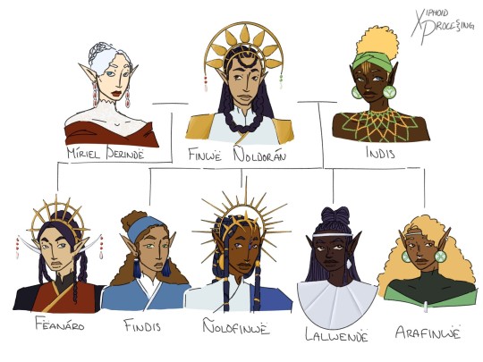
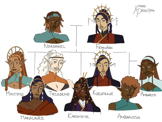

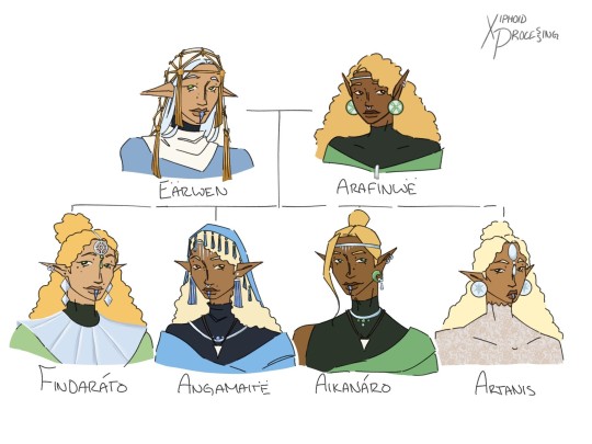
Noldorin family trees again bc its been like. 2 years and ive changed everything
design notes below readmore
-originally wanted finwe to be very blue bc of the color coding w miriel and indis, and then their children ie: Fëanor wearing red bc he's Míriel's son, Fingolfin wearing blue bc he's Finwë's son, and Finarfin wearing green bc he's Indis' son. scrapped it bc he looks good in white and gold but the idea stuck around with the silver n gold
-little gemstones on Finwë's headdress are red and green for his wives. similarly Fëanor's are only red for his mother
-Finarfin wears a more vanyarin style compared to his siblings, aside from Lalwen but her style is also like. noldorinized
-the different clans of elves have different resting positions for their ears! the vanyar have theirs almost straight up, the noldor have the classic kinda-up, and the teleri have theirs parallel to the ground
-thr large headdresses Finwë, Fëanor, Fingolfin, Maedhros, Curufin, and Fingon wear are like noldorin royalty stuff for eldest sons/heirs. While i do think Maedhros is Fëanor's heir i do think some favoritism let Curufin wear one too. i dont think i need to explain why Curufin's looks so similar to Fëanor's
-fingolfins headdress is meant to resemble the sun bc he's the first High King under the Sun and Moon
-the nose ring that some of them have is a Vanyarin style indicating that they are married
-Maedhros and Fingon have similar headdreses bc i think it would have pissed tf out of their fathers. also i think its funny
-Nerdanel and Anairë are noldor, but to me they are from noldorin minorities which have slightly differing cultures frm the majority which is why their clothing is slightly different
-that boob window thing Nerdanel, Maedhros, and Celegorm have are specific and iconic to the Aulendil
-Maglor is channeling Míriel’s clothing style here, which although might come off as a bit feminine is not. i dont think elves would be very strict abt that kinda stuff
-Míriel and Celegorm have albinism
-the Ambarussa aren't quite identical and i think they have very dif personalities and styles. to me. Amras (short hair) is more mainstream noldorin while Amrod is more of their mothers style
-while he and his siblings r very noldorin in style, Argon is channeling his mothers style more than the majority
-the mark on Eärwen and her children's lower lips are a coming of age kinda thing. dont ask me how they get those vibrant colors bc i dont know
-the gold on Eärwen’s headscarf is meant to resemble fishing nets
-each of Finarfin and Eärwen’s children channel a dif. part of their heritage in their clothing--Finrod is a noldorinized vanyarin style, Angrod and Aegnor are different kinds of Telerin, and Galadriel is Noldorin
-the like. shark tooth necklaces that Angrod and Aegnor have is a symbol of being an accomplished fisherman
-everyone born in Valinor has light in the center of their eyes which correspond to the light of the trees they were born under. this doesnt really matter and you cant really see it but its important to me that you know
-on that note, Finarfin has both lights in his eyes bc he was born during the mingling. bc i can do whatever i want
#Indis and Finarfin's clothes are based on Xhosa and Ndebele styles so if i got anything wrong PLEASE tell me#also idk why i wrote all their names in quenya but i was too lazy to go back and change it so. rip.#tolkien#xiphoids art#silmarillion#the silmarillion#only tagging the elders n heads of house bc thats way too many names otherwise#miriel therinde#miriel#finwe#indis#feanor#fëanor#fingolfin#finarfin#nerdanel#anaire#anairë#Eärwen#earwen#maedhros#fingon#finrod#i spent a lot of time on the details please please look at them. if tunglr didnt fuck my shit up
260 notes
·
View notes
Text
Crafting update: new projects are still delayed, I'm doing better and have mostly recovered from the allergic reaction, but I have messed up my ribs in new and exciting ways. I'm okay, the electrolyte issues just mean I'm prone to pretty strong muscle cramps and spasms, and sometimes that combines badly with existing structures like ribs lol
#the person behind the yarn#I am prone to costochondritis (inflammation of the cartilage in the rib cage) but this is the first time#I've had my xiphoid process affected??? it's less bad today than it was yesterday#but it was ridiculously inflamed yesterday and it turns out a lot of muscles connect around there#also very funny (perhaps only to me): my mom was over yesterday and I was like hey weird favor#can you feel my sternum and tell me if it really is inflamed or if its all in my head?#and she was like I think it's okay and I had to go mom that's the knot in the muscle under my sternum#and moved her hand to my sternum and she was like OH MY GOSH#and then like. visibly realized that was perhaps not a helpful reaction. paused. recalibrated.#and said yes I think it is very swollen and you should probably but some ice on it (in a much calmer voice) lol#so I am very sore and moving my arms too much makes it worse which means I am crocheting a bit#but I keep getting arm cramps from the electrolyte issues so I have to stop. so no craft updates#just lots of rest and electrolytes and water in my near future lol
17 notes
·
View notes
Text
i love human anatomy class because all our papers look like the teacher is just really opinionated

5 notes
·
View notes
Text
youtube
The Xiphoid Process performing live in Denver, Colorado. Full set. Video courtesy of Denver Heavy Metal Society.
2 notes
·
View notes
Note
favorite word?
off the top of my head, xiphoid. If I think of more I’ll add them…..
#like the xiphoid process :] which means sword shaped apparently!#sorry I don’t have too many! I don’t think abt it much
2 notes
·
View notes
Text
If you've ever seen this book recommended
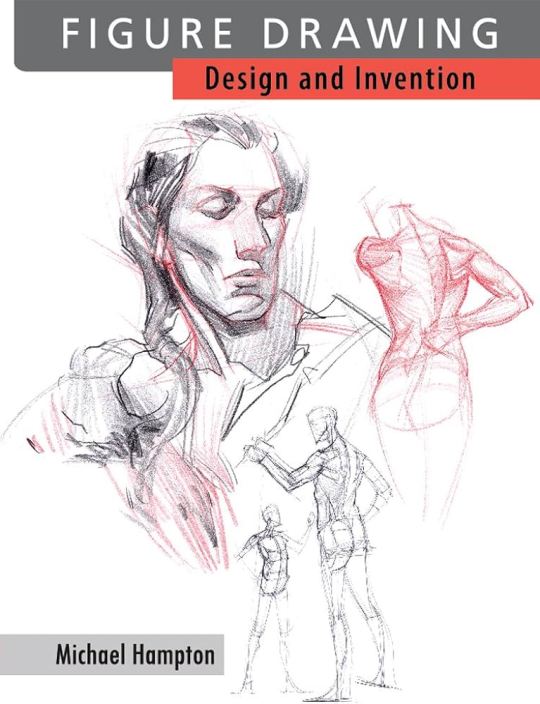
you'll be glad to know that the artist has a youtube channel where he goes through different anatomy concepts!
I also highly recommend the Fat Stacks series by Jesse Thompson; he's also an actual art professor and his series reviews different art books, which can be great for breaking down these texts if you're overwhelmed by the (very useful) info they give!
Can't afford art school?
After seeing post like this 👇

And this gem 👇
As well as countless of others from the AI generator community. Just talking about how "inaccessible art" is, I decided why not show how wrong these guys are while also helping anyone who actually wants to learn.
Here is the first one ART TEACHERS! There are plenty online and in places like youtube.
📺Here is my list:
Proko (Free)
Marc Brunet (Free but he does have other classes for a cheap price. Use to work for Blizzard)
Aaron Rutten (free)
BoroCG (free)
Jesse J. Jones (free, talks about animating)
Jesus Conde (free)
Mohammed Agbadi (free, he gives some advice in some videos and talks about art)
Ross Draws (free, he does have other classes for a good price)
SamDoesArts (free, gives good advice and critiques)
Drawfee Show (free, they do give some good advice and great inspiration)
The Art of Aaron Blaise ( useful tips for digital art and animation. Was an animator for Disney)
Bobby Chiu ( useful tips and interviews with artist who are in the industry or making a living as artist)
Second part BOOKS, I have collected some books that have helped me and might help others.
📚Here is my list:
The "how to draw manga" series produced by Graphic-sha. These are for manga artist but they give great advice and information.
"Creating characters with personality" by Tom Bancroft. A great book that can help not just people who draw cartoons but also realistic ones. As it helps you with facial ques and how to make a character interesting.
"Albinus on anatomy" by Robert Beverly Hale and Terence Coyle. Great book to help someone learn basic anatomy.
"Artistic Anatomy" by Dr. Paul Richer and Robert Beverly Hale. A good book if you want to go further in-depth with anatomy.
"Directing the story" by Francis Glebas. A good book if you want to Story board or make comics.
"Animal Anatomy for Artists" by Eliot Goldfinger. A good book for if you want to draw animals or creatures.
"Constructive Anatomy: with almost 500 illustrations" by George B. Bridgman. A great book to help you block out shadows in your figures and see them in a more 3 diamantine way.
"Dynamic Anatomy: Revised and expand" by Burne Hogarth. A book that shows how to block out shapes and easily understand what you are looking out. When it comes to human subjects.
"An Atlas of animal anatomy for artist" by W. Ellenberger and H. Dittrich and H. Baum. This is another good one for people who want to draw animals or creatures.
Etherington Brothers, they make books and have a free blog with art tips.
As for Supplies, I recommend starting out cheap, buying Pencils and art paper at dollar tree or 5 below. For digital art, I recommend not starting with a screen art drawing tablet as they are more expensive.
For the Best art Tablet I recommend either Xp-pen, Bamboo or Huion. Some can range from about 40$ to the thousands.
💻As for art programs here is a list of Free to pay.
Clip Studio paint ( you can choose to pay once or sub and get updates)
Procreate ( pay once for $9.99)
Blender (for 3D modules/sculpting, ect Free)
PaintTool SAI (pay but has a 31 day free trail)
Krita (Free)
mypaint (free)
FireAlpaca (free)
Libresprite (free, for pixel art)
Those are the ones I can recall.
So do with this information as you will but as you can tell there are ways to learn how to become an artist, without breaking the bank. The only thing that might be stopping YOU from using any of these things, is YOU.
I have made time to learn to draw and many artist have too. Either in-between working two jobs or taking care of your family and a job or regular school and chores. YOU just have to take the time or use some time management, it really doesn't take long to practice for like an hour or less. YOU also don't have to do it every day, just once or three times a week is fine.
Hope this was helpful and have a great day.
#reference#taking an anatomy class rn and other texts my prof recced are michael hampton and mattesis force#and also Bammes anatomy + stephen rogers Peck#bridgeman was also recced#really interesting to take the class because the prof had criticisms for all of them and theyre each individually useful for diff purposes#pecks one he said was good for looking at the musculature names but the super rendered vers arnt that useful it more the ones that denote#movemen & functio that were useful. bammes is extremely structural and so is good for like learning th planar rs of the body & construction#force is good for muscle breakdowns also and how the body crunches and stretches#bridgeman is very good for simplifyig stuff but prof pointed out how he shortenes the ribcage such that if you follow him you need to draw#the xiphoid process on lower third rather than centre of it#so all the illsutrators having their own ways n mistakes n benefits makes it better to like study as a collective rather thsn take any of#them as pure gospel#i rlly love this class so much#i love art classes i love art sch i wish i could continue going to these classes forever until i die#** by force i meant michael hampton is good for muscle breakdown oops#force i havent read yet maybe its also good for that
105K notes
·
View notes
Text
added an exercise at the advice of my massage therapist that's supposed to help strengthen the muscles keeping the ribs from flaring out. and you know it wasn't weighted or anything, I didn't think I was doing that many reps, but oh my GOD I'm unbelievably sore. right around that darn xiphoid 🗡️🩻
#xiphoid is Greek for sword-like and hoo buddy do i feel like I'm being stabbed rn. lol#🩻 on android is the ribcage so i cant wait to see how that translates on other systems#gotta check emojipedia....
0 notes
Text

cheering myself up...i also edited all the posts with the snippets from my comic and added all the characters' names :) so you can look up "missed connection" on my blog
#arter#neo sarajevo#missed connection#zina frenzy#luis n. fabula#shirin alkonost#xi xiphoid#paloma metalloloma#varvara#kdp
54 notes
·
View notes
Text
Hello Thoracic Ribcage, we meet again
Why are you so complicated to draw???
You too, Spine Lumbar!
Fucking why sooo fucking hard to draw ahhhhhhhhhh.
#mystuff#rage#do I need to know the bones while drawing? the inner me that supports my fleshy body#No they are hidden inside inssssssssssssssssssssssside we do not need to see inside the body#fuck skeletons man it's not even holloween#the fuck is a xiphoid process??
0 notes
Text
youtube
Denver death/doom/stoner metal band The Xiphoid Process live at 3 Kings Tavern in Denver, CO. Video courtesy of Denver Heavy Metal Society.
#The Xiphoid Process Denver#death metal#doom metal#stoner metal#Denver Heavy Metal Society#3 Kings Tavern#Colorado metal#Denver metal#Youtube
1 note
·
View note
Text
Med-student!Satoru who comes home late after spending the entire day cramming for his upcoming exam.
Don't get me wrong; he is insanely smart, and he doesn't need a lot of time to progress and understand the given material. He does, however, want to get to the bottom of things, understanding them beyond whatever the professor had taught them.
He took pleasure into understanding and getting down to a t about the different concepts. It's no surprise that he loves the complexity of neurology, neoplasia and the immune system.
However, something as simple as anatomy has had his heart ever since the beginning of his degree. Especially because it was something he could share with you.
—
"Nd this," he had whispered out, index finger softly pushing down on the little slope that was right between your collarbones, "this is what we call the manubrium. It's the first part of your sternum."
Satoru had learnt this in his first year, remembering the very few classes he had gotten about anatomy in the first quarter. How he had practiced on Suguru's chest to find the manubriosternal joint.
Now, he was trying to find it on you.
His finger trailed a little more downwards, just above the cleavage of your breasts, "then there is a thin line in between the first part, the manubrium, and the middle part, which we call the corpus of the sternum."
This wasn't the first time Satoru had laid in your sheets, hand resting in the palm of his hand, which he held up by leaning on his elbow in bed, half his body turned to you. You had loved it from the very first time he had started doing it. It felt intimate, and yet so meaningful.
Satoru chose your body to describe something he had an interest in, something he wanted to pursue a career in. He explained it in simple terms, making sure you could always follow along and understand what he was saying or illustrating.
"The thin line is known as the manubriosternal joint, an identification mark for doctors to find the second costa, which is latin for rib, as it is immediately attached to the manubriosternal joint." You could feel how his finger would move a little more tot the right, in search of your second rib. Once he could feel the bone underneath his fingertip, he smiled softly before going back to the very middle, trailing downwards in between your breasts.
When it fell right underneath your costal arch, you felt your breath hitch in your throat, eying his face, only to find his eyes completely focused on his fingertip, "The xiphoid process is the last part of your sternum, divided from the corpus by the xiphisternal joint."
You knew exactly what would happen next, already opening your arms widely to let him settle his head on top of your chest, right on the apex of your heart.
"To listen to your heartbeat," he had admitted once, after a very long day at his univeristy.
So, without keeping him up any longer to make sure he'd be rested for tomorrow, you had placed your lips against his temple, murmuring his favourite sentence against his soft skin, "I love you, 'toru."
#gojo x reader#gojo x you#gojo fluff#satoru gojo x you#jjk x reader#gojo satoru#jjk x you#gojo satoru fluff#gojo satoru x reader#jjk gojo#jujutsu gojo#jjk fluff#gojo jjk#this is self-indulgent but I hope you are able to enjoy it anyway <3
1K notes
·
View notes
Text
📲 YOU JUST GOT A MESSAGE FROM HNUH TRAUMA CENTRE!
while being a doctor in cardiology, the new arrogant but brilliant trauma doctor asked you and Yang Jae Won to be his fellow.
or in which— this is your day after entering the chaotic trauma center

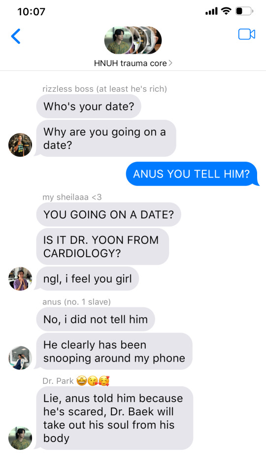


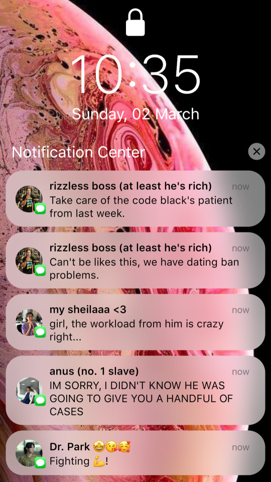
🚑 HNUH TRAUMA CENTRE 🏨
The antiseptic smell hits you as soon as you walk outside your room, The night shift really made you look like hell because you can only get 2 hours of sleep and last night you walk like a zombie to your room.
"Doctor Ji, you wake up already? it's only been 2 hours, and your patient is stable right now," one of the nurses said when she saw you walk through the trauma center door.
You gave her a light smile, "Emergency usually calls on this hour, need to stand by because the soon we get them to the operation, their life percentage will also be bigger."
She nodded at your words, and not even 5 minutes later, a patient surged through the hall with a stab wound, and he's unconscious.
You ran as fast as you could to the TICU, and there you saw Jae Won with his EKG, checking the heart of the patient.
"He's having a cardiac tamponade. Let's do puncture first! Call on the cardiologist, tell them we have a cardiac tamponade patient!" You said, and the nurse gave you a syringe to take the blood from the heart.
Xiphoid Process, 45 degrees to the left.
"Doctor! They can't schedule an operation. They said it's already full," You can visibly see the desperate on Jaewon's face, "Tell them it's cardiac tamponade patient"
But the nurse have already asked that before, "They have a heart rupture patient," She said with worried attached on her face.
You sigh, "Well then, call Doctor Baek." The nurse acknowledged your words with a nod.
All of you tried your best not to disturb Doctor Baek kang hyuk because he's on a meeting with the whole department on hospital.
"BP is getting too low! It's 40!" That words make Kang hyuk take the stairs, after running with his whole strength out of the meeting room.
He instructed the nurse to call an anesthesiologist— Park Gyeong won, of course, and prepare the operation room.
[⋆✴︎˚。⋆]
"You look like a zombie," Gyeong won said after you get out of the operating room.
You let a light chuckles, "Expect that when you only have 2 hours of sleep," you answer while washing both of your hands.
"Doctor Ji, why are you still doing here? i thought i told you to check the code black patient from last week?" You sigh at yourself, wanting to punch the man beside you until he passes out, but instead, you just smile at him and answer him with a quiet 'yes'.
Jae won only looked at your back with pity, "Don't you think it's too much? it's only a date"
Kang hyuk ignore him and just give both of them a sly smirk.


#baek kang hyuk x reader#the trauma code: heroes on call#baek kang hyuk#cheon jang mi#yang jae won#park gyeong won#the trauma code : hoc#jiu writes ���
95 notes
·
View notes
Text
Writing Notes: Autopsy
Autopsy - dissection and examination of a dead body and its organs and structures.
The word autopsy is derived from the Greek autopsia, meaning “the act of seeing for oneself.”
Also known as: necropsy, postmortem, postmortem examination
Why is an autopsy done?
To determine the cause of death
When a suspicious or unexpected death occurs
To observe the effects of disease; when there's a public health concern, such as an outbreak with an undetermined cause
To establish the evolution and mechanisms of disease processes
When no doctor knows the deceased well enough to state a cause of death and to sign the death certificate
When the doctor, the family or legally responsible designee of the deceased person requests an autopsy
Who does the autopsy?
Autopsies ordered by the state can be done by a county coroner, who is not necessarily a doctor
A medical examiner who does an autopsy is a doctor, usually a pathologist
Clinical autopsies are always done by a pathologist
How is an autopsy done?
After the patient is pronounced dead by a physician, the body is wrapped in a sheet or shroud and transported to the morgue, where it is held in a refrigeration unit until the autopsy.
Autopsies are rarely performed at night.
Autopsy practice was largely developed in Germany, and an autopsy assistant is traditionally honored with the title "diener", which is German for "helper".
The prosector and diener wear fairly simple protective equipment, including scrub suits, gowns, gloves (typically two pair), shoe covers, and clear plastic face shields.
The body is identified and lawful consent obtained.
The procedure is done with respect and seriousness.
The prevailing mood in the autopsy room is curiosity, scientific interest, and pleasure at being able to find the truth and share it.
Most pathologists choose their specialty, at least in part, because they like finding the real answers.
Many autopsy services have a sign, "This is the place where death rejoices to help those who live." Usually it is written in Latin ("Hic locus est ubi mors gaudet succurrere vitae").
EXTERNAL EXAMINATION
The prosector checks to make sure that the body is that of the patient named on the permit by checking the toe tag or patient wristband ID.
The body is placed on the autopsy table.
Experienced dieners, even those of slight build, can transfer even obese bodies from the carriage to the table without assistance.
Since the comfort of the patient is no longer a consideration, this transfer is accomplished with what appears to the uninitiated a rather brutal combination of pulls and shoves, not unlike the way a thug might manhandle a mugging victim.
The body is measured.
Large facilities may have total-body scales, so that a weight can be obtained.
The autopsy table is a waist-high aluminum fixture that is plumbed for running water and has several faucets and spigots to facilitate washing away all the blood that is released during the procedure.
Older hospitals may still have porcelain or even marble tables.
The autopsy table is basically a slanted tray (for drainage) with raised edges (to keep blood and fluids from flowing onto the floor).
After the body is positioned, the diener places a "body block" under the patient's back. This rubber or plastic brick-like appliance causes the chest to protrude outward and the arms and neck to fall back, thus allowing the maximum exposure of the trunk for the incisions.
Abnormalities of the external body surfaces are then noted and described, either by talking into a voice recorder or making notes on a diagram and/or checklist.
OPENING THE TRUNK
The diener takes a large scalpel and makes the incision in the trunk. This is a Y-shaped incision. The arms of the Y extend from the front of each shoulder to the bottom end of the breast bone (called the xiphoid process of the sternum). In women, these incisions are diverted beneath the breasts, so the "Y" has curved, rather than straight, arms. The tail of the Y extends from the xiphoid process to the pubic bone and typically makes a slight deviation to avoid the umbilicus (navel). The incision is very deep, extending to the rib cage on the chest, and completely through the abdominal wall below that.
With the Y incision made, the next task is to peel the skin, muscle, and soft tissues off the chest wall. This is done with a scalpel. When complete, the chest flap is pulled upward over the patient's face, and the front of the rib cage and the strap muscles of the front of the neck lie exposed. Human muscle smells not unlike raw lamb meat in my opinion. At this point of the autopsy, the smells are otherwise very faint.
An electric saw or bone cutter (which looks a lot like curved pruning shears) is used to open the rib cage. One cut is made up each side of the front of the rib cage, so that the chest plate, consisting of the sternum and the ribs which connect to it, are no longer attached to the rest of the skeleton. The chest plate is pulled back and peeled off with a little help of the scalpel, which is used to dissect the adherent soft tissues stuck to the back of the chest plate. After the chest plate has been removed, the organs of the chest (heart and lungs) are exposed (the heart is actually covered by the pericardial sac).
Before disturbing the organs further, the prosector cuts open the pericardial sac, then the pulmonary artery where it exits the heart. He sticks his finger into the hole in the pulmonary artery and feels around for any thromboembolus (a blood clot which has dislodged from a vein elsewhere in the body, traveled through the heart to the pulmonary artery, lodged there, and caused sudden death. This is a common cause of death in hospitalized patients).
The abdomen is further opened by dissecting the abdominal muscle away from the bottom of the rib cage and diaphragm. The flaps of abdominal wall fall off to either side, and the abdominal organs are now exposed.
REMOVING THE ORGANS OF THE TRUNK
The most typical method of organ removal is called the "Rokitansky method." This is not unlike field dressing a deer. The dissection begins at the neck and proceeds downward, so that eventually all the organs of the trunk are removed from the body in one bloc.
The first thing the diener does is to identify the carotid and subclavian arteries in the neck and upper chest. He ties a long string to each and then cuts them off, so that the ties are left in the body. This allows the mortician to more easily find the arteries for injection of the embalming fluids.
A cut is them made above the larynx, detaching the larynx and esophagus from the pharynx. The larynx and trachea are then pulled downward, and the scalpel is used to free up the remainder of the chest organs from their attachment at the spine.
The diaphragm is cut away from the body wall, and the abdominal organs are pulled out and down.
Finally, all of the organs are attached to the body only by the pelvic ligaments, bladder, and rectum.
A single slash with the scalpel divides this connection, and all of the organs are now free in one block. The diener hands this organ bloc to the prosector. The prosector takes the organ bloc to a dissecting table (which is often mounted over the patient's legs) and dissects it. Meanwhile, the diener proceeds to remove the brain.
Another method is called Virchow method, which entails removing organs individually.
EXAMINATION OF THE ORGANS OF THE TRUNK
At the dissection table, the prosector typically dissects and isolates the esophagus from the rest of the chest organs. This is usually done simply by pulling it away without help of a blade (a technique called "blunt dissection"). The chest organs are then cut away from the abdominal organs and esophagus with scissors. The lungs are cut away from the heart and trachea and weighed, then sliced like loaves of bread into slices about one centimeter thick. A long (12" - 18"), sharp knife, called a "bread knife" is used for this.
The heart is weighed and opened along the pathway of normal blood flow using the bread knife or scissors. Old-time pathologists look down on prosectors who open the heart with scissors, rather than the bread knife, because, while the latter takes more skill and care, it is much faster and gives more attractive cut edges than when scissors are used. The coronary arteries are examined by making numerous crosscuts with a scalpel.
The larynx and trachea are opened longitudinally from the rear and the interior examined. The thyroid gland is dissected away from the trachea with scissors, weighed, and examined in thin slices. Sometimes the parathyroid glands are easy to find, other times impossible.
The bloc containing the abdominal organs is turned over so that the back side is up. The adrenal glands are located in the fatty tissue over the kidneys (they are sometimes difficult to find) and are removed, weighed, sliced, and examined by the prosector.
The liver is removed with scissors from the rest of the abdominal organs, weighed, sliced with a bread knife, and examined. The spleen is similarly treated.
The intestines are stripped from the mesentery using scissors (the wimpy method) or bread knife (macho method). The intestines are then opened over a sink under running water, so that all the feces and undigested food flow out. As one might imagine, this step is extremely malodorous. The resultant material in the sink smells like a pleasant combination of feces and vomitus. The internal (mucosal) surface of the bowel is washed off with water and examined. It is generally the diener's job to "run the gut," but usually a crusty, senior diener can intimidate a young first- year resident prosector into doing this ever-hated chore. Basically, whichever individual has the least effective steely glare of disdain is stuck with running the gut.
The stomach is then opened along its greater curvature. If the prosector is lucky, the patient will have not eaten solid food in a while. If not, the appearance of the contents of the stomach will assure the prosector that he will not be eating any stews or soups for a long time. In either case, the smell of gastric acid is unforgettable.
The pancreas is removed from the duodenum, weighed, sliced and examined. The duodenum is opened longitudinally, washed out, and examined internally. The esophagus is similarly treated.
The kidneys are removed, weighed, cut lengthwise in half, and examined. The urinary bladder is opened and examined internally. In the female patient, the ovaries are removed, cut in half, and examined. The uterus is opened along either side (bivalved) and examined. In the male, the testes are typically not removed if they are not enlarged. If it is necessary to remove them, they can be pulled up into the abdomen by traction on the spermatic cord, cut off, cut in half, and examined.
The aorta and its major abdominal/pelvic branches (the renal, celiac, mesenteric, and iliac arteries) are opened longitudinally and examined.
Most of the organs mentioned above are sampled for microscopic examination. Sections of the organs are cut with a bread knife or scalpel and placed in labeled plastic cassettes. Each section is the size of a postage stamp or smaller and optimally about three millimeters in thickness. The cassettes are placed in a small jar of formalin for fixation. They are then "processed" in a machine that overnight removes all the water from the specimens and replaces it with paraffin wax. Permanent microscopic sections (five microns, or one two-hundredth of a millimeter thick) can be cut from these paraffin sections, mounted on glass slides, stained, coverslipped, and examined microscopically. The permanent slides are usually kept indefinitely, but must be kept for twenty years minimum.
Additional small slices of the major organs are kept in a "save jar," typically a one-quart or one-pint jar filled with formalin. Labs keep the save jar for a variable length of time, but at least until the case is "signed out" (i.e., the final written report is prepared). Some labs keep the save jar for years. All tissues that are disposed of are done so by incineration.
A note on dissection technique: All of the above procedures are done with only four simple instruments -- a scalpel, the bread knife, scissors, and forceps (which most medical people call "pick-ups." Only scriptwriters say "forceps"). The more handy the prosector, the more he relies on the bread knife, sometimes making amazingly delicate cuts with this long, unwieldy-looking blade. The best prosectors are able to make every cut with one long slicing action. To saw back and forth with the blade leaves irregularities on the cut surface which are often distracting on specimen photographs. So the idea is to use an extremely sharp, long blade that can get through a 2000-gram liver in one graceful slice. Some old-time purist pathologists actually maintain their own bread knives themselves and let no one else use them. Such an individual typically carries it around in his briefcase in a leather sheath. This would make an excellent fiction device, which, to my knowledge, has not been used. Imagine a milquetoast pathologist defending himself from a late-night attacker in the lab, with one desperate but skillful slash of the bread knife almost cutting the assailant in half!
Note on the appearance of the autopsy suite: Toward the end of the autopsy procedure, the room is not a pretty sight. Prosectors vary markedly in how neat they keep the dissection area while doing the procedure. It is legendary that old-time pathologists were so neat that they'd perform the entire procedure in a tux (no apron) right before an evening at the opera (pathologists are noted for their love of classical music and fine art). Modern prosectors are not this neat. Usually, the autopsy table around the patient is covered with blood, and it is very difficult not to get some blood on the floor. We try to keep blood on the floor to a minimum, because this is a slippery substance that can lead to falls. The hanging meat scales used to weigh the organs are usually covered with or dripping with blood. The chalk that is used to write organ weights on the chalkboard is also smeared with blood, as may be the chalkboard itself. This is an especially unappetizing juxtaposition.
Another example using the Virchow method:
After the intestines are mobilized, they may be opened using special scissors.
Inspecting the brain often reveals surprises. A good pathologist takes some time to do this.
The pathologist examines the heart, and generally the first step following its removal is sectioning the coronary arteries that supply the heart with blood. There is often disease here, even in people who believed their hearts were normal.
After any organ is removed, the pathologist will save a section in preservative solution. Of course, if something looks abnormal, the pathologist will probably save more. The rest of the organ goes into a biohazard bag, which is supported by a large plastic container.
The pathologist weighs the major solid organs (heart, lungs, brain, kidneys, liver, spleen, sometimes others) on a grocer's scale.
The smaller organs (thyroid, adrenals) get weighed on a chemist's triple-beam balance.
The next step in the abdominal dissection will be exploring the bile ducts and then freeing up the liver. The pathologist uses a scalpel or other similar tool.
After weighing the heart, the pathologist completes the dissection. There are a variety of ways of doing this, and the choice will depend on the case. If the pathologist suspects a heart attack, a long knife may be the best choice.
In the example: The liver is removed. The pathologist finds something important. It appears that the man had a fatty liver. It is too light, too orange, and a bit too big. Perhaps this man had been drinking heavily for a while.
The pathologist decides to remove the neck organs, large airways, and lungs in one piece. This requires careful dissection. The pathologist always examines the neck very carefully.
The liver in this example weighs much more than the normal 1400 gm.
The lungs are almost never normal at autopsy. In the example, the lungs are pink, because the dead man was a non-smoker. The pathologist will inspect and feel them for areas of pneumonia and other abnormalities.
The liver is cut at intervals of about a centimeter, using a long knife. This enables the pathologist to examine its inner structure.
The pathologist weighs both lungs together, then each one separately. Afterwards, the lungs may get inflated with fixative.
The rest of the team continues with the removal of the other organs. They may decide to take the urinary system as one piece, and the digestive system down to the small intestine as another single piece. This will require careful dissection.
One pathologist holds the esophagus, stomach, pancreas, duodenum, and spleen. He opens these, and may save a portion of the gastric contents to check for poison.
Another pathologist holds the kidneys, ureters, and bladder. Sometimes these organs will be left attached to the abdominal aorta. The pathologist opens all these organs and examine them carefully.
Dissecting the lungs can be done in any of several ways. All methods reveal the surfaces of the large airways, and the great arteries of the lungs.
Most pathologists use the long knife again while studying the lungs. The air spaces of the lungs will be evaluated based on their texture and appearance.
Before the autopsy is over, the brain is usually suspended in fixative for a week so that the later dissection will be clean, neat, and accurate.
If no disease of the brain is suspected, the pathologist may cut the brain fresh.
The kidneys are weighed before they are dissected.
It is the pathologist's decision as to whether to open the small intestine and/or colon. If they appear normal on the outside, there is seldom significant pathology on the inside.
One pathologist prepares the big needle and thread used to sew up the body.
When the internal organs have been examined, the pathologist may return all but the tiny portions that have been saved to the body cavity. Or the organs may be cremated without being returned.
The appropriate laws, and the wishes of the family, are obeyed.
The breastbone and ribs are usually replaced in the body.
The skull and trunk incisions are sewed shut ("baseball stitch").
The body is washed and is then ready to go to the funeral director.
These notes do not show all the steps of an autopsy, but will give you the general idea.
During the autopsy, there may be photographers, evidence technicians, police, hospital personnel, and others.
In the example, the pathologists submit the tissue they saved to the histology lab, to be made into microscopic slides.
When these are ready, they will examine the sections, look at the results of any lab work, and draw their final conclusions.
The only finding in this sample autopsy was fatty liver. There are several ways in which heavy drinking, without any other disease, can kill a person. The pathologists will rule each of these in or out, and will probably be able to give a single answer to the police or family.
CLOSING UP AND RELEASING THE BODY
After all the above procedures are performed, the body is now an empty shell, with no larynx, chest organs, abdominal organs, pelvic organs, or brain. The front of the rib cage is also missing. The scalp is pulled down over the face, and the whole top of the head is gone. Obviously, this is not optimal for lying in state in public view. The diener remedies this problem. First, the calvarium is placed back on the skull (the brain is not replaced), the scalp pulled back over the calvarium, and the wound sewn up with thick twine using the type of stitch used to cover baseballs. The wound is now a line that goes from behind the ears over the back of the skull, so that when the head rests on a pillow in the casket, the wound is not visible.
The empty trunk looks like the hull of a ship under construction, the prominent ribs resembling the corresponding structural members of the ship. In many institutions, the sliced organs are just poured back into the open body cavity. In other places, the organs are not replaced but just incinerated at the facility. In either case, the chest plate is placed back in the chest, and the body wall is sewn back up with baseball stitches, so that the final wound again resembles a "Y."
The diener rinses the body off with a hose and sponge, covers it with a sheet, and calls the funeral home for pick- up. As one might imagine, if the organs had not been put back in the body, the whole trunk appears collapsed, especially the chest (since the chest plate was not firmly reattached to the ribs). The mortician must then remedy this by placing filler in the body cavity to re-expand the body to roughly normal contours.
Ultimately, what is buried/cremated is either 1) the body without a brain and without any chest, abdominal, or pelvic organs, or 2) the body without a brain but with a hodgepodge of other organ parts in the body cavity.
FINISHING UP
After the funeral home has been called, the diener cleans up the autopsy suite with a mop and bucket, and the prosector finishes up the notes and/or dictation concerning the findings of the "gross exam" (the part of the examination done with the naked eye and not the microscope; this use of the term "gross" is not a value judgement but a direct German translation of "big" as opposed to "microscopic").
For some odd reason, many prosectors report increased appetite after an autopsy, so the first thing they want to do afterwards is grab a bite to eat.
The whole procedure in experienced hands, assuming a fairly straightforward case and no interruptions, has taken about two hours.
Complicated cases requiring detailed explorations and special dissections (e.g., exploring the bile ducts, removing the eyes or spinal cord) may take up to four hours.
AFTER THE AUTOPSY
Days to weeks later, the processed microscopic slides are examined by the attending pathologist, who renders the final diagnoses and dictates the report.
A final report is ready in a month or so. The glass slides and a few bits of tissue are kept forever, so that other pathologists can review the work.
Only the pathologist can formally issue the report, even if he or she was not the prosector (i.e., the prosector was a resident, PA, or med student).
The report is of variable length but almost always runs at least three pages. It may be illustrated with diagrams that the prosector draws from scratch or fills in on standard forms with anatomical drawings.
The Joint Commission for the Accreditation of Healthcare Organizations (JCAHO), which certifies hospitals, requires the final report to be issued within sixty days of the actual autopsy.
The College of American Pathologists, which certifies medical laboratories, requires that this be done in thirty days.
Nevertheless, pathologists are notorious for tardiness in getting the final report out, sometimes resulting in delays of years.
Perhaps the non-compensated nature of autopsy practice has something to do with this. Pathologists are otherwise very sensitive to turnaround times.
THE BRAIN-CUTTING
The examiner returns to the brain left suspended in a big jar of formalin for a few weeks. After the brain is "fixed," it has the consistency and firmness of a ripe avocado.
Before fixation, the consistency is not unlike that of three-day- old refrigerated, uncovered Jello.
Infant brains can be much softer than that before fixation, even as soft as a flan dessert warmed to room temperature, or worse, custard pie filling. Such a brain may be difficult or impossible to hold together and can fall apart as one attempts to remove it from the cranium.
Assuming good fixation of an adult brain, it is removed from the formalin and rinsed in a running tap water bath for several hours to try to cut down on the discomforting, eye-irritating, possibly carcinogenic formalin vapors.
The cerebrum is severed from the rest of the brain (brainstem and cerebellum) by the prosector with a scalpel.
The cerebellum is severed from the brainstem, and each is sliced and laid out on a tray for examination.
The cerebrum is sliced perpendicularly to its long axis and laid out to be examined.
Sections for microscopic processing are taken, as from the other organs, and a few slices are held in "save jars."
The remainder of the brain slices is incinerated.
Sources: 1 2 3 4
If these notes help with your poem/story, do tag me, or leave a link in the replies. I would love to read them!
#writing notes#spilled ink#dark academia#writing prompt#writers on tumblr#poetry#poets on tumblr#literature#writeblr#fiction#creative writing#writing reference#studyblr#langblr#linguistics#words#writing resources
236 notes
·
View notes
Text
238 notes
·
View notes
Text
Had a dream that Xiph was randomly included in a dating sim as an option. And it was really funny because the "route" (as far as i got) was like: you run into Xiph at the store and you go "oh hey ur the sexy robot, could we have sex? ;)" And then its just choice after choice of Xiph asking preliminary questions about how you want to have sex, to the point that the player character got bored and gave up.
#cfr talks ick#xiphoid#honestly if i did make a dating sim with my robots. that is exactly how it would go down with xiph. my subconscious is correct.
12 notes
·
View notes