#Antonie van Leeuwenhoek
Explore tagged Tumblr posts
Visit Tumblr Blog
Explore Tumblr blogs with no restrictions, modern design and the best experience.
Last Seen Tumblr Blogs


Fun Fact
Women make up for the other 50% of Tumblr’s audience.
Text
𝗟𝗘𝗘𝗨𝗪𝗘𝗡𝗛𝗢𝗘𝗞 𝗙𝗜𝗥𝗦𝗧 𝗢𝗕𝗦𝗘𝗥𝗩𝗘𝗗 𝗕𝗔𝗖𝗧𝗘𝗥𝗜𝗔 🔍

On this day in 1676, Antonie van Leeuwenhoek (24 October 1632 – 26 August 1723) was able to observe bacteria for the first time.
He initially called the tiny organisms swimming around his pepper water "animalcules."
In the 1600s, Dutch businessman Antonie van Leeuwenhoek turned his hobby of lens-making into groundbreaking science.
With handmade microscopes that magnified over 200 times, he peered into a whole new world and eventually became the "Father of Microbiology."
In 1676, after leaving a flask of pepper water on a shelf for three weeks, he noticed it had turned cloudy. Curious, he took a closer look and made history.
Through his lens, Leeuwenhoek saw thousands of tiny creatures swimming in a single drop of water — so small they were just 1 to 2 micrometers wide, about one-hundredth the width of a human hair!
He called them "animalcules," believing they possessed tiny organs, such as hearts and stomachs.
He formally communicated his findings to the Royal Society in 1683, following a series of experiments and scrutiny by fellow scientists.
However, it took nearly 200 years more before his findings on bacteria were formally recognized.
With nothing more than a simple lens and a curious mind, Leeuwenhoek opened humanity’s eyes to the microbial world, long before we even knew what microbes were.
#on this day#science#discovery#Antonie van Leeuwenhoek#bacteria#animalcules#microscopes#Father of Microbiology#Royal Society#microbes#microbiology#curiosity#microscopy#netherlands#1600s#17th century#1700s#18th century
3 notes
·
View notes
Text


A Manga History of Medicine
by Tamotsu Ibaraki
Chapter 14
[Chapter 12] [Chapter 13]
This is the third and final part I'll be doing. Once again, I apologize for the one year delay since the last chapter, but 2024 was a brutal year that threw multiple spanners into the works.
Just a minor note for page 81: While he did live in the same house during his entire adult life, it's not exactly true that he never left Delft or the Netherlands. He did travel occasionally, even visiting London in 1668 and Antwerp in 1698.
Special thanks to Lens on Leeuwenhoek for being an invaluable resource on this topic!
#a manga history of medicine#robert hooke#antoni van leeuwenhoek#regnier de graaf#scanlation#translation#history#manga
5 notes
·
View notes
Text
3311 De beller
Is het nou al een verregende zomer of is het gewoon miezerig rotweer? Ik mopper eigenlijk nooit op het weer, maar je kunt het ook te gek maken. Het is bijna midden juni, de gemiddelde temperatuur zou 21 graden per dag moeten zijn, maar in plaats daarvan is het 11 graden, max 15 min 10 en er wordt regen verwacht. Nou, die is er al, hoor! Van die lekkere druilerige regen die de hele dag duurt. Die…

View On WordPress
0 notes
Photo

Cochineal
Cochineal is a brilliant red dye extracted from the crushed bodies of parasitic insects which prey on cacti in the warmer parts of the Americas. The dye was an important part of trade in ancient Mesoamerica and South America and throughout the colonial era when its use spread worldwide. Even today, cochineal continues to be used in foodstuffs and cosmetics.
Cacti & Bugs
The insects required to make cochineal red dye are females of the Dactylopius coccus which feed on the nopal cactus (aka the prickly pear cactus) in tropical and subtropical areas of the American continent and in some highlands in South America. A massive amount of insects is required, some 25,000 live insects or 70,000 dried ones to make around 450 grammes or one pound of dye. Only a few millimetres in length, the insects were so small that there was confusion over what they actually were, most thought them a worm that derived from a berry turning rotten. Not until the arrival of the microscope and the work of Nicolaas Hartsoeker in 1694 and Antoni van Leeuwenhoek in 1704 did science shed its light on the subject of just what was the source of this brilliant red dye.
The insects are collected from cacti and then subjected to extreme heat before being crushed, the precise method and temperature used dictate the colour shade of the resulting dye, produced by the presence of carminic acid. Cochineal red dye ranges in colour shades from orange to scarlet. Pure cochineal dye was also used to make other red-based colour pigments such as lake and carmine. Cochineal dye is particularly effective at bonding with natural animal fibres like silk, rabbit hair, feathers, and the wool of sheep, llamas, and alpaca.
Continue reading...
29 notes
·
View notes

Text
biology !
✧ THE MICROSCOPE !
microscope - tool used by scientists to view objects to small to be seen with the naked eye
allows view of a magnified version of microscopic organisms or cells
most cells studied under microscopes are measured in micrometers (μm) equal to 0.001 millimeter
details of cells can be studied
mid-1660s - dutch scientist antonie van leeuwenhoek invented first practical microscope
used this to examine protozoans, bacteria, and other microscopic organisms
compound light microscope
most common microscope used in laboratories
two lenses - objective and ocular
monocular microscope - one ocular lense
binocular microscope - two ocular lense
has several objective lenses with different magnification power
scanner - 4x
low power objective (LPO) - 10x
high power objective (HPO) - 40x
oil immersion objective (OIO) - 100x
can be used to examine both living and nonliving specimens

parts and functions include:
mechanical
draw tube - holds ocular lens
body tube - connects ocular lens to the revolving nosepiece
coarse adjustment knob - moves either the body tube or stage upward or downward in greater increments to bring specimen into initial focus and should only be used with the scanner or lpo
fine adjustment knob - moves either the body tube or stage upward or downward in lesser increments to bring the specimen into sharp focus and used with hpo or oio
arm - supports body tube and is used to carry microscope
revolving nosepiece - circular and revolving part holding objectives
stage - flat platform to hold specimen slide
stage clips - secures specimen slide
inclination joint - attaches arm to pillar
pillar - provides support above base
base - provides firm and steady support
magnifying
ocular lens/eyepiece - detachable cylinder, capable of magnifying objects to 10x
objective lens - used to magnify the specimen under study, has LPO, HPO, and OIO
illuminating
condenser - concentrates light onto specimen
iris diaphragm - regulates how much light passes through the specimen
mirror - reflects light and directs it to the object
light source - sometimes replaces mirror, usually a small electric lamp
✧ CELLS !
1839 - matthias schieden, theodor schwann formulated cell theory
all living things are made of cells
new cells come from pre-existing cells
energy flows within cells
cells contain dna passed on from parent cell/s
all cells have basically the same chemical composition and metabolic activities
cell activity depends on the activities of subcellular structures within the cell
two main cell types :
prokaryotic cells
lack nucleus and membrane-bound organelles
eukaryotic cells
have both a nucleus and membrane-bound organelles
includes both animal and plant cells
three main parts of a cell include:
cell membrane
nucleus
cytoplasm
cell membrane
outermost boundary
separates cells
regulates movement of materials in/out of cell (semipermeable)
nucleus
membrane-bound structure at the center of the cell
contains dna (deoxyribonucleic acid)
nuclear membrane: gateway to nucleus and separates nucleus from cytoplasm
chromosomes: carriers of the genes, made up of dna
nucleolus: center for ribosomal activity, usually contains protein and ribonucleic acid (rna)
cytoplasm
largest part of the cell in terms of volume
composed of membrane-bound organelles
supports and suspends organelles and cellular molecules
protein synthesis
organelles
endoplasmic reticulum: channel for transport
quality control unit of cell
rough er: presence of ribosomes
smooth er: no ribosomes, site of lipid and steroid synthesis
number of ribosomes depend on activities of the cell
golgi body: packaging of secretory materials
proteins from ribosomes are chemically modified, packed, and sealed off in vesicles
lysosome: breakdown & degradation of substances by digestive enzyme + aptosis (cell suicide)
peroxisome: detoxification of harmful compounds + helps with oxygen
mitochondria: energy generators
produces adenosine triphosphate
number of mitochondrion varies depending on function/cell type
vacuole: for storage and support, filled with fluids and soluble molecules, bigger in plant cells
organelles found in specific cells
plastid: found in only plant cells, double-membrane-bound organelles
called chloroplast if containing chlorophyll
centriole: formation of spindle fibers, only in animal cells
cell wall: for protection and coverage and defines shape of plant cells
cell junction: for cell connections in only animal cells
other cytoplasmic inclusions
ribosome: protein factory, site of protein synthesis in all cells
recieve genetic info from rna and translates into amino acids
cytoskeleton: cellular strength and matility
plant cell

animal cell

✧ LEVELS OF ECOLOGICAL ORGANIZATION !
most cells look the same in early stages
new cells become specialized to carry out specific functions
tissues - cells grouped together to perform certain functions
animal tissues:

epithelial
cover the outermost part of the body
found lining the walls of digestive and respiratory tubes
protect underlying tissues
absorb nutrients
secretion of wastes
classified in number of layers:
simple
composed of one layer of cells
stratified
composed of two or more layers
muscular
ability to contract and relax
enables body movement
give shape to the body
classified according to structure:
striated
found in skeletal ad cardiac muscles
unstriated
smooth muscles found in some internal organs
classified according to movement:
voluntary
if its movement can be controlled
involve skeletal muscles
involuntary
when movement is beyond conscious control
controlled by involuntary muscles (e.x smooth muscles of the heart, esophagus, and urinary bladder)
connective
connect, bind, and pack body parts together
classified as loose and dense:
loose connective tissues
most common type
usually elastic
e.x areolar, adipose, and reticular tissues
dense connective tissues
have fibers as main matrix
composed of inelastic collagenous fibers
compactly arranged
designed for strength and support
e.x tendons, ligaments, cartilages
specialized connective tissues (e.x blood and bone) are not classified
nervous
responsible for the reception of stimuli and conduction of impulses in the body
neuron - nerve cell, basic functional unit of the nervous system
three types:
sensory neuron
reveive impulses from the different sense organs of the body
carry these impulses toward the central nervous system
motor neuron
carry impulses away from the central nervous system to muscle tissues and glands
located in the peripheral nervous system
interneuron
also called associative neurons
link the sensory neurons to motor neurons
plant tissues:
meristematic or embyronic tissues
made up of young, actively dividing cells
small, six-sided, and boxlike
transform into many different shapes and sizes according to function during maturity
usually found at the tips of roots and stems/shoots
types of meristematic tissues:
apical meristems
located at the plant shoot and root tips
produce three tissues:
protoderm
gives rise to the epidermis, the outer protective covering of a plant
ground meristem
produces ground tissues that form the bulk of the interior of a plant
procambium
produces vascular tissues that are responsible for the transport of water and nutrients
lateral meristems
located on the sides of a plant’s stem and in most plant’s roots
cause secondary growth, characterized by an increase in diameter
have two types:
vascular cambium
produces secondary tissues that induce secondary growth
extends throughout plant axis
cork cambium
produces the cork cells of the bark
located in the outer layer of the stems of woody plants and originates under the epidermis
intercalary meristems
located at the internodes or bases of leaves
help increase the length of internodes by pushing newly produced cells upward
nonmeristematic or permanent tissues
composed of mature (nondividing) and differentiated cells
two types:
simple nonmeristematic tissues
composed of only one kind of cell
perform several functions (e.x support, protection and secretion)
complex nonmesterimatic tissues
two types of complex tissues
xylem
aid in upward distribution of water and minerals
phloem
transport the food produced during photosynthesis to other parts of the plant
organs - a group of tissues working together to perform a particular function
made up of cells and tissues which carry out processes that keep animals alive


organ system - several organs working together to perform particular tasks


organ systems of plants are classified as the shoot and root systems
shoot system - usually above ground, and comprised of several organs that work together to perform specific functions (e.x buds, leaves, stems, flowers, and fruits)
root system - comprised of the roots and other associated parts, and is usually found underground, function is to anchor the plant in soil and absorb water and nutrients
organism - life-form composed of related parts that maintain different essential processes
population - group of organisms that belong to the same species living in the same geographical area
community - different populations of different species interacting with one another and their environment
ecosystem - smallest functional unit of ecology, consisting of all living organisms in a given area and nonliving factors in the environment
biome - major ecosystem type characterized by its distinct flora and fauna, which are adapted to their particular environment
biosphere - considered the highest level of ecological hierarchy, as it includes the xones of land, air and water where organisms live
✧ DOMAINS OF LIFE !
swedish taxonomist carolus linnaeus proposed only two kingdoms: plant and animal
the new six kingdoms include:
archaebacteria
eubacteria
fungi
protista
plantae
animalia
1990 - american microbiologist car woese introduce three domains of life:
archaea
consists of extremophiles or prokaryotic microorganisms that live in harsh conditions, such as extreme temperature, pH, and salinity
diverse microorganisms that can adapt to harsh environments
proteins on the gene sequences found in archaebacteria are mostly stable at extreme temperatures
classified based on their habitat and metabolism
archaebacteria - similar to eubacteria, is prokaryotic, but is more similar to eukaryotic cells, and lacks peptidogylcan made of carbohydrates, and cell membranes contain different kinds of lipid
methanogens
strictly anaerobic organisms (don’t need oxygen) whose metabolic activities produce methane
found in lake and swamp sediments, where decompose dead vegetation
found in the rumens of some herbivores where they help digest cellulose
found deep in the oceans where they thrive in undersea volcanic vents, where they synthesize organic molecules from carbon dioxide and hydrogen gas
abundant in dumpsites and raw sewage
e.x methanosarcina
methanotrix
halophiles
live in environments where salt concentration is high
found in tidal pools that have dried up over time
e.x halobacterium, halococcus, and salinivibrio
thermophiles
lack cell walls and a nucleus
usually live in hot and highly acidic conditions (e.x coal refuse piles)
also found in sulfur-rpoducing envronments such as sulfur vents or hydrothermal vents
survival in anaerobic environments depends on sulfur respiration
e.x thermoplasma acidophilum and thermoplasma volcanium
hyperthermophiles
thrive in the deep parts of the ocean
temperature and pressure are high
reduce sulfate to hydrogen sulfide in oil-rich environments
also live in locations rich in sulfide deposits, hot springs, and thermal vents
these places are acidic due to the production of sulfuric acid by hyperthermophiles
bacteria
consists of proteobacteria, cyanobacteria, spirochetes, and gram-positive bacteria
eubacteria - the true bacteria
organisms under this domain are almost everywhere
one of the oldest and most abundant organisms on earth due to their ability to survive in various environments or conditions
has existed for more or less 3.5 billion years
classified based on shape:

can live as a single cell or in colonies
colony - group of identical bacterial cells closely associated with one another
may join together in clusters or chains
prokaryotic
cell wall
rigid, outermost protective covering of a bacterial cell and is composed of peptidoglycan
peptidoglycan - molecule made of polysaccharides (carbohydrates) and amino acids (proteins)
maintains the shape and structure of the cell
site where viruses and antibiotics attach
cell membrane
thin, flexible, and semipermeable material that regulates the flow of materials into and out of the cells
has inward-folding membranes called mesosomes where cellular respiration takes place
nucleoid
where the genetic material is located
usually at the of the cell
not bound by a nuclear membrane
easily distinguished from the other parts of the bacterial cell
cytoplasm
occupies largest part of the cell
semiliquid portion that houses all the cytoplasmic inclusions such as the plasmid
site of essential processes needed for the cell growth, metabolism, and replication
ribosome
same function as those found in other cells
usually smaller
protein factories that translate the genetic codes from messenger rna (mrna) into proteins
pilus or fimbria
hollow hairlike structures made of proteins located on the surface of most species of bacteria
fimbriae allow bacteria to attach themselves to their host
pili are used for bacterial conjugation
flagellum
long appendage that propels the cell by spinning in a corkscrew motion
located at the terminal end of the cell membrane
capsule
serves as the outermost protective covering of some species of bacteria
composed of a slimy gelatinous substance
shields pathogenic bacteria from harsh environments and phagocytosis (also known as cell eating)
endospores
formed by some bacteria
highly resistant to harsh conditions
bacteria are nourished via the same food we eat
they spoil the food, and a change in smell and taste usually indicates their presence
some can make their own food through:
photosynthesis - a special type of chlorophyll is used to produce food (cyanobacteria)
chemosynthesis - converting inorganic compounds from their surroundings into organic materials
some bacteria are decomposers, who break down wastes and dead organisms into usable forms of energy
eukarya
includes most of the living organisms on earth, such as animals, plants, protists, and fungi
protists
eukaryotic organisms that can be unicellular or multicellular
grouped into:
plantlike or algae
diverse and widely distributed
range from single-celled to colonial and multicellular forms
not considered plants due to lack of protective structures for their reproductive cells
not classified as animals because they do not use cilia or flagella for movement
not considered fungus for lack of chitin in cell wall
some phytoplankton are protists
unicellular photosynthetic organisms that float on the surface of fresh water and salt
responsible for 50 to 85% of the oxygen in atmosphere
golden algae - diatoms
brown algae - kelp
red algae - gracilaria verrucosa
dinoflagellates - have two flagella in each cell and are found in marine environments
can grow bloom rapidly, turning water along coasts reddish-brown (red tide)
produces large amount of neurotoxins that harm marine organisms
animallike or protozoans
heterotrophic (depend on other organisms for food)
include zooflagellates
amoeba - one of the simplest known protozoans
consists of a nucleus and all other cellular parts present in a eukaryotic cell
crawls using pseudopodia (false feet)
powered by a protein called actin
eats tiny food particles
amoebiasis/amoebic dysentery - usually acquired in crowded and unsanitary areas
funguslike or slime molds
saprophytic (derive food and energy by breaking down dead organic matter)

fungi
includes wide variety of species
four major groups of fungi:
zygomycetes
rapid growers in soil/dead plants
asomycetes
parasitic and takes over their brains
basidiomycetes
spores develop in basidia
chytridiomycetes
live in soil, fresh water, and saline estuaries
found in dark, moist and humid environments
have eukaryotic cells with chitin cell walls
lack chlorophyll and procures food from other sources
some fungi obtain nutrients through parasitism
a few fungi are unicellular, most are multicellular
hyphae - threadlike filaments present in some species of fungi
elongated, tubular, and have branching filaments from the mycelium
the hyphae of the mycelium secrete enzymes that digest the food
horizontal hyphae grow across the surface of moldy food, making it appear moldy
rhizoids grows into the food
molds can reproduce asexually by growing vertical hyphae, where sporangium disperse individual spores that can develop into a new mycelium
other parts include:
cap - top part of the mushroom that protects the gills and spores
gills - found on the underside of the cap where the special structures called basidia are found
basidia - found on the gills, microscoping club-shaped reproductive structures that bear spores
stalk/stipe - supports the cap
annulus - ringlike structure along the stipe
mycelium - a mass of branching hyphae below the soil or growth medium that absorbs nutrients


✧ REPRODUCTION !
NTO DONE WIP DOSRYR GUS IM NOT DONE WITH MY NTOES
10 notes
·
View notes
Text
Leeuwenhoek Lore Dump
since I am doing this now appearently, Have some Leeuw.
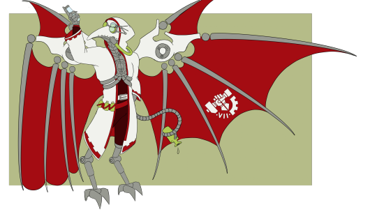
Welcome to "How the fuck has Nurgle not claimed this idiot yet" Name: Leeuwenhoek, after Antoni van Leeuwenhoek, who is considered the father of microbiology. literally means lion's corner but that is unrelated. Pronounce Lay-wun-hook. History and things: early life is still WIP up to the point where Leeuwenhoek was recruited into the Legiones skitarii. Being naturally lithe, he was made into a Pteraxius and put through all the nessecary augmentations: he was given wings, had his limbs replaced, eyelids removed and all that good stuff, as well as linked to a magos for recieving command orders. He served as such for a good couple of years, but his overseers very quickly found out that their new pteraxii recruit displayed signs of incredible intelligence and natural curiosity. Going against their protocols for the potential of this recruit, the young Leeuwenhoek was pulled from the Legiones and inducted into the cult mechanicus. and Leeuw took to this so hard... He quickly joined the ordo biologis and his fascination and obsession blossommed. Life as a Magos: If it's a pathogen, Leeuwenhoek is studying it. And he's crossbreeding different strains, just to see what will happen. Do not go into his lab unannounced unless you no longer have airways to worry about, whatever is airborne in there should NOT be inhaled. Oh and, for the love of the machine god, don't spook him! His reflexes have dulled a bit since his time as a pteraxii but the augmentations are still in place, and if you scare him he will instinctively fly up, dropping whatever is in his hands at that moment, which is likely a vial of some variety. however, If you do announce that you're coming over, Leeuw will sanitise the place first and welcome you happily, though keeping him from his work is hard and he's very likely to start infodumping about his latest projects. Many of his peers are uncomfortable around him for this reason, not really wanting to hear someone getting super excited about the latest strain of tyranid cancer, which in turn results in Leeuw being somewhat lonely. He has minor beef with the magos dominus that used to be his overseer, as the augmentations brain-linking them are still in place, much to their mutual distate. They don't dislike each other personally, personally, they just dislike the situation. Being a former pteraxii, a disease specialist and a loyal servant of the imperium, leeuwenhoek is occasionally deployed like a human cropduster, a tool in biological warfare. He is uncanny when observing the results of his work, he gets all giddy and excited and will forget to eat or sleep. Luckily for him he has a loving partner that will remind him of such things, and her presence also forces him to keep himself clean and on top of his lab safety, he does not want to risk harming her. notes on his augmentations: He used to be a pteraxii sterylisor. He has replaced his knife-feet with a model that at least allows him to walk. He sleeps perched like a bird, the wingpack making it impossible to lay down comfortably. the "beak" of his face is the result of the many, many airfilters and sensors that have been placed in front of his airways. He cannot remove these and has to eat by means of injections. feel free to ask things about him :)
22 notes
·
View notes
Text
just read that the first guy to ever view microorganisms under a microscope looked at his own sperm under his microscope too. antonie van leeuwenhoek found out that there were tiny animals unseen to the human eye in all corners of nature and immediately decided to chuck his own jizz under there like guys there’s gotta be something in there i just know it. and he jerked off onto a brass plate and he was right. what is that if not the true spirit of scientific inquiry
7 notes
·
View notes
Text
"As important as food fermentation has been for most of human history we've had no idea what these creatures looked like, nor that they even existed. It wasn't until the late 1600s that a Dutch scientist by the name Antonie van Leeuwenhoek got to see the kind of zoomed-in picture of microbes that we just saw in our sauerkraut. As a textile merchant, Leeuwenhoek had perfected the art of grinding lenses to help him see the quality of fabrics in detail. One day he decided to put a drop of Delft canal water under the lens. What he saw must have been astounding--multitudes of tiny creatures moving around! A new world, just on a smaller scale. And not so different from the world in our mead and sauerkraut."
-Michael Brenner, Pia Sörensen, and David Weitz, Science and Cooking (2020)
7 notes
·
View notes
Text
In JumpStart Adventures 3rd Grade: Mystery Mountain, one of the missions involves finding the Dutch scientist Antonie van Leeuwenhoek. Whenever Botley says Leeuwenhoek's name during the Time Machine segment, it sounds as though the same voice clip was used multiple times and stitched into the rest of the Botley's speech, which is rather unusual given that this sort of thing doesn't seem to happen at any other point in the game. This video includes the lines in question isolated from music/sound effects.
#jumpstart 3rd grade#jsfotd#the first two sound identical to me#the last two sound like they were only pitched up/down
5 notes
·
View notes
Text


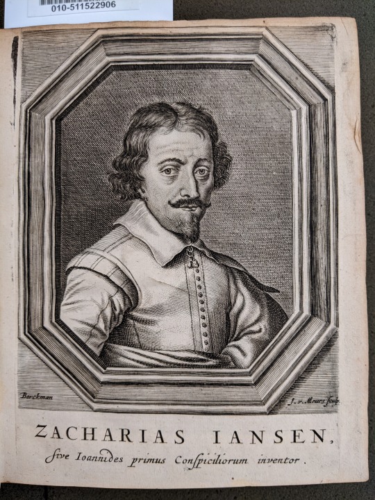
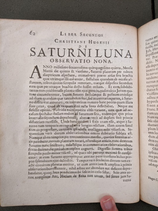

We have an exciting new acquisition to feature this Friday! Last week, a generous donor gave the Libraries a copy of Pierre Borel's De Vero Telescopii Inventore (1655). This work is a history of the invention of the telescope from antiquity to the time of writing. At the time, both Hans Lippershey and Zacharias Janssen claimed to be the inventor of the telescope, and Pierre Borel found Janssen's claims to be more convincing. Present-day historians have tended to recognize Lippershey instead, but they do give Janssen credit for inventing the compound microscope.
Borel also printed a letter from Christiaan Huygens' in which he details his observations of Titan, one of the moons of Saturn. This letter also contained Huygens' identification of the rings of Saturn in an anagram - a sly way to sneak the idea into print without giving it away before Huygens' own publication of Systema Saturnium in 1659.
This volume is bound with several others: Johann Franz Griendel's Micrographia nova (1687); Anton van Leeuwenhoek's Ontledingen en ontdekkingen van levende dierkens in de teel-deelen van verscheide dieren, vogelen en visschen (1696); and Govard Bidloo's Brief aan Antony van Leeuwenhoek (1698).
Many thanks to donor Rick Hardin for this newest addition to the collection.
De vero telescopii inventore : cum brevi omnium conspiciliorum historia : ubi de eorum confectione, ac usu, seu de effectibus agitur, novaque quaedam circa ea proponuntur : accessit etiam centuria observationum microcospicarum / Authore Petro Borello, regis Christianissimi consiliario, & medico ordinario. VAULT QB88 .B72 1655
#history of science#histsci#astronomy#history of astronomy#newacq#bookhistory#rare books#mizzou#special collections#university of missouri#libraries#kelli h
34 notes
·
View notes
Text


[Leewenhoek rolling in his grave]
...Now, to be fair: Has anyone ever seen the two of them in the same room together?
7 notes
·
View notes
Text
3299 Bijna pijnvrij
Maandag begin ik met de oxycodon. Nu zal dan eindelijk mijn pijn verlicht worden. Helaas voel ik geen enkele verlichting, geen spirituele verlichting, geen verlichting van de pijn, ik voel nog geen fietsverlichting. Niet na twee en niet na vier dagen. Ik lees de bijsluiter, want misschien ben ik te ongeduldig, maar overal wordt gesproken van een paar uur tot zelfs een half uur. Hier klopt iets…

View On WordPress
0 notes
Photo

The Microscope & the Scientific Revolution
The microscope was one of the most significant inventions of the Scientific Revolution, opening up completely new and miniaturised worlds. The first microscopes were invented in the first quarter of the 17th century in the Netherlands, but soon scientists across Europe were using the instrument to make new and often bewildering discoveries in the fields of botany, entomology, and anatomy.
The First Microscopes
The first optical microscopes appeared in the early 17th century, shortly after the invention of the telescope, which is generally credited to the Flemish spectacle-maker by Hans Lippershey (c. 1570 to c. 1619). A year or two later, Galileo (1564-1642) made a superior telescope, with which he observed the heavens in great detail, publishing his findings in Sidereus Nuncius (The Starry Messenger) in 1610. The microscope also originated in the Netherlands, its invention is usually credited to Cornelius Drebbel (1572-1635) or Hans Janssen. Like the telescope, the microscope used two lenses set in a hollow tube. Drebbel's model followed the telescope design not of Galileo, which had a concave and a convex lens, but of Johannes Kepler (1571-1630) who used two convex lenses in his instruments. Although in this latter arrangement, the image was inverted, it was also much clearer.
Soon there were specialist makers of microscopes, one highly respected manufacturer was John Marshall. A Marshall-designed compound microscope, which has three lenses (eyepiece, field lens, and objective lens) and the possibility to add extra light using a candle under the base, can be seen today in the Science Museum in London. One notable private maker was Antonie van Leeuwenhoek (see more below), who made over 500 microscopes, including examples that had an impressive magnification of 270 using a tiny glass bead instead of a larger glass lens. Further adaptations to improve the instrument were made, such as adding a small mirror to the base whose angle could be adjusted to direct more light to the specimen under view. The instrument maker Edward Culpeper (1670-1737) made this mirror concave, increasing the light made available in his microscopes. It was not enough to have an excellent instrument, though, preparing specimens for viewing was a skill in itself and could make the difference between gaining a new scientific discovery or seeing nothing at all.
Scientists soon put the new device to good use and began to investigate what had previously been indistinct or invisible to the naked eye. Anatomists, entomologists, and botanists were particularly keen to use this new invention to further their understanding of the natural world. In 1625, for example, Francesco Stelluti examined in detail the bodies of bees and published his research as The Apiarium, the first study based on microscopic science. Many other discoveries and academic papers soon followed, and by the second half of the 17th century, beautifully illustrated works were being published to reveal to interested readers exactly what could be seen through the latest microscopes. It now became clear that a tiny insect could be just as complex in its structure as a large mammal. The view through a microscope also raised some perplexing questions, such as if a parasitic flea itself has fleas, might not these fleas also have fleas and so on ad infinitum? The microscope had revealed new worlds, but where did they end? The invention seemed to pose more questions than the current technology could answer.
Continue reading...
30 notes
·
View notes
Text

That moment when the work of printing forbidden potato chips biocompatible hydrogels gets so crazy specific that you go full Antonie Van Leeuwenhoek and construct a crude micro vise from microscopic slides
yes i used a glass cutter
2 notes
·
View notes
Text
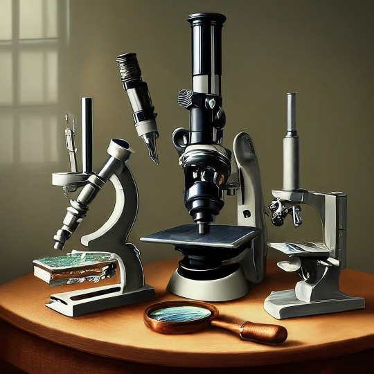
A Journey into the World of Microscopy: From Humble Beginnings to High-Tech Magnification
The science of looking into the hidden invisible Microscopy has transformed our understanding of the world around us. It can explore the universe beyond the reach of our naked eyes, with complex cellular structures, red blood cells, viruses and other viruses and microorganisms taking on amazing perspectives
The history of the microscope is a fascinating story of human curiosity, scientific genius, and relentless exploration. From the humble beginnings of simple magnifying glasses to the sophistication of modern electronic microscopes, the invention of microscopes has shaped our understanding of the microscopic world
In the 1600s, Dutch opticians such as Hans and Zachary Janssen are credited with inventing the first microscope. Known for this hybrid microscope, many lenses were used to magnify objects up to 30 times.At the end of the 17th century, Antony van Leeuwenhoek, Dutch draper some changed our perception of thumbnails. Armed with a well-made single-lens microscope, and explored the hidden reaches of nature. In 1674, Leeuwenhoek discovered microorganisms in lake water, which he aptly named “animalcules”. His discovery laid the foundations of biology and inspired generations of scientists. This incredible feat allowed him to uncover a hidden universe – the first sightings of bacteria, red blood cells, and other microorganisms.
Formation of the scientific environment (17th-19th centuries): Leeuwenhoek’s discoveries boosted scientific research. Robert Hooke, an English scientist, established these developments. In 1665, his book "Micrographia" recorded his observations with a compound microscope. Notably, the term "cell" was coined by Hooke when he examined cork tissue, laying the foundation for cell biology.Microscope systems flourished throughout the 18th and 19th centuries Joseph Lister and other scientists addressed the limitations of the early lenses, introducing improvements that reduced image distortion.
Beyond the Limits of Light: The Beginning of the New Age (19th-20th century): As the 19th century progressed, the limitations of optical microscopy became apparent and scientists yearned for a tool which can go deeper into cells. This research culminated in the development of the electron microscope in the 1930s. The 20th century was revolutionary with the invention of the electron microscope. Unlike light microscopes, which use visible light, electron microscopes use electron beams to achieve much higher magnification.Formation of the scientific environment (17th-19th centuries): Leeuwenhoek’s discoveries boosted scientific research. Robert Hooke, an English scientist, established these developments. In 1665, his book "Micrographia" recorded his observations with a compound microscope. Notably, the term "cell" was coined by Hooke when he examined cork tissue, laying the foundation for cell biology.Microscope systems flourished throughout the 18th and 19th centuries Joseph Lister and other scientists addressed the limitations of the early lenses, introducing improvements that reduced image distortion.
Beyond the Limits of Light: The Beginning of the New Age (19th-20th century): As the 19th century progressed, the limitations of optical microscopy became apparent and scientists yearned for a tool which can go deeper into cells. This research culminated in the development of the electron microscope in the 1930s. The 20th century was revolutionary with the invention of the electron microscope. Unlike light microscopes, which use visible light, electron microscopes use electron beams to achieve much higher magnification.
In the 1930s, German experts Max Knoll and Ernst Ruska made the first electron microscope. This tool let us see tiny things like cells and even atoms by using electron beams, not light, getting images many times bigger. This cool invention showed us the tiny parts inside cells, viruses, and stuff too small to see before. The 1900s brought even more cool microscopes. New kinds like phase-contrast and confocal microscopy let scientists look at live cells without using stuff that could hurt them. Now, the world of looking at tiny things is getting even better. Today, we have high-tech microscopes that use computers and lasers. These let us see and even change tiny things in ways we never could before.
Modern Microscopy's Diverse Arsenal - Today, the field of microscopy boasts a diverse range of specialized instruments, each tailored to address specific scientific needs. Here's a glimpse into some remarkable examples:
Scanning Electron Microscope (SEM): Imagine a high-tech camera that captures images using a beam of electrons instead of light. That's the essence of a SEM. By scanning the surface of a sample with a focused electron beam, SEMs generate detailed information about its topography and composition. This makes them ideal for studying the intricate structures of materials like insect wings, microchips, and even pollen grains.
Transmission Electron Microscope (TEM): While SEMs provide exceptional surface detail, TEMs take us a step further. They function by transmitting a beam of electrons through a very thin sample, allowing us to observe its internal structure. TEMs are the go-to instruments for visualizing the intricate world of viruses, organelles within cells, and macromolecules like proteins.
Confocal Microscopy: Ever wished to focus on a specific layer within a thick biological sample and blur out the rest? Confocal microscopy makes this possible. It utilizes a laser beam to precisely illuminate a chosen plane within the sample, effectively eliminating information from out-of-focus regions. This allows researchers to create sharp, three-dimensional images of cells, tissues, and even small organisms.
Atomic Force Microscopy (AFM): This technique takes a completely different approach, venturing into the realm of physical interaction. AFM employs a tiny cantilever, akin to a microscopic feeler, to physically scan the surface of a sample. By measuring the minute forces between the cantilever and the sample's surface, AFM can map its topography at an atomic level. This provides invaluable insights into the properties of materials at an unimaginable scale, making it crucial for research in fields like nanotechnology and surface science.
Fluorescence Microscopy: Imagine illuminating a sample with specific wavelengths of light and observing it glowing in response. That's the essence of fluorescence microscopy. This technique utilizes fluorescent molecules or tags that bind to specific structures within a cell or tissue. When excited by light, these tags emit their own light, highlighting the target structures with remarkable clarity. This allows researchers to visualize specific proteins, DNA, or even pathogens within biological samples.
Super-resolution Microscopy (SRM): Overcoming the limitations imposed by the wavelength of light, SRM techniques like STED (Stimulated Emission Depletion) and PALM (Photoactivated Localization Microscopy) achieve resolutions surpassing the diffraction limit. This allows researchers to visualize structures as small as 20 nanometers, enabling the observation of intricate cellular machinery and the dynamics of individual molecules within living cells.
Cryo-Electron Microscopy (Cryo-EM): This powerful technique takes a snapshot of biological samples in their near-life state. Samples are rapidly frozen at ultra-low temperatures, preserving their native structure and minimizing damage caused by traditional fixation methods. Cryo-EM has been instrumental in determining the three-dimensional structures of complex molecules like proteins and viruses, providing crucial insights into their function and potential drug targets.
Correlative Microscopy: Combining the strengths of multiple microscopy techniques, correlative microscopy offers a comprehensive view of biological samples. For instance, researchers can utilize fluorescence microscopy to identify specific structures within a cell and then switch to electron microscopy to examine those structures in high detail. This integrated approach provides a deeper understanding of cellular processes and their underlying mechanisms.
Light Sheet Microscopy (LSM): Imagine illuminating a thin slice of a sample within a living organism. LSM achieves this feat by focusing a laser beam into a thin sheet of light, minimizing photobleaching and phototoxicity – damaging effects caused by prolonged exposure to light. This allows researchers to observe dynamic processes within living organisms over extended periods, providing valuable insights into cellular behavior and development.
Expansion Microscopy (ExM): This innovative technique physically expands biological samples by several folds while preserving their structural integrity. This expansion allows for better resolution and visualization of intricate cellular structures that would otherwise be difficult to distinguish using traditional microscopy methods. ExM holds immense potential for studying the organization and function of organelles within cells.
Scanning Near-Field Optical Microscopy (SNOM): This innovative technique pushes the boundaries of resolution by utilizing a tiny probe that interacts with the sample at an extremely close range. SNOM can not only image the surface features of a sample with exceptional detail but also probe its optical properties at the nanoscale. This opens doors for research in areas like material science and photonics, allowing scientists to study the behavior of light at the interface between materials.
X-ray Microscopy: Stepping outside the realm of light and electrons, X-ray microscopy offers unique capabilities. By utilizing high-energy X-rays, this technique can penetrate deep into samples, making it ideal for studying the internal structure of dense materials like bones and minerals. Additionally, it allows for the visualization of elements within a sample, providing valuable information about their distribution and composition.
From revealing the building blocks of life to aiding in the development of new medicines, the microscope has played an undeniable role in shaping our scientific understanding. As technology continues to evolve, one can only imagine the future breakthroughs this remarkable invention holds in unveiling the secrets of our universe, both seen and unseen. These advancements hold the potential to revolutionize our understanding of biological processes, develop new materials with extraordinary properties, and ultimately pave the way for breakthroughs in medicine, nanotechnology, and countless other fields. As we continue to refine and develop novel microscopy techniques and the future holds immense promise for further groundbreaking discoveries that will undoubtedly revolutionize our perception of the world around us.
#science sculpt#life science#science#molecular biology#biology#biotechnology#artists on tumblr#microscopy#microscope#Scanning Electron Microscope#Transmission Electron Microscope#Confocal Microscopy#Atomic Force Microscopy#Fluorescence Microscopy#Expansion Microscopy#X-ray Microscopy#Super-resolution Microscopy#Light Sheet Microscopy#illustration#illustrator#illustrative art#education#educate yourself#techniques in biotechnology#scientific research#the glass scientists#scientific illustration#scientific advancements
7 notes
·
View notes