#Ex-situ characterization
Link
1 note
·
View note
Text
Meta-omics profiling of full-scale groundwater rapid sand filters explains stratification of iron, ammonium and manganese removals
Rapid sand filters (RSF) are an established and widely applied technology for groundwater treatment. Yet, the underlying interwoven biological and physical-chemical reactions controlling the sequential removal of iron, ammonia and manganese remain poorly understood. To resolve the contribution and interactions between the individual reactions, we studied two full-scale drinking water treatment plant configurations, namely (i) one dual-media (anthracite and quartz sand) filter and (ii) two single-media (quartz sand) filters in series. In situ and ex situ activity tests were combined with mineral coating characterization and metagenome-guided metaproteomics along the depth of each filter. Both plants exhibited comparable performances and process compartmentalization, with most of ammonium and manganese removal occurring only after complete iron depletion. Within each compartment, the homogeneity of the media coating and genome-based microbial composition highlighted the effect of backwashing on filter media mixing. In stark contrast, intra-compartment contaminant removal was highly stratified following decreasing substrate availability along the filter height. This apparent and long-standing conflict was resolved by quantifying the expressed proteome at different filter heights, revealing a consistent stratification of proteins catalysing ammonia oxidation and protein-based relative abundances of nitrifying genera. This implies that microorganisms adapt their protein pool to the available nutrient load at a faster rate than the backwash mixing frequency. Ultimately, these results show the unique and complementary potential of metaproteomics to understand metabolic adaptations and interactions in highly dynamic ecosystems. http://dlvr.it/Sf2Gfz
0 notes
Photo

Tracking the atomic pathways by in-situ liquid cell TEM
Recently, platinum-containing core-shell structures with tunable magnetic and catalytic properties have attracted intensive attentions and offered a wide range of applications. To date, their synthetic routes are mostly based on galvanic replacement, co-reduction, thermal decomposition and seed-mediated method. But the detailed formation mechanisms of core-shell structures in solution, especially, at gas-liquid interface are still not completely clear, which is mostly achieved based on post reaction studies or ex situ characterizations. In this regard, it is worthwhile but still very challenging to directly visualize the complicated and delicate dynamic processes.
Technical advantages of in-situ liquid electron microscopy (TEM) allow us to monitor the growth trajectory of pure metal nanoparticles in liquid media, including nucleation growth, nanorod self-assembly, and electrochemical deposition. Compared with pure metal nanocrystals, the growth pathway for alloy and its oxide core-shell structures is more complicated. It is noteworthy that little is known about the atomic growth pathway of Pt based-oxide core-shell structures and structural stability in solution, especially, at the gas-liquid interface. Due to the lack of direct observation methods with high spatial resolution, some intermediate states may be easily missed.
Read more.
#Materials Science#Science#Materials Characterization#TEM#In situ liquid electron microscopy#Core shell structures#Platinum#Nanoparticles#Liquids#Metals#Nanotechnology
3 notes
·
View notes
Text
Iris Publishers_Journal of Textile Science & Fashion Technology (JTSFT)
Brief Overview of Developments in BC-Based Biosynthesized Nano-Fabrics Production: Challenges and Future Perspectives
Authored by Muhammad Awais Naeem

Abstract
Apparel industry is infamous for causing water pollution, carbon dioxide emissions, soil erosion and huge waste generation. There is a need to find novel, sustainable and green manufacturing methods, which are cost effective and production efficient. In this regard, this mini-review discusses the recently reported techniques to biosynthesize BC and BC-based hybrid fabrics which can ecologically affect the textiles industry and the environment.
Keywords: Bacterial cellulose; Nano-fabrics; In-situ fermentation; Biosynthesis
Abbreviations: BC: Bacterial Cellulose; H-Bonding: Hydrogen Bonding
Introduction
Fashion trends are rapidly changing and consumers’ demand for affordable apparel is rising. Traditional textiles production process involves several stages and growing production has raised various environmental concerns, including the enormous amounts of harmful processing wastes produced, landfill consumption, carbon dioxide emissions, air/water pollution and so on [1-5]. Hence eco-friendly biomaterials [6-8] and green production techniques are being actively sought after [9-12]. Nature has the best sustainability model as it creates no harmful wastes and natural biomaterials like polysaccharides, cellulose, chitosan etc. [13-16], are sustainable resources which have biocompatible, biodegradable, and hazard-free features. Bacterial cellulose (BC) is a promising biodegradable natural polymer, possessing high modulus and strength [17,18], secreted by Acetobacterxylinum through a hierarchical cell-directed self-assembly process [19]. BC is harmless to humans and the environment and free of impurities, such as lignin and hemicellulose. Its unique physical characteristics and cultivation properties have demonstrated a great potential to achieve zero-waste design for apparels (20). BC based fabrics are highly porous, biocompatible, with exceptional environmental biodegradability and can be easily customized for size, shape, and thickness [21-23].
Recent research works have reported innovative ways to produce BC-based fabrics by using natural renewable resources and techniques, instead of using conventional manufacturing methods. Hence apparel production can be redefined for cost effectiveness, labor friendliness, low environmental impact, and biodegradability [12]. Shape and dimensions of cultivation containers are the factors which limit the customized growth of BC at air-liquid interface. Genetically modified bacteria have been successfully grown on three dimensional templates to produce seamless fabrics. Such biological fabrication technique will eradicate a considerable amount of material wastage at various production stages and enhance the overall process efficiency [24]. Cultivation of bacterial cellulose in form of sheets has also been reported, in order to make clothing through traditional cut-and-sewn method. Production of self-grown seamless 3D BC fabrics was also realized, to prepare tailor-shaped clothing based on zero-waste design [20]. Other studies analyzed the bleaching and dyeing of BC with help of direct and reactive dyes, by using in-situ and ex-situ approaches [25,26]. However, BC alone lacks the durability required for everyday use, considering sustainable apparel applications [27-30]. Hybrid fabrics can be produced by incorporating other materials of interest (natural fibers, polymers, nano-fillers etc.) during BC fermentation. Numerous hydroxyl groups present on the surfaces of hydrophilic substrates and BC result into strong interaction (H-bonding) between the components combined [31-34]. Several recent studies report the localized growth of BC nanofibers on electrospun membrane support, to form a nano-fabric structure [35-39]. Seamless tubular hybrid fabrics were prepared by using controlled bioreactors which consisted of rectangular containers, equipped with micro-fluidics system to inject additional nutrients as needed. Cellulose acetate (CA) electrospun membrane was used as support material for BC growth. A conveyer-roller arrangement used to expose pristine membrane on air-liquid interface enabled the formation of seamless tubular fabrication. Another recent study has reported the formation of functional fabrics to be used for biomedical application [37]. The characterization results for water holding, vertical wicking, contact angle and mechanical analyses suggested the use of biologically prepared hybrid fabrics for wound dressing application. Future research works can help to investigate the possibility of incorporating biodegradable hydrophobic medicine-loaded nanofibrous membranes (e.g. polylactic acid/ poly glycolic acid) to achieve specific clinical objectives. With aforementioned self-synthesizing and tailor-shaped characteristics, the biosynthesized BC-based fabrics have great potential to be new sustainable textile materials for the apparel industry. However, the cost and pace of production; handling, laundering and aesthetics related issue still remain a challenge to be addressed.
For More Open Access Journals in Iris Publishers Please click on:
https://irispublishers.com/
For More Articles in Journal of Textile Science & Fashion Technology
https://irispublishers.com/jtsft/
For More Information:https://irispublishers.com/jtsft/fulltext/brief-overview-of-developments-in-bc-based-biosynthesized-nano-fabrics-production.ID.000580.php
#Iris Publishers#Iris Publishers LLC#Open Access Fashion Journals#Open Access Textile Journals#Journal of Textile science and Fashion Technology
0 notes
Text
Endogenous Adult-Derived Telomerase-Positive Stem Cells: OAJBS

Endogenous Adult-Derived Telomerase-Positive Stem Cells by Henry E Young* in Open Access Journal of Biomedical Science (OAJBS)
Discovered in 1975 Located in the connective tissues throughout the body, ~ 10% of all stem cells in the body Found in 16 different species: terrestrial salamander, Komodo dragon, chicken, waddle crane, mouse, rat, rabbit, cat, dog, sheep, goat, pig, cow, speckled bear, horse, and human They are the natural healing cells of the body, they do NOT decrease in number with increasing age of the individual Cloned from single cells by repetitive serial dilution clonogenic analysis and then extensively characterized Consist of the following. Totipotent stem cells-form any cell type in the body, including spermatogonia and oogonia. Pluripotent stem cells-form any cell type in the body, except spermatogonia and oogonia Ectodermal stem cells-will only form cells of the ectodermal embryonic lineage• Mesodermal stem cells-will only form cells of the mesodermal embryonic lineage. Endodermal stem cells- will only form cells of the endodermal embryonic lineage Utilized for treatments. Will NOT form teratomas (cancerous cells) in situ or ex vivo. Normally quiescent, unless activated. Inside the body, activation requires about 2 weeks Outside the body, activation takes 2 hours. Can be expanded to significant numbers inside the body (in situ) using combinatorial nutraceuticals and outside the body (ex vivo) in culture using proprietary culture media: Essentially unlimited proliferation potential. Transplanted either as autologous (self) and/or allogeneic (donor)Donor needs to be matched for gender, ABO blood group (or O-negative), infectious disease-free and deleterious genetic mutation-free.
https://biomedscis.com/fulltext/endogenous-adult-derived-telomerase-positive-stem-cells.ID.000139.php
#Biomedical Science#oajbs#open access journal of biomedical Science#open access journal of biomedical science and research studies
0 notes
Text
300+ TOP ONCOLOGY Objective Questions and Answers
ONCOLOGY Multiple Choice Questions :-
1. Cytotoxic T cells (CTL) are capable of recognizing:
A. Peptide antigens associated with major histocompatibility complex (MHC) molecules.
B. Membrane-bound antigens.
C. Cytoplasmic antigens.
D. Nuclear antigens.
E. All of the above.
Answer: E
2. Adoptive immunotherapy with lymphokine activated killer cells (LAK) and tumor infiltrate (TIL) cells are characterized by:
A. Nonspecific stimulation of effector cells.
B. Expansion ex vivo of large numbers of lymphocytes.
C. Infusion with interleukin 2 (IL-2).
D. Significant toxicity at high doses.
E. All of the above.
Answer: E
3. Previous clinical studies with cancer vaccines have:
A. Clearly demonstrated induction of tumor-specific immune response.
B. Repeatedly demonstrated clinical response to large tumor burden.
C. Not clearly demonstrated induction of tumor-specific immune response.
D. Not been performed to date.
Answer: C
4. Which of the following statements is/are true of the epidemiology and etiology of melanoma?
A. Most patients are diagnosed after age 60 years.
B. Skin color has no association with risk of melanoma.
C. Sun exposure is the only risk factor for melanoma.
D. The per capita incidence of melanoma is highest in Australia.
Answer: D
5. Which of the following variables best predicts prognosis for patients with a recent diagnosis of cutaneous melanoma and no clinical evidence of metastatic disease?
A. Breslow thickness.
B. Clark's level.
C. Ulceration.
D. Gender.
E. Celtic complexion.
Answer: A
6. A 38-year-old man presents with a melanoma on the skin of the right calf measuring 5 mm. in thickness. Several large nodes are palpable in the right inguinal region. Which of the following statements about the appropriate management of this clinical problem is false?
A. In the absence of systemic disease, the primary melanoma of the right calf should be excised with at least a 2-cm. margin.
B. Complete right inguinal node dissection should be performed if there is no evidence of systemic metastasis.
C. If further work-up reveals multiple lung metastases of melanoma, they should be excised as soon as possible.
D. Chemotherapy for melanoma is primarily palliative; so surgical therapy is preferred if there is no evidence of metastatic disease beyond the inguinal region.
E. If the nodes do not contain metastatic disease but are simply reactive, the chance of 5-year survival is 50% or less.
Answer: C
7. A 42-year-old woman presents with an 8 cm. × 6 cm. × 4 cm. mass in the posterior thigh. Incisional biopsy reveals a high-grade liposarcoma. Her management should include:
A. High thigh amputation.
B. Extracompartment excision with negative margins.
C. Complete excision with negative margins.
D. Adjuvant radiation therapy.
E. Adjuvant chemotherapy.
Answer: CD
8. Biologic features of adult soft tissue sarcomas include the following:
A. Mutations of p53 in metastatic liposarcoma.
B. A low (less than 1%) risk of metastasis for small, low-grade lesions.
C. Recurrent disease in at least 33% of patients.
D. Lymph node metastasis in less than 3% of patients.
E. Mutations of p53 in Li-Fraumeni syndrome.
Answer: BCDE
9. Which of the following statements describes an ideal tumor marker?
A. The ideal tumor marker should be tumor specific; that is, in the normal population or patients with benign diseases, false-positive test results are rare.
B. The ideal marker must have a low false-negative rate; that means that all patients with a particular type of cancer should test positive.
C. The circulating level of an ideal tumor marker should correlate directly with the amount of viable tumor and be a measure of the response to therapy.
D. The ideal tumor marker should act as a prognostic indicator.
E. All of the above.
Answer: E
10. A marker for the diagnosis of pancreatic cancer is:
A. CA 15-3.
B. CA 19-9.
C. Alphafetoprotein (AFP).
D. Carcinoembryonic antigen (CEA).
E. CYFRA 21-1.
Answer: B

ONCOLOGY MCQs
11. Which of the following tumors may cause elevated CEA levels?
A. Breast cancer.
B. Colorectal cancer.
C. Gastric cancer.
D. Lung cancer.
E. All of the above.
Answer: E
12. The presence of which marker is a significant poor prognosis variable for patients with breast cancer:
A. CEA.
B. C-erb B-2.
C. AFP.
D. Human chorionic gonadotropin (hCG).
E. RB-1.
Answer: B
13. The most useful circulating marker for patients with hepatocellular carcinoma is:
A. CA 50.
B. Levels of vitamin B 12.
C. CEA.
D. AFP.
E. hCG.
Answer: D
14. In patients with colorectal cancer the serum CEA level is a clinically useful measure for all reasons except:
A. Prognosis.
B. Detection of recurrence.
C. Guiding second-look operations.
D. Following treatment response.
E. Early diagnosis.
Answer: E
15. Which serum markers are useful in the management of patients with testicular cancer?
A. hCG.
B. AFP.
C. CA 15-3.
D. Two of the above.
E. None of the above.
Answer: D
16. Which tumor marker is useful for the management of patients with breast cancer?
A. CA 125.
B. Inhibin.
C. CA 19-9.
D. CA 15-3.
E. CEA.
Answer: D
17. A new marker that has possible utility in the management of patients with non–small-cell lung cancer (NSCLC) is:
A. Calcitonin.
B. Neuron-specific enolase.
C. CYFRA 21-1.
D. Glucagon.
E. Chromogranin A.
Answer: C
18. A circulating marker that may be useful in the management of patients with any neuroendocrine tumor is:
A. Chromogranin A.
B. Neuron-specific enolase.
C. hCG.
D. Two of the above.
E. None of the above.
Answer: D
19. A 65-year-old man is seen two years following right hemicolectomy for a Duke’s B-2 carcinoma of the cecum. Although asymptomatic, the CEA level has risen four-fold from a value obtained six months previously. Computed tomography reveals a single, 3 cm lesion in the right hepatic lobe. There is no evidence of extra-hepatic metastatic disease and the patient undergoes right hepatic lobectomy. Which of the following correctly represents the chance of overall 5 year survival?
a. 15%
b. 33%
c. 50%
d. 66%
Answer: b
20. The most serious long-term side effect of bleomycin therapy is which of the following?
a. Pulmonary fibrosis
b. Cataract formation
c. Cardiomyopathy
d. Aplastic anemia
Answer: a
21. Which of the following malignancies have declined in incidence in the United States over the past two decades?
a. Breast carcinoma
b. Gastric carcinoma
c. Endometrial carcinoma
d. Prostate cancer
e. Carcinoma of the uterine cervix
Answer: b, c, e
22. Oncogenes have been implicated in the development of a number of human neoplasms. Oncogene activation is believed to be required for oncogenesis. Which of the following potential mechanisms are relevant to these processes?
a. Chromosome translocation
b. DNA point mutation
c. Amplification
d. Gene deletion
Answer: a, b, c
23. A 45-year-old woman undergoes excision of a 1 cm breast mass. Histologic examination reveals invasive ductal carcinoma. Flow cytometric analysis is also performed which determines that a fraction of the tumor cells are “aneuploid”. The patient asks for an explanation of this term. Which of the following is/are correct?
a. The cells have a DNA content 1 times the baseline content
b. The cells have hyperchromatic nuclei
c. The cells have a DNA content 2 times the baseline content
d. The cells have squamous morphology
e. The cells have a DNA content not an even multiple of baseline content
Answer: e
24. A 45-year-old man with long-standing gastroesophageal reflux undergoes upper endoscopy that reveals patchy areas of epithelium resembling gastric mucosa extending 5 cm proximal to the esophagogastric junction. Biopsies are obtained. The pathologic report describes “Barrett’s epithelium”. Which of the following processes does this finding represent?
a. Cellular hyperplasia
b. Cellular hypertrophy
c. Metaplasia
d. Carcinoma in situ
Answer: c
25. Which of the following statements regarding the inherited form of retinoblastoma is/are correct?
a. Retinoblastoma results from amplification of the H-ras oncogene
b. Clinical disease results after chromosomal loss in a retinal cell after birth
c. Retinoblastoma results from the loss of a tumor suppressor gene
d. Clinical disease results from chromosomal translocation
Answer: b, c
ONCOLOGY Objective type Questions with Answers
26. Analysis of metastatic tumor cells has revealed expression of factors promoting tissue invasion. Which of the following is/are among such factors?
a. Collagenase
b. Plasminogen activator
c. Fibroblast growth factor
d. Interleukin-2
Answer: a, b
27. The most common complication that requires alteration of planned chemotherapy regimens is which of the following?
a. Pulmonary fibrosis
b. Gastrointestinal ulceration
c. Hematologic suppression
d. Hepatotoxicity
Answer: c
28. Resistance of tumors to multiple chemotherapeutic agents is often due to the MDR gene. This gene encodes a protein that acts by which of the following mechanisms?
a. As a transmembrane efflux pump for chemotherapeutic agents
b. As a DNA repair molecule
c. As an isoform of superoxide dismutase
d. As a membrane stabilizer
Answer: a
29. The high incidence of hepatitis B infection in Africa and parts of Asia is thought to be causally associated with increased incidence of which of the following malignancies?
a. Hepatocellular carcinoma
b. Esophageal cancer
c. Burkitt’s lymphoma
d. Gastric carcinoma
Answer: a
30. Workers exposed to asbestos are at increased risk for which of the following tumors?
a. Thoracic mesothelioma
b. Bladder carcinoma
c. Laryngeal carcinoma
d. Testicular carcinoma
e. Non-Hodgkin’s lymphoma
Answer: a, b, c
31. The Lynch Syndrome is also known as hereditary non-polyposis colorectal cancer. Which of the following is/are features of this syndrome?
a. Left sided colon cancers
b. Autosomal dominant inheritance
c. Multiple polyps beginning in adolescence
d. Multiple cutaneous nevi
Answer: b
32. A number of clinical factors have been noted to decrease sensitivity of tumors to the effects of ionizing radiation. Which of the following is most important in this regard?
a. Increased tissue vascularity
b. High tumor mitotic rate
c. Tissue hypoxia
d. Subcutaneous tumor location
Answer: c
33. Brachytherapy involves the delivery of radiation therapy locally via specially designed catheters placed in direct apposition to the treated tissue. The most common radioisotope used in this application is which of the following?
a. 125I
b. 14C
c. 3H
d. 34P
Answer: a
34. Patients that have acquired immunodeficiency syndrome are at increased risk for which of the following neoplasms?
a. Colorectal cancer
b. Meningioma
c. Kaposi’s sarcoma
d. Hepatocellular carcinoma
e. Esophageal carcinoma
Answer: c
35. DNA viruses have been implicated as etiologic agents in several human tumors. Evidence for a causative role exists for which of the following neoplasms?
a. Burkitt’s lymphoma
b. Testicular carcinoma
c. Cervical carcinoma
d. Osteogenci sarcoma
e. Esophageal carcinoma
Answer: a, c
36. When a chemotherapeutic agent is stated to have caused a partial response this implies what degree of reduction in measurable tumor volume?
a. 0–9%
b. 10–29%
c. 30–49%
d. 50–99%
Answer: d
37. Which of the following represent obstacles to the use of retroviruses in therapeutic gene transfer?
a. Viral receptors may not be present on target cell membranes
b. For integration, the host cell must undergo mitosis
c. Virus particles are labile
d. Viral purification is difficult
Answer: a, b, c, d
38. Which of the following statements regarding alpha-1-antitrypsin deficiency is/are correct?
a. Alpha-1-antitrypsin is a plasma elastase inhibitor
b. Most homozygous patients develop chronic obstructive pulmonary disease
c. The spleen is the primary site of alpha-1-antitrypsin synthesis
d. Intracellular accumulation of abnormal protein occurs in hepatocytes
Answer: a, b, d
39. Which of the following statements regarding retroviruses is/are correct?
a. The genetic material contained within a retrovirus is RNA
b. Inside the host cell the viral RNA is converted to single-stranded DNA
c. Proviral DNA is integrated into the host chromosome
d. Retroviruses can be used to transfect both replicating and non-replicating cells
Answer: a, c
40. Which of the following statements relating to adenoviruses is/are correct?
a. Adenoviral infection is a common cause of upper respiratory tract infection
b. Adenoviral genetic material consists of double-stranded DNA
c. Adenovirus can be produced in large quantity and easily purified
d. Adenoviral infection requires host cell mitosis
Answer: a, b, c
41. Which of the following statement relating to cystic fibrosis is/are correct?
a. Cystic fibrosis is inherited as an X chromosome-linked recessive trait
b. Cystic fibrosis is caused by a defective chloride channel
c. Cystic fibrosis is caused by defective acetylcholine receptors
d. Cystic fibrosis is inherited as an autosomal recessive trait
Answer: b, d
42. Hemophilia B has been treated in a pre-clinical model by gene transfer for which deficient clotting factor?
a. Factor II
b. Factor VII
c. Factor IX
d. Factor X
Answer: c
43. Familial hypercholesterolemia has been proposed as a disease to be treated by gene therapy. The molecular basis of familial hypercholesterolemia is which of the following?
a. Absence of hepatic low density lipoprotein receptors
b. Overproduction of high density lipoprotein
c. Absence of lipoprotein lipase
d. Overproduction of hepatic ornithine transcarbamylase
Answer: a
44. Antisense oligodeoxynucleotides have been proposed as agents for cancer-directed gene therapy. When delivered intracellularly, antisense molecules act to block which of the following?
a. Transcription
b. Translation
c. Post-translational processing
d. Ribosylation
Answer: b
45. Which of the following viruses is/are considered to be neurotropic?
a. Adenovirus
b. Herpes simplex virus
c. Retrovirus
d. Adeno-associated virus
Answer: b
ONCOLOGY Questions and Answers pdf Download
Read the full article
0 notes
Link
1 note
·
View note
Text
Manila Ocean Park
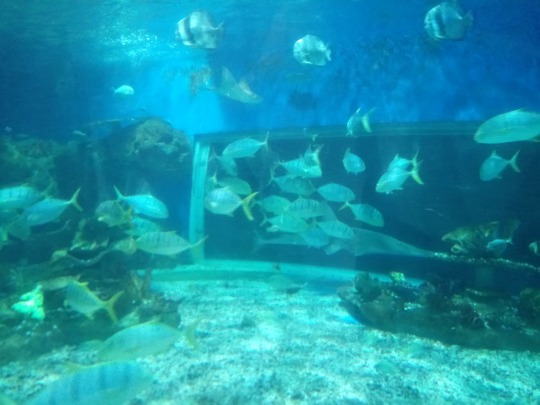
General History of Ex-Situ Conservation site:
The park is owned by China Oceanis Philippines Incorporated, which is a subsidiary of China Oceanus Group Ltd (COG). Specializations of the company include: investment, putting up, and operation of various oceanarium attractions. After several years of experience, the group responsible for running this company now comprises of an experienced team of specialists that are experts on providing services to the developers as well as operators of different public aquarium facilities.
Conservation Strategy and Management Practices:
1. Endangered species are banned in the area.
2. Coastal resource management is being employed in places where the species found in the park are retrieved from.
3.Energy-efficiency is considered to be a priority in the running of the park’s operations
4. Those working in the park are constantly being updated regarding the threatened and endangered organisms in the country.
Some freshwater and marine organisms in the area:

1. Large Tooth Sawfish- member of a group of fish called elasmobranchs, whose skeletons comprise of cartilage. These organisms can grow up to 23 feet long and may weigh up to 1,300 pounds. They have a life span of up to 80 years.
2. Sharks- also a member of the elasmobranchs. Distinct five to seven gill slits are found to be present near the side of the head of these organisms. There are more than 465 identified species living in the marine systems as of today.
3. Stringrays- These are flattened fishes that are closely related to sharks. They have no bones present in the body. Instead, their bodies are made up of flexible cartilage. They are oviparous creatures, which means that they lay eggs and these eggs are enclosed and protected by what is known as a “mermaid’s purse.”
3. Sea Lions- sea mammals that are quadrupedals (can walk on all fours). They have external ear flaps, elongated foreflippers, short and thick hair, and a big abdomen. They are categorized under family Otariidae.
4. Jellyfish- were already present before dinosaurs even strived to live on Earth. These are jellylike creatures that move along the ocean currents and are usually abundant in cold as well as warm ocean water and even along the coastlines. They are characterized by their tiny stinging cells found in their tentacles which serve to stun the prey before devouring them.
5. Glassy Perchlet- found mainly in Asia and can grow up to 3.9cm in length. These are categorized under family Ambassidae of order Perciformes, which is the only species in the genus Chanda. They usually are found in coastal and brackish waters, and may even strive in freshwaters.
6. Common seahorse- these organisms can easily blend into their surroundings and are among the smallest seahorse species found all over the globe. What appears to be crowns located on the top of the heads of the seahorses are actually very distinct in each organism and can be likened to fingerprints of humans.
7. Starfish- They can also be called as “sea stars” and are star-shaped echinoderms under class Asteroidea. There are up to 2,000 living species of sea stars identified all over the world. Most species have five arms but some may have up to 40.
8. Clownfish- Also known as anemonefish. They belong to subfamily Amphiprioninae in the family Pomacentridae. They are known to have symbiotic mutualism relationship with the sea anemones. A popular animated movie entitled: “Nemo” is inspired by this marine organism.
9.Saddleback butterflyfish- has the scientific name: Chaetodon ephippium and resides along the Indian and Pacific Oceans from Sri Lanka, north to southern Japan, and New South Wales in Australia. What makes them unique is the presence of a large black “saddle” with white border found on its upper body.
10. Pajama Cardinalfish- also known as pajama cardinalfish, spotted cardinaldish, coral cardinalfish, and polkadot cardinalfish. This is actually a very common and popular aquarium fish. Its body comprises of a rainbow of colors and has a greenish-yellow colored face.
Trivia:
Water coming from Manila Bay is actually being filtered and used as the water source for the park’s Oceanarium. The special filtering process is being conducted so as to ensure that the quality of water is suitable for marine life.

0 notes
Photo


Sardinian waste management. . . . . . . . This week we learned a lot about Sardinian waste management. Our first guest lecturer was an member of the team who developed the waste management plan in Sardinia, put into place in 2004. He discussed that the major focus on the plan is reducing the amount of waste produced by the country and recovering or recycling as much waste as possible. There are several government guidelines for the regional waste management plans in Italy including limiting land filling to residual waste, limiting energy recovery to non-recyclable materials, safely managing hazardous waste, eradicating the illegal waste export of regions, reducing food waste production, and using recycled waste as a raw material source. A major way that Sardinia works towards these goals is by implementing separate collection in the regions of Sardinia. This means that glass, plastic, metal, and organic waste (food and yard waste) are all collected in separate bins and managed differently. Organic waste is composted. Plastics are either recycled or burned for waste to energy recovery. Glass and metal are reused or recycled. At the end of this management process any leftover waste, generally ash from waste to energy plants, is landfilled as inert material. Our second guest lecturer discussed general methods of contaminated site remediation, the first of many lecturers on this topic though future ones will be discussing specific remediation examples. We were taught about the objectives of site remediation and how, in Italy, whether remediation efforts are needed is based upon site specific characterization, risk assessment, and threshold levels. Contaminated sites can be above the government defined thresholds for different contaminants if the specific site study determines the risk factor to humans and the environment is low enough. We discussed difference between ex situ and in situ treatment methods and how in situ (on site) treatment methods are always preferred to reduce the potential spread of the contamination. Then he presented the different kinds of contaminants that generally pose a risk to groundwater and human populations and they methods of both containing and treatment methods. Methods of containing pollutants are either physical or based on the hydrodynamics of the groundwater. Containment methods are generally cheaper than treatment methods, though when the two are used in tandem the best results can often be achieved. The final portion of our Sardinia waste management knowledge journey this week was the Arborea Waste Management Complex. Here dry mixed waste not suitable for recycling, organic material, and plastics are are sorted and broken down for different purposes and let me tell you did it smell great, not. The compost piles smelled like mulching season back in Ohio, but the mixed dry waste smelled like dying animals. The mixed dry waste is mostly collected from areas with separated collection, but some areas in Sardinia still don't have separated collection so there is some organic material mixed in with waste. This waste (photo 1: mixed waste receiving area) is shredded to create even sizes since this material is mostly going to a waste to energy plant for recovery. After being shredded it passes under a magnet to remove any metal, which won't burn in the incinerator, before being sieved, dried, and baled. This final material is baled using plastic to ensure that it burns in the incinerator (photo 2: baled mixed dry waste). There is a landfill on site where they store the bales of this waste temporarily if the waste to energy plants are closed for maintenance; however, unlike traditional landfills they simply place the baled material and cover it with traps to then be removed and sent to the plants when they are open again (photo 3: landfill area). Organic materials, comprised of food waste in biodegradable bags and yard waste, is shredded into even sizes and then weighed before mixing to make sure that the end product had the right percentages of Carbon and Nitrogen (photo 4: organic material shredder). Once weighed out to the proper amounts, the material is mixed and often wood chips or other structural support is added to increase the structural stability of the compost piles. It also increased the air flow in the piles, essential for healthy compost, by creating void space. After two weeks of being stored the material is sifted to remove the added support material and aged again for 3 months at which point it is considered mature and can be sold to local farmers as fertilizer. The complex is actually working to develop a packaging plant for the compost so it can be sold in retail. Plastics received at the complex (photo 5: plastics receiving area) are separated but huge conveyor belts and air blasters into first PET plastics and non-PET plastics and then those two are separated by color. Since different color plastics are made from different materials they need to be separated before they can be baled for sale. In Italy, there are several national recycling consortiums that purchase different recyclable materials to resell for use. The plastic is baled together using a compressor and steel wire, not plastic like the dry waste since this material isn't being burned (photo 6: baled plastic ready for sale). The fumes from inside the complex are vacuumed out every day and go through scrubbers to remove some of the odor and particulates before entering biofilters next to the complex. These are pretty much just huge piles of wood chips where the scrubbed air is pushed through the pile from underneath to be filtered. This area also doesn't smell great but it smells way better than the gust of wind that hit us from the leachate tanks which hold the liquid pumped out from below the landfill area. Even though it was a smelly day, and our bus broke down, it was a really interesting week educationally and I'm so excited to be learning more about the intricacies of solid waste management.
1 note
·
View note
Text
Engineered Protein Adhesive Gel as an Osteo Conductive Material for Bone Healing Abstract The present study explores the bone fracture repairing efficacy of an engineered protein in detail. Engineered protein prepared using phenolic acids and gelatin demonstrated adhesive property under in-situ condition while subjected to oxidation. The experimental groups include control and sample treatment and all the experiments were conducted in Albino rats in Tibia. Followed by the fracture, the glue was applied and subjected to casting. Healing or rejoining of bone was assessed using X-ray, SEM and H&E sectioning of repaired bone tissue at zero and 30th day. Results revealed that engineered protein supports bone fracture repair at faster rate compared to control. Both SEM and H&E sections also corborates well with the repairing pattern observed. The presence of minerals and the in-situ gelation property of the engineered protein promote the repair as expected. Keywords: Bioadhesive; Hydrogel; Gelatin; Caffeic acid; Fracture bone repair Go to Introduction The current sceanrio in the development of health care materials introduced numerous biological materials [1,2]. Most of the biological materials target skin and wounds and only few materials focused the internal organs [3]. Bone, an important organ and if there is any critical injury or fracture it needs immediate attention. To fix a bone fracture surgeons use metal screws, pins, rods or plates to hold the bones in the place [4]. However, these materials needs to be removed (by another surgery) once the bone got repaired. In recent years, tissue regeneration, an important research area paves a way to have remediation through supporting materials in the form of biomaterials. Since, most of the biomaterials are from synthetic followed by natural, the biocompatibility and immunological issues restrict the benefits [5]. Our recent research on the development of tissue approximation agent for cut wounds demonstrated interested results for both external and internal tissue approximation [6,7]. The tissue approximation agent used in the present study is an engineered protein which under in- situ condition acts as a glue and fix the fracture. Since, there is no biological glue is available for fixing the bone fracture, in the present study, an attempt was made to explore the bone fracture repairing efficacy of the engineered protein glue in small animals models. Go to Experimental Methods Preparation of the PAMG and PAMG-gel Phenolic acid modified gelatin (PAMG) prepared through EDC/NHS chemistry as per the reported literature [6,7]. To prepare PAMG-gel, in brief, 2ml of 16%(w/v) PAMG solution was prepared and treated with 50-100μl of 2%(w/v) sodium meta–periodate. The transformation of PAMG solution to the gel state was termed as PAMG-gel. In-vivo bone healing studies: animal model To access the bone fracture repair efficacy, in the present study we used tibia bone fracture model [8]. All the studies were performed upon Institute ethical approval. Six male albino rats (wistar strain) were segregated into two groups: (i) untreated (Control) and (ii) PAMG-gel treated group. The animals were housed in standard animal cages and fed with pelletized feed and surplus water. The house temperature was maintained at 25 °C with a 12 hour light-dark cycle. The rat was anesthetized by the intra-peritoneal injection of ketamine: Xylazine (60:20mg/kg body weight). The left hind tibia was taken for the bone fracture study. The operative site was dehaired and sterilized with provide on iodine solution. Th bone was fractured using a side cutting diamond disk and high speed micro motor under an abundant irrigation with sterile solution. Experimental animals receiving sample treatment was indented with PAMG-gel on site to fix the fracture and the control animals are left as such by simple fixing. Both the animals groups and the injured skin site was approximated with restorable sutures and provided with orthopaedic casting. All the animals were undisturbed until the end of the experiment. Upon healing, X-ray imaging was performed to analyze the rejoining efficacy. Further, all the animals were sacrificed and the experimental bone was removed for further SEM and H&E analysis. Go to Results And Discussion In general, bone has an intrinsic healing capacity and it does not require any healing agent to rejoin upon fracture [9]. However, rejoining of bone requires proper guiding and requires additional supporting factors for the faster rejoining and strengthening of the bone. With regard to complex bone fracture repair, conventional screws and plates are not helpful and the patients undergo number of surgery. Rejoining the bone fracture with suitable adhesives of either synthetic or natural origin with high compatibility is always a welcoming idea for the orthopaedic surgeons [10]. However, till now there are no specific adhesives have been designed as per the clinical requirement. Decades before several synthetic adhesive were in the clinical markets and now excluded for its biocompatible and biodegradable properties. Though natural materials like fibrin and gelatin were taken into consideration, the inherent low mechanical property restricts its applications. Our previous report on the mussel mimicking biological adhesives for skin approximation widens the scope, for several other 3 applications [6,7]. PAMG-gel is a composition of gelatin conjugated with phenolic acid. Gelatin, the hydrolyzed form of collagen is an integral protein in bone formation. Reported literature suggests that mechanically stable hydrogel act as osteo conductive materials for bone tissue regeneration [11]. The present study exploits bone fracture repairing efficacy of the PAMG-gel as an inherent osteo conductive protein and as a bone adhesive. Preparation and characterization of the PAMG were discussed in our earlier paper. Further PAMG-gel was prepared by oxidizing the PAMG solution with mild oxidizing agents. The phenolic acids conjugated to the gelatin (From Fish scale collagen) backbone further undergoes Michael's addition reaction to form inter and intra molecular cross linking with the protein and forms a gel like material. This reaction was performed on-site to imitate the approximation the bones and tissues. In-vivo tibial fracture model was performed to access the bone healing efficacy of the PAMG-gel. During the course of the study all the animals remained healthy and observed for 30 days for the complete joining of the bone. At scheduled time interval, the walking pattern and speed of the rat was analyzed physically It has been found that from third week onwards the PAMG- gel treated group can able to give a normal walking posture, whereas for the untreated wound, the walking pattern were not the same as earlier. On, day 30 X-ray imaging was performed for all the rats, from the results (Figure 1) we can able to conclude that the PAMG-gel treated rat shows improved repair pattern on comparing with the untreated group. Though the untreated fractured bone shows the rejoined structure in the X-ray imaging, the morphology of the rejoined bone was different. Further the animals were sacrificed to excise the fractured bone. The ex-vivo images of both the untreated and PAMG-gel treated were shown as a representative image in the Figure 1. The untreated wound shows calcification in the rejoined bone area whereas the PAMG- gel treated wound was very clean and equal to that of that of the normal bone. Further, the SEM images of the ex-vivo bone show a clean repair pattern for the PAMG-gel treated bone, whereas for the untreated bone, due to calcification, the bone appears porous and minor cracks were seen. Similarly, results from the H&E section of the cross sectioned bone shows high porous structure for the untreated group were as for the PAMG-gel treated group shows dense packed bone structure. Click here to view Large Figure 1 Go to Conclusion The present studies evidently proves that in-situ gelation of phenolic acid engineered protein repair bone fracture without any calcification and thus avoid the use of metal screws and plates. For more Open Access Journals in Juniper Publishers please click on: https://juniperpublishers.com/ for more details click on the juniper publishers material science
#Juniper Publishers#juniper publishers group#material science#composite materials#biological materials
0 notes
Text
Biologic Treatments for Sports Injuries II Think Tank—Current Concepts, Future Research, and Barriers to Advancement, Part 1
Biologic Treatments for Sports Injuries II Think Tank—Current Concepts, Future Research, and Barriers to Advancement, Part 1 Biologic therapies, including stem cells, platelet-rich plasma, growth factors, and other biologically active adjuncts, have recently received increased attention in the basic science and clinical literature. At the 2015 AOSSM Biologics II Think Tank held in Colorado Springs, Colorado, a group of orthopaedic surgeons, basic scientists, veterinarians, and other investigators gathered to review the state of the science for biologics and barriers to implementation of biologics for the treatment of sports medicine injuries. This series of current concepts reviews reports the summary of the scientific presentations, roundtable discussions, and recommendations from this think tank.
There has long been an interest in biologics for treatment of sports medicine injuries, although the past few decades of research have largely focused on anatomic, biomechanical, and clinical outcome studies of surgical treatments for ligament, tendon, rotator cuff, and cartilage injuries. Biologic therapies may augment healing by improving the biomechanical quality of healing tissue and helping to restore native tissue. However, there are still many critical gaps in understanding the basic science, translational use, and optimal clinical applications of biologics. The incorporation of biologics into routine clinical practice may result in a shift in the care of sports injuries, similar to that observed when sports medicine adopted the use of the arthroscope, advancing the care of both athletes and the general population. This will require the development of analytic tools with a high sensitivity, specificity, and selectivity to assess healing, tissue quality, and clinical outcomes.50 Patient-based outcomes data are critical to prove safety and efficacy and will be essential in acquiring US Food and Drug Administration (FDA) approval, establishing procedural reimbursement codes, and facilitating widespread use in clinical care.
The purpose of this current concepts review is to present the findings of the 2015 AOSSM Biologics II Think Tank, synthesizing the current state of the literature and future direction of both laboratory and clinical studies on the use of biologics for treatment of sports medicine injuries. Part 1 of this series includes an overview of mesenchymal stem cells (MSCs), growth factors and cytokines, and platelet-rich plasma as well as the regulatory environment. The use of biologic therapies in the treatment of ligament injuries and tendinopathy is also reviewed. Parts 2 and 3 (published in the Orthopaedic Journal of Sports Medicine) focus on the use of biologics in the treatment of rotator cuff and articular cartilage pathology, respectively.
CURRENT STATUS OF STEM CELLS IN REGENERATIVE APPLICATIONS IN SPORTS MEDICINE
MSCs have the potential to contribute to tissue regeneration directly by differentiation into damaged cell types or indirectly by stimulating angiogenesis, limiting inflammation, and recruiting local tissue-specific progenitors. MSCs are adult stem cells and are believed to be present within almost every tissue in the body.70 Minimum criteria to define MSCs were provided in a consensus statement by the International Society for Cellular Therapy (ISCT).31 The ISCT criteria stated that cells must be plastic adherent, express certain cell surface antigens (CD105, CD73, and CD90) but not others (CD45, CD34, CD14, CD11b, CD79a, CD19, or human leukocyte antigen–antigen D related), and have the capacity to differentiate into osteoblasts, adipocytes, and chondroblasts in vitro. The following is an overview of MSCs for use in orthopaedic surgery, and Table 1 provides a summary of targeted areas for future research and barriers to clinical implementation.
MSC Sources and Purification
A range of MSC preparations are available, which vary in tissue source, whether the MSC populations within preparations have been enriched through culture or machine purified, and typical yield. Table 2 summarizes the major groups of MSC preparations being studied in the field of orthopaedics. Irrespective of tissue source, and by definition, MSCs can be driven to a chondrogenic, osteogenic, or adipogenic fate among other lineages. This is routinely achieved in laboratory culture by supplementation with lineage-specific growth factor combinations. For example dexamethasone, b-glycophosphate, and ascorbic acid are used to promote osteogenic differentiation.74 Whether the desired lineage is induced before MSC delivery is an area of ongoing research. MSCs were first isolated from bone marrow, which remains the most common clinical source because of its accessibility to surgeons and the extensive laboratory characterization of bone marrow–derived MSCs.72 Small-volume bone marrow aspirates (usually less than 4-5 mL) are preferred for obtaining MSCs because further volume extraction results in hemodilution, likely owing to mixing with peripheral blood.2 Although MSCs make up a small minority of cells within bone marrow (less than 1/10,000 cells), unpurified preparations (eg, concentrated bone marrow aspirate) have been used directly with the aim of harnessing the potential of containedMSCs. 15 However, available studies demonstrate that these heterogeneous populations, including inflammatory cells, hematopoietic cells, endothelial cells, and nonviable cells, may result in poor and inconsistent tissue formation compared with enriched MSC preparations. Currently, clinical-grade bone marrow–derived or adipose derived MSCs are grown and expanded in serum-based media; the use of serum-free media with necessary growth factors to minimize both potential immunologic responses and the risk of contamination remains an area needing further investigation. However, there is evidence that long-term culture is associated with genetic instability and a reduction in therapeutic potency.87 Production of clinically utilized MSCs requires facilities that comply with good manufacturing practices. However, cell expansion in culture is considered ‘‘manipulation,’’ which currently renders this technique as not viable for clinical practice in the United States.7 Adipose tissue is the other main clinical source of MSCs, referred to as adipose-derived stem cells. They have a higher yield than bone marrow–derived MSCs and are harvested from adipose aspirates or liposuction.9,103 The infrapatellar fat pad has also been identified as a source for adipose-derived stem cells.33 Methods for separation of cells have been designed and are commercially available, including several centrifugation systems and other mechanical systems. Raposio et al83 described a system that utilized vibration as a means to separate cells. Ultrasound-based devices have also been described, although there is concern for cell death due to thermal energy, which may be addressed using pulsed systems. A filtration-based system has also been described for cell isolation.14
Perivascular MSCs (Pericytes and Adventitial Cells)
It was recently demonstrated that 2 populations of perivascular cells can adopt an MSC-like phenotype.73 Microvascular pericytes and adventitial cells that reside within the tunica adventitia of larger vessels fulfil all aspects of ISCT criteria defining MSCs and can be purified to homogeneity using fluorescence-activated cell sorting (FACS).26,27 Unlike conventionally derived MSCs (adipose-derived stem cells or bone marrow–derived MSCs), the processes used to isolate perivascular MSCs do not require extended periods of laboratory culture.74 It is not yet clear whether all MSC populations, including those isolated from laboratory culture, are actually derived from perivascular cells.18 A major theorized advantage of isolating perivascular MSCs using FACS is the high yields that can be purified and delivered immediately without any of the delays and risks associated with laboratory culture.72 Up to 31 million MSCs may be yielded from just 200 mL of lipoaspirate.45
Tissue-Specific Stem Cells
In addition to use of bone- and adipose-derived stem cells, some investigators have evaluated the characteristics of tissue-specific stem cells. Matsumoto et al60 studied human anterior cruciate ligament (ACL)–derived vascular stem cells; they identified that the ACL septum region contains a population of stem cells and theorized that these cells may play a role in healing. Randelli et al82 harvested samples of rotator cuff and proximal biceps tendons during rotator cuff surgery; resident cells were identified to have adult stem cell characteristics, were cultured in vivo, and were able to undergo differentiation into different cell types. However, it has been reported that diseased rotator cuff tissues have a lower number of resident MSCs, which may limit the potential for in situ activation or ex vivo cell culture.43
Clinical Use of MSCs
The clinical use of MSCs and associated outcomes of treatment are being investigated and are reviewed in the subsequent sections on ligament injury, tendinopathy, rotator cuff tears, and articular cartilage defects. However, there remain several unanswered questions regarding the clinical use of MSCs, and defining the specific growth factors, local cellular interactions, and the local survival and degree of differentiation of MSCs will be required to allow observations of clinically or objectively measurable improvements.
GROWTH FACTORS AND CYTOKINES FOR REGENERATIVE APPLICATIONS IN SPORTS MEDICINE
Growth factors are one of the key regulators of the normal response to injury, tissue regeneration, and healing. Harnessing the capacity of growth factors to promote cellular proliferation, migration, survival, and differentiation while contributing to angiogenesis may form an integral part of future therapies in orthopaedic sports medicine. The majority of growth factors are pleiotropic, causing multiple biological effects, with some stimulating changes in numerous cell types. Growth factors tend to exist in families of structurally related proteins binding with large, specific transmembrane receptor molecules present on the surface of target cells. As such, the presence or absence of specific receptors defines a cell’s ability to respond to any given factor.
Growth factors can be delivered individually or as synergistic combinations directly to sites of injury, where they act directly on host cells to bring about their therapeutic effect. In addition, growth factors are increasingly being used in combination with MSCs, whose ability to differentiate into bone, fat, muscle, and cartilage while beneficially modifying local immune environments and creating a regenerative microenvironment has made them a promising substrate for musculoskeletal regeneration.19 Concomitant delivery of growth factors may augment the regenerative potential of transplanted MSCs while optimizing a regenerative microenvironment through actions on cells within target tissues. In addition, growth factors are playing an increasing role in the preparation and preconditioning of MSCs in laboratory culture before delivery. There is currently great interest from basic scientists and translational researchers in the use of growth factor–supplemented (serum-free) media for MSC culture to end the reliance on animal products such as fetal bovine serum, which have a theoretical risk of immune reactions and infection.52,86
CURRENT STATUS OF PRP IN REGENERATIVE APPLICATIONS IN SPORTS MEDICINE
Autologous PRP has become increasingly utilized in clinical applications as a theoretical adjunct to musculoskeletal tissue healing because of the presence of several growth factors that may promote healing. PRP is defined as a sample of autologous blood with platelet concentrations above baseline produced by the centrifugal separation of whole blood.59 In addition to platelets, PRP contains varying levels of leukocytes (namely, monocytes and neutrophils) that may either positively or negatively affect the repair process. The concentration of platelets and leukocytes in individual PRP preparations may be variable depending on the system utilized21 and there are significant variations reported even within an individual patient over a 2-week time period.61 An overview of PRP contents (Table 4), preparations, and basic science is provided. In addition, Table 5 includes a summary of targeted areas for future research and barriers to clinical implementation.
PRP Contents
PRP contains platelets, plasma, leukocytes, and erythrocytes (although in small numbers). To date, more than 300 distinct molecules have been detected in platelet releasates. 25 The major components of PRP and their selected contents/releasates relevant to orthopaedic regeneration are summarized in Table 4. PRP contains several important growth factors that can enhance tissue healing by serving as chemoattractants and stimulators of cell proliferation, such as TGFb, platelet-derived growth factor, insulin-like growth factor, and vascular endothelial growth factor (VEGF). Once activated, near-complete release of growth factors from platelets occurs within 1 hour and the half-life is on the order of minutes to hours. This underscores the importance of appropriate timing of PRP application and may support a series of injections.
PRP also contains varying concentrations of leukocytes depending on the method of preparation. Leukocyte concentration in PRP may be compared with the concentration in whole blood and categorized as leukocyte rich (LR) or 4 LaPrade et al The American Journal of Sports Medicine leukocyte poor (LP).84 In general, preparations with higher concentrations of platelets also include more extraneous cells. As such, the systems with the highest concentrations of platelets tend to be LR.77 Leukocytes have been associated with increased interleukin-1 and tumor necrosis factor-a, both of which are inflammatory cytokines as outlined in Table 4. Further clarification of the role of leukocytes in PRP and selection of LP versus LR PRP for certain clinical conditions is needed. In addition to platelet concentration, studies must also control for inclusion/ exclusion of leukocytes to allow for comparison.
Although there are many important growth factors in PRP, it may also contain inflammatory cytokines and matrix metalloproteinases (MMPs) that can increase tissue damage. It has also been reported that PRP contains growth factors that may be beneficial for healing for one tissue and may be deleterious for another. For example, TGFb1 has been reported to be beneficial for healing of tendon and ligament injuries,13 whereas it has been shown to be deleterious to muscle due to fibrosis44 and may negatively affect articular cartilage. VEGF has been noted to promote angiogenesis and thereby tissue healing; however, it has been found to negatively affect articular cartilage healing. Further research is recommended to categorize the growth factors present in PRP and determine methods of preparation to allow customization of PRP to be tissue specific by the removal of deleterious growth factors.
BIOLOGIC OPTIONS TO AUGMENT HEALING IN LIGAMENT RUPTURE
The ACL has been studied extensively with respect to its anatomy, biomechanics, treatment options for rupture, and clinical outcomes and serves as a good model for the study of ligament injuries because of its high injury incidence and importance for knee biomechanics. Historically, ACL repair was initially associated with early favorable outcomes; however, midterm follow-up revealed a high incidence of recurrent symptoms and meniscal injury.35 Furthermore, it has been demonstrated that a complete ACL rupture does not undergo healing (ie, restoration of functional stability) with nonsurgical treatment.11 With improved understanding of anatomy as well as the development of multiple graft and fixation options, arthroscopically assisted reconstruction has become the current standard surgical treatment for active patients with ACL tears and knee instability. Aided by biologic augmentation, improved healing of reconstruction grafts and options for repair have received greater research attention in recent years with in vitro studies, preclinical animal models, and some early clinical studies. Strategies to improve graft healing in biologically impaired conditions may facilitate earlier and more aggressive postoperative rehabilitation programs. The following is an overview of biologic options to augment ligament healing, and Table 6 presents targeted areas for future research and barriers to clinical implementation.
ACL Reconstruction Graft Maturation
Current ACL reconstruction techniques rely on a tendon graft that undergoes a maturation process termed ligamentization. Arnoczky et al8 reported necrosis of deepfrozen allografts in a canine study, whereas necrosis was not observed in a sheep study using hamstring autografts performed by Goradia et al.39 A recent systematic review highlighted the relatively limited number of human studies on graft maturation and suggested that the process is a continuum and may take more than 2 years.23 Slower graft maturation, such as can occur with allograft tissue, may result in ACL graft elongation or failure over time.
ACL Reconstruction Graft-Tunnel Healing
Integration of an ACL reconstruction graft within its bone tunnel is also believed to be an important aspect of ACL graft healing. Grana et al41 reported early formation of collagenous fibers that provided early fixation of a hamstring graft to bone in a rabbit model and were similar to the appearance of Sharpey fibers. The application of BMP2 to the bone-tendon interface has been reported to improve healing of the interface and improve pullout strength through improved osseous ingrowth.5 TGFb also enhanced bone formation within the tunnel wall at the graft-bone interface.97 Improved means for healing of the bone-tendon interface may allow earlier rehabilitation progression and an earlier return to work and sporting activities.
Biologics and ACL Reconstruction
The use of PRP after ACL reconstruction has also been investigated. ACL reconstructions are among the most frequently performed surgeries in the United States.37 Despite the reported generally good outcomes after an ACL reconstruction, patients have a 3- to 5-fold greater risk of the development of posttraumatic osteoarthritis compared with the uninjured contralateral control group.3,10 It has been proposed that the early administration of PRP postoperatively may accelerate or potentiate the healing cascade and lead to earlier ACL graft healing.81 In fact, the authors of a systematic review of the use of PRP postoperatively concluded that its use may have a 20% to 30% beneficial effect on earlier graft maturation.94 In addition, the use of a PRP gel at the patellar tendon graft harvest site was found to accelerate patellar tendon donor site healing and its antiinflammatory effects were also thought to decrease postoperative pain.28 A recent study that utilized a bone–patellar tendon– bone autograft canine model reported that TGFb1 application inhibited the natural deterioration of the ACL graft and also enhanced healing and remodeling of the tendon reconstruction graft.98 In addition, a synergistic beneficial healing effect has been reported when TGFb1 is used concurrently with VEGF.95
Vascular-derived Stem Cells, Angiogenesis
Although reconstruction with a tendon graft continues to be the predominant treatment choice for ACL tears in active patients, there is laboratory evidence of an intrinsic ACL healing capability. The blood vessels in the septum between the 2 bundles of the ACL contain cells expressing CD34 and CD146 surface markers, and these cells were found to exhibit stem cell characteristics and may contribute to healing and regeneration of the injured ACL.60 Takayama et al91 recently evaluated the effect of inhibiting angiogenesis on ACL healing. It was found that VEGF promotes angiogenesis for ACL healing, whereas inhibiting VEGF (with soluble fms-like tyrosine kinase-1) led to reduced graft maturation and biomechanical strength.91
ACL Reconstruction Bioaugmentation With Cell Sheets
Cell sheet technology has recently been developed for stem cells for improved delivery to affected tissues. Mifune et al64 investigated the use of a cell sheet impregnated with CD34-expressing vascular-derived stem cells obtained from the central septal region of the ACL to augment ACL reconstruction in a rat model. They reported enhancement of healing at the bone-tendon junction by the deposition of greater numbers of collagen fibers connecting the graft to the bone tunnel, quicker graft maturation, and increased ACL graft biomechanical strength compared with injection of the same cells intra-articularly. This technique has been suggested to result in improved cell incorporation into the grafted tendon compared with direct intra-articular cell injections.
ACL Repair With Biologic Augmentation: Preclinical Studies, Clinical Trial
ACL bio enhanced repair and ACL reconstruction had no biomechanical differences in a porcine study.94 The authors of a study of 64 minipigs with 4 groups (bio enhanced ACL repair, bio enhanced ACL reconstruction, traditional ACL reconstruction, and ACL transection) reported no difference in the biomechanical properties of an ACL repair versus reconstruction.75 Of note, there was a decreased incidence of chondral degeneration at 12 months for the bio enhanced ACL repair compared with both ACL transection and reconstruction. Supported by preclinical studies,75,93 Murray et al recently initiated a prospective study of bio enhanced ACL repair in a select patient group (‘‘Bridge-Enhanced ACL Repair (BEAR) Clinical Trial’’; ongoing study).
BIOLOGIC OPTIONS TO AUGMENT HEALING IN TENDINOPATHY
Tendinopathy Overview, Basic Science, and Imaging Overview. The clinical condition of tendinopathy encompasses subjective pain and patient-reported dysfunction with objective histologically identified pathologic characteristics of tendons and has been characterized as a failed healing response with multiple suggested origins.54 The presence of a continuum of tendon pathology has been proposed,24,47,62 although this concept has not been fully accepted into clinical use. Correlation of histology with the presence of pain requires further investigation, although inflammation and neurovascular ingrowth have been implicated.62
Tendinopathy represents a significant proportion of overuse injuries and may lead to disability and prolonged time away from athletic training or work. Furthermore, underlying tendinopathy has been implicated in up to 97% of acute tendon ruptures.46 An improved understanding of the basic science of tendinopathy and an objective means to diagnose tendinopathy is crucial in evaluating treatment methods. The following is an overview of biologic options to augment healing in tendon disorders, and Table 7 provides a summary of targeted areas for future research and barriers to clinical implementation.
Basic Science of Tendinopathy. Tendon healing has been evaluated experimentally using transected animal tendon models and it occurs acutely in 3 overlapping phases: inflammation, proliferation, and remodeling.68,88 The inflammatory phase is characterized by increased vascular permeability and a local influx of inflammatory cells that release chemotactic agents to recruit blood vessels, fibroblasts, and intrinsic tenocytes. During the proliferative phase, fibroblasts produce collagen and matrix with concomitant angiogenesis. During the remodeling phase, which commences at approximately 6 weeks, total cellularity decreases and type I collagen content increases. The collagen orients more parallel to the axis of the tendon and forms cross-links with adjacent healthy matrix as the healing response matures over several months.
The healing of transected tendons has been described in a relatively clear progression of events; however, the presence of a clear progression of histological events for tendinopathy is debated. Cook and Purdam24 described a continuum starting with normal tendon and progressing through reactive tendinopathy, tendon disrepair, and finally degenerative tendinopathy. The role of inflammation in early tendinopathy has been reported by Millar et al,65 although inflammation is not believed to play a role in disease progression.55 However, inflammatory mediators may play a role in tendinopathy whether or not inflammatory cells are found near the lesion.47
Reactive tendinopathy is characterized by synthesis of large proteoglycans and a subsequent increase in bound water; this results in a fusiform swelling on imaging, including ultrasonography and magnetic resonance imaging (MRI). There may be an inflammatory component with early tendinopathy.1,62 During the tendon disrepair stage, there is matrix disorganization and separation of collagen. Degenerative tendinopathy is characterized by areas of cellularity, apoptosis,53 disorganized matrix, and areas with limited collagen.24
Animal models of chronic tendinopathy have utilized induced injury from incline/decline treadmill running (‘‘overuse’’ injury), partial laceration, and collagenase injection.92 However, models that replicate the biological processes in human chronic tendinopathy are lacking and further research is necessary.
Tendon histologic properties have been evaluated in diseased and adjacent normal tendon.80 In tendinopathic regions, there was reported to be an increase in the ratio of collagen type III to type I fibers, buckling of the collagen fibrils in the extracellular matrix, buckling of the tenocytes and nuclei, increased lipid deposition, calcification, and decreased large-diameter fibers. No inflammatory cells were identified in the chronic tendinopathic biopsy specimens. However, a recent systematic review by Dean et al30 suggested that inflammatory cells including macrophages, mast cells, and T cells were present in intact tendinopathic tissue.
The possible uses of biologic agents to enhance or restore healing in this phase will require different treatment strategies than acute or subacute tendon pathologies and thus need further investigation. In addition, improvement of animal models of tendinopathy that more closely replicate the process in humans is necessary.
Imaging Modalities for Tendinopathy. The use of noninvasive imaging modalities, including ultrasonography and MRI, allows for the potential diagnosis and monitoring of patients with specific stages of tendinopathy. Improved methods to detect pathologic changes in soft tissues are necessary to allow for the optimal timing and monitoring of treatment. Ultrasonography has been used to evaluate the patellar tendon in jumping athletes. In a prospective study of volleyball players, ultrasonography was used to correlate the onset of pain with findings of neovascularity in patellar tendinopathy.57,58 Studies have also focused on the use of quantitative MRI to evaluate tendon properties and specifically the effect of cyclic loading on T2 * values.48 The use of specific imaging protocols may ultimately allow for the assessment of tissue organization and the effects of biologics using a quantitative metric.
Use of PRP for the Treatment of Tendinopathy
Preclinical Studies on PRP for Modulation of Inflammatory Processes. PRP has also been evaluated for its role in the reduction of inflammatory mediators and its ability to improve healing in tendinopathy. An in vitro experiment on rabbit tendon cells and an in vivo experiment on a mouse Achilles tendon injury model were performed to investigate the effect of PRP containing hepatocyte growth factor (HGF).101 Investigators reported that PRP with HGF resulted in the suppression of cyclooxygenase-2 (COX-2) expression and reduced prostaglandin E2 (PGE2) production, both known inflammatory cytokines, supporting its role as an anti-inflammatory mediator. PRP is also reported to have a positive effect on healing in an in vitro study using rabbit patellar tendon stem cells.102 In this study, an anabolic effect was found with PRP, resulting in increased collagen production and number of activated tenocytes. HGF, along with PRP, was also reported to suppress tendon inflammation and to decrease PGE2 production. Studies have also reported that LR PRP lead to an increased acute inflammatory response, whereas LP PRP resulted in less inflammation. 16,32 In concert with the in vitro PRP anti-inflammatory effects reported to date, further investigation is needed to ascertain whether these effects can decrease pain and improve function clinically in patients with tendon pathology. In addition, the use of a PRP gel at the patellar tendon graft harvest site was found to accelerate patellar tendon donor site healing and its anti-inflammatory effects were also thought to decrease postoperative pain.28
Clinical Use of PRP for Tendinopathy. Anatomic sites of tendon overuse injuries have been described, including most commonly the Achilles tendon, patellar tendon, and wrist extensors,76 although the posterior tibial tendon, iliotibial tract, hamstring tendons, and rotator cuff tendons may also be involved.56 The use of PRP to treat tendinopathy has been reported in prospective clinical studies, including those on lateral epicondylar tendinopathy, 67 Achilles tendinopathy, 29 and patellar tendinopathy.34,79 The clinical condition of lateral epicondylar tendinopathy has been histologically described as angiofibroblastic dysplasia.49 Mishra et al67 performed a double-blind randomized controlled trial of needling with or without leukocyte-enriched PRP for chronic lateral epicondylar tendinopathy. At 24 weeks, the investigators found significant improvement for the PRP-treated cohort compared with the control group for both lateral elbow tenderness and overall treatment success. Midportion Achilles tendinopathy can affect both running athletes and the sedentary population. The use of PRP to augment the treatment of chronic Achilles tendinopathy was evaluated by de Vos et al.29 All study participants underwent an eccentric exercise program and investigators randomized 27 patients to the PRP group and 27 to the placebo (saline) group. There was no reported difference in pain or activity scores between the 2 groups. Dragoo et al34 studied the role of PRP in the treatment of patellar tendinopathy in a recent double-blind randomized control trial. Standardized eccentric exercises were performed by both groups; one group received dry needling alone and the other received dry needling along with LR PRP. The PRP group had greater early clinical improvement at 12 weeks. A likely cause for the reported inconsistent effect of PRP on healing tissues is that studies have varied in the concentration of platelets, leukocytes, and other factors. Future studies need to better define the type of PRP and the leukocyte concentration to best determine its ability to augment tissue healing.66 PRP likely has a role in the treatment of chronic tendon pathology, although the indications are still evolving. PRP may also have potential to enhance the healing of acute soft tissue injuries, although rigorous scientific evidence is limited.63 Its use early after injury may be beneficial because of the presence of growth factors, such as plateletderived growth factor (PDGF) for angiogenesis and TGFb for collagen synthesis, although the optimal timing is not yet known. The optimization of the use of PRP in vivo and the use of adjuvant growth factors requires further advanced and collaborative scientific investigation among research centers. Clarifying the role of PRP in accelerating ligament and tendon healing is important and an objective measure of its ability to improve tissue structure and overall joint function is essential. Customization of PRP for specific pathology and specific patient populations warrants further study, and it is likely that this will allow realization of more clearly defined indications for use.
CONCLUSION
The continued laboratory and preclinical investigation of biologic treatments for sports injuries has led to increased clinical use of biologics by orthopaedic surgeons. However, significant knowledge gaps still exist and must be addressed before expanded clinical use. Expanded use of MSCs in the clinical setting will depend on a continued dialogue between clinicians, scientists, and regulators. To support clinical use of PRP in the treatment of acute and chronic soft tissue injuries, further clarification and standardization of its contents and the optimal use for various clinical conditions will be essential. Although some biologic treatments have shown great promise for benefiting tissue healing, many areas remain unstudied and the true efficacy of specific treatments must now be clarified and clinical indications defined. Further rigorous and objective studies are necessary before widespread clinical use.
ACKNOWLEDGMENT
The authors acknowledge the significant contributions to the Biologics II Think Tank by Bart Mann, PhD (deceased), director of research for AOSSM. His work to advance the field of sports medicine was greatly appreciated, and he will be missed by all.
0 notes
Link
0 notes
Text
Preserving Kalinga's Heirloom Corn Varieties
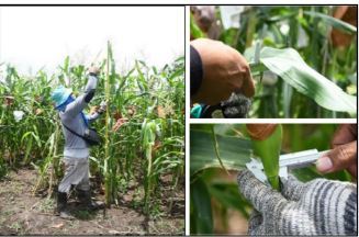
Abstract
Heirloom corn is one of the distinctive and significant cultivars grown in Kalinga Province. Factors such as shifting to GM corn, reduction of corn area, entry of OPV varieties, and the age of farmers impact heirloom corn varieties in Kalinga. This study looked into the ex-situ characterization of heirloom corn varieties and the effect of fertilizer on the growth and yield of heirloom corn. The genetic base of heirloom corn consisted of twenty-four accessions in ex-situ characterization with eighty-four descriptors. Quantitative and qualitative characteristics of white and purple heirloom corn were determined. The cultivars possess a unique qualitative character to distinguish their traits. The ex-situ characterization shows a slight variation in both quantitative and qualitative data for the cultivars tested. Field trials of heirloom corn planted in the two experiment areas showed no significant difference in the variables tested except in the plant height of heirloom corn under the Tabuk City condition. Sole ammonium phosphate (16-20-0) significantly affected the weight of the kernel and the yield of purple corn. The combined Ultimax organic and ammonium phosphate fertilizer did not yield significantly. Ammonium phosphate application is more effective in a sloped area than organic fertilizer. This phenomenon may be due to the faster release of inorganic fertilizer than organic fertilizer, which has a slow release; moreover, combining inorganic and organic fertilizers significantly improved heirloom corn. Farmers in Kalinga province must continuously cultivate and bequeath these cultivars to the young generation to preserve heirloom corn germplasm.
Introduction
Heirloom corn is one of the unusual and essential varieties in Kalinga Province. The study collected existing heirlooms, recorded them, and conducted field tests to conserve them. Zea mays, sometimes known as corn, is a significant food crop in the Philippines and is used as a staple meal by 20% of the country's citizens (World Food Program, 2012), particularly in the southern Visayas and Mindanao islands (Logroo et al., 1996). The product provides a livelihood for five million Filipinos. Corn comes in third overall regarding gross value added (GVA) in agriculture, behind only rice and coconut (PCARRD, 2006). The crop has significant economic value as a primary component of animal and poultry diets and as a raw material for various industrial goods, including starch oil, artificial sweeteners, and organic liquids (Siopongco et al., 1999). In 2010, Php70 billion, or US$1.7 billion, was spent on corn production (Bureau of Agricultural Statistics 1).
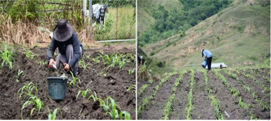
The two most common kinds of maize grown in the Philippines are yellow and white. In times of rice shortage, white maize is the most important staple food, especially for those living in rural regions (Gerpacio et al., 2004). Kalinga has flint and glutinous white, yellow, and purple heritage corn varieties. Although it does not receive much notice, purple corn has been commonly grown and enjoyed in other regions worldwide. White maize is eaten as a vegetable or a snack in regions where rice is the primary food. "Poor man's rice" is frequently used to describe white maize. Purple corn and other colored maize are not commonly grown in the Philippines (Greenpeace). Around 22 percent of the world's maize supply, according to data from 1990 to 2005,
Eighty-one countries agree that genetic deterioration in crops is mainly caused by replacing native species and varieties with better or foreign ones (FAO, 1997). Population pressure, environmental deterioration, legislation/policy, pests/weeds/diseases, shifting agricultural systems, and species overexploitation are additional factors contributing to genetic erosion. According to Greenpeace (2012), farmers may soon lose access to the traditional OPV of white corn for planting since genetically modified organism (GMO) contamination in maize types is already occurring among varieties patented and held by agrochemical corporations. It demonstrates the ineffectiveness of RA 7308, often known as the "Seed Industry Development Act," which requires the government to "conserve, maintain, and develop the plant genetic resources of the nation and supply the local communities with the genetic resources they need." It is essential to preserve landraces and crop wild relatives to preserve genetic resources for future crop improvement (Ford–Lloyd et al., 2011). Crop wild relatives and landraces offer valuable genetic material for breeding current improved lines, reducing the susceptibility of inbred crops to diseases and pests, enhancing performance, and adding distinctive features (Lopes et al., 2015). The heirloom corn of Kalinga is no different from other corn varieties in danger of being lost. Hence this study aims to conserve the Kalinga heirloom corn varieties. Furthermore, it looks into the ex-situ characterization (qualitative and quantitative) of the two heirloom corn varieties. The field trials aim to look into the effect of organic and inorganic fertilizers on the heirloom corn cultivars’ growth and yield production.
Source : Saving the heirloom Corn varieties of Kalinga Province
1 note
·
View note
Link
1 note
·
View note
Link
1 note
·
View note
Link
1 note
·
View note
Last Seen Blogs

uveneal
Uveneal

nemomonga-blog
Aya Log.

izumi-alastor
Untitled

myraelvira
Mysterious Things

mrketosis2525-blog
Untitled