#Ophthalmology open access journals
Text
International Journal of Clinical Images and Medical Reviews

International Journal of Clinical Images and Medical Reviews (ISSN 2771-6309) is a peer reviewed journal dedicated to publishing clinical images, Case Reports, Researches, Reviews, Mini Reviews, Short communications etc, from all sectors of science and medicine. The goal of this magazine is to disseminate information about new discoveries and treatments in science and medicine and accepts topics such as surgery, histology and cytology, oncology, dentistry, immunology, diagnostic method, clinical case, transplantation, ophthalmology, forensic science and all medicine-related fields.
International Journal of Clinical Images and Medical Reviews is open access journal, a peer reviewed journal with a large intellectual impact. Before publishing a manuscript, it goes through a rigorous editorial review procedure. The authors are encouraged to provide the manuscripts in accordance with the guidelines. The work can be submitted online using an online submission system. The manuscripts are peer-reviewed before being verified by the editors' panels. Finally, in order to preserve the highest quality of the information in this journal, only the quality contents are published.
For more details: https://ijcimr.org/
#ijcimr#short communicatiom#case reports#editorial#review article#research article#ISSN:2771-6309#mini review#clinical image
2 notes
·
View notes
Text
NEW ARTICLE The Covert Borderline
Psychology & Psychological Research International Journal (PPRIJ)
ISSN: 2576-0319
Title: The Covert Borderline
Authors: Vaknin S*
DOI: 10.23880/pprij-16000400
Volume 9, Issue 1
Abstract
I propose a new clinical entity, a hybrid between narcissistic and borderline personality disorders. It is not the comorbidity which it quite common in clinical settings. It is a personality disorder that seamlessly integrates features of both NPD and BPD.
Keywords: Personality Disorders; Covert Borderline
0 notes
Text
Read Journal Of Ophthalmology And Advance Research Online at Athenaeum Pub
Open access, a peer-reviewed online magazine called Journal of Ophthalmology and Advance Research, contributes to the scientific community by publishing high-calibre submissions about the most recent developments in the field of ophthalmology research. Researchers can post their fresh ideas or discoveries on the open access platform of the Journal of Ophthalmology and Advance Research. The Journal of Ophthalmology and Advance Research focuses on eye disorders such as myopia, neuro-ophthalmology, ocular migraine, ocular microbiology, and refractive surgery. Read Journal Of Ophthalmology And Advance Research Online at Athenaeum Pub. Visit now!
0 notes
Text
Journal of Clinical Images

Clinical Image Journal: Journal of Clinical Images accepting articles in the form of images, image case, clinical images journal, image of journal, clinical image illustrations journal, clinical research image journal, clinical research imaging journal etc. Journal paves a great platform to access the recent developments of the clinical and medical world and is employed for publishing the varied case reports & clinical images that pertains to many clinical and medical conditions. Clinical Image Journal: Journal of Clinical Images is an international peer reviewed open access journal focused on publishing the most complete, reliable source of research information, current developments, and clinically interesting, trainees and researchers in all surgical subspecialties, as well as clinicians in related fields. Clinical Image Journal: Journal of Clinical Images is internationally peer reviewed and provides major understanding of diagnosis of many diseases, their management and their therapeutic strategies that aims in improving health outcomes globally. Case reports and clinical images are required altogether areas of medicine and involves research using the human volunteers who are intended to contribute to the clinical and medical knowledge.
Journal Homepage: https://www.literaturepublishers.org/
Manuscript Submission
Authors are requested to submit their manuscript by using Online Manuscript Submission Portal:
(or) also invited to submit through the Journal E-mail Id: [email protected]
The mission of Clinical Image Journal: Journal of Clinical Images is to publish, in a timely manner, the very best clinical research around the world with special attention to the impact of medical imaging on patient care. Clinical Image Journal: Journal of Clinical Images publications cover all imaging modalities, radiology issues related to patients, policy and practice improvements, and clinically-oriented imaging physics and informatics. Clinical Image Journal: Journal of Clinical Images is a valuable resource for practicing radiologists, radiologists-in-training and other clinicians with an interest in imaging.
Papers are carefully peer-reviewed and selected by our experienced subject editors who are leading experts spanning the range of imaging sub-specialties, which include: Body Imaging- Breast Imaging- Cardiothoracic Imaging- Imaging Physics and Informatics- Molecular Imaging and Nuclear Medicine- Musculoskeletal and Emergency Imaging- Neuroradiology- Practice, Policy & Education- Pediatric Imaging- Vascular and Interventional Radiology.
Clinical Image Journal: Journal of Clinical Images Scope
Case reports / Clinical Images can be prospective or retrospective and examine the effects of an intervention in more than one patient. All case reports and clinical Images submitted need to comply with the relevant reporting criteria. It is dedicated to publishing Medical Case Reports, Clinical Images, Case Series and Clinical Videos. The following classifications and topics related to it will be considered for publication in the Journal but not limited to the following fields.
Neurology Image Journal, Oncology Image Journal, Dentistry Image Journal, Surgery Image Journal, Cardiology Image Journal, Nutrition and Dietetics Image Journal, Ophthalmology Image Journal, Gastroenterology Image Journal, Internal Medicine Image Journal, Nephrology Image Journal, Palliative Care Image Journal, Physiotherapy Image Journal, Radiation Oncology Image Journal, Sleep Disorders Image Journal & Sleep Studies Image Journal, Anesthesiology Image Journal, Emergency Medicine Image Journal and Critical, Forensic Image Journal and Legal Medicine Image Journal, Infectious Disease Image Journal, Infertility Case reports, Neurological Surgery Image Journal, Obstetrics Image Journal and Gynecology Image Journal, Otolaryngology Image Journal, Pharmacology Image Journal and Therapeutics Image Journal, Physical Medicine Image Journal & Rehabilitation Image Journal, Radiology Image Journal, Dermatology Image Journal, Endocrinology Image Journal, Diabetes Image Journal & Metabolism Image Journal, Orthopedics Image Journal & Rheumatology Image Journal, Pathology- Anatomic & Clinical Image Journal, Pulmonary Image Journal Disease, Preventive Medicine Image Journal, Respiratory Medicine Image Journal, Urology Image Journal, Oral Medicine Image Journal, ENT Image Journal, Geriatric Medicine Image Journal, Maxillofacial Surgery Image Journal, Neonatology Image Journal, Nuclear Medicine Image Journal, Pain Management Image Journal, Pediatrics Image Journal, Psychiatry Image Journal, Sexual Health Image Journal, Vascular Medicine Image Journal, Family Medicine Image Journal and Public Health Image Journal, Allergy Image Journal & Immunology Image Journal, Diabetology Image Journal, Hematology Image Journal.
0 notes
Text
Journal of Clinical and Medical Case Reports

Journal of Clinical and Medical Case Reports publishes clinical case reports, medical case series, medical case studies, medical case reports and clinical images for publication that fall under the scope of all clinical and medical studies. Journal of Clinical and Medical Case Reports mainly focuses on symptoms, signs, diagnosis, treatment, and follow-up of patient disease in different areas of the journal in diagnostic case report and treatment.
Journal Homepage: https://www.literaturepublishers.org/
Journal of Clinical and Medical Case Reports is a peer-reviewed open access high impact factor indexed Journal that publishes highly cited research work conducted as case reports in the medical field on various types of diseases, covering their respective clinical journal case reports, medical journal case report, clinical reports, medical case reports, clinical images, clinical case reports, journal of medical case reports and diagnosis issues.
Scope and Keywords: Journal of Clinical and Medical Case Reports, Open Journal of Clinical and Medical Case Reports, Journal of Medical Case Reports, Clinical and Medical Case Reports, Journal of Clinical Images and Medical Case Reports, Journal of Clinical Studies & Medical Case Reports, Journal of Clinical and Medical Case Studies, International Journal of Clinical and Medical Cases, Journal of Clinical Medicine, Clinical Case Reports, International Medical Case Reports Journal, Archives of Clinical and Medical Case Reports, Case Reports - A journal for medical case reports, International Journal of Clinical Case Reports and Reviews, Japanese Journal of Clinical and Medical Case Reports etc.
Journal of Clinical and Medical Case Reports
Journal publishes only high quality articles from all over the world. Journal of Clinical and Medical Case Reports follows double blinded peer review process. All Editors are active and Editorial Board Members belonging to reputed institutions from abroad. They are senior faculty members, doctors, scientist and research fellows etc. Journal regularly releasing issues with good number of articles in the form of clinical images and case reports.
Scope of Clinical and Medical Case Reports Journal
Authors can also find this journal in their scope on the basis these keywords: medical case reports journals, journal of medical case reports, clinical image, cardiology case reports, case reports cardiology, case reports in cardiology, case reports pediatrics, pediatrics case reports, ent journal, case reports hematology, hematology case reports, journal of otolaryngology head and neck surgery, orthopaedics & traumatology, case reports gastroenterology, case reports in gastroenterology, gastroenterology case report, clinical case report journal, International journal of surgery case reports, case images, dermatology case reports, case report in ophthalmology, case report ophthalmology, case reports in surgery, clinical image journal, journal of surgical case reports, ophthalmology case report, journal of clinical imaging science, literature publishers, cardiology case report journals, journal of traumatology, case reports in nephrology, case reports nephrology, nephrology case reports, clinical images in medicine, journal of pediatric surgery case reports, medical image analysis journals, journal of medical case reports impact factor, otolaryngology case reports, clinical case reports impact factor, case reports otolaryngology, surgical case reports, journal of orthopedic case reports, case reports in neurological medicine, best case report journals, journal of otology and laryngology and, clinical imaging impact factor etc.
Medical Case Report Journal scope also includes medical advancements with an aim towards special techniques that are implementing in all aspects of the human anatomy journal. The body image journal is running with a strong desire to provide knowledge on recent scientific research and advances in the field of Clinical and Medical Studies. The aim of the clinical imaging journal is to collect an article in the Journal of Clinical and Medical Case Reports across all clinical imaging science, medical imaging science and clinical fields, thereby integrating international medical case reports and clinical knowledge.
We feel honored to associate with and invite scientists and researchers to submit their original research/ medical case report journal/ body imaging journal/ clinical imaging journal/ clinical imaging science in International journal of clinical and medical images and case reports work for publication in literature publishers: journal of clinical and medical case studies and reports. This journal considers articles in the form of a research article, review article, short communication, opinion, Image, Case reports and commentary.
Journal of Clinical and Medical Case Reports covers all the areas of Medical Science Journal that includes: case reports in oncology, oncology case reports journal, case reports in cardiology, journal of cardiology case reports, international journal of surgery case reports, case reports in surgery, journal of surgery case reports, general surgery case report, surgical case reports journal, surgery case reports journal, journal of dermatological case reports, case reports in dermatology, case reports in endocrinology, case reports endocrinology, case reports in gastroenterology, gastroenterology case report journals, case reports in hematology, case reports in nephrology, orthopedic surgery case reports, journal of orthopedic case reports, case reports in pediatrics, journal of pediatric surgery case reports, case reports in microbiology, clinical microbiology case reports, case reports in genetics, case reports in toxicity, case reports in neuroscience, case reports in ophthalmology, case reports in andrology and gynecology, case reports in dentistry, case reports in odontology, case reports in otolaryngology, case reports in ENT, case report in head and neck surgery etc.
Journal of Clinical and Medical Case Reports encourages authors and scientists all over the world to submit their work related to various diseases, clinical trials, radiology, surgery, basic research, epidemiology, and palliative care. At a time when the research on drug delivery is taking place at a tremendous phase.
Manuscript Submission
Authors are requested to submit their manuscript by using Online Manuscript Submission Portal:
(or) also invited to submit through the Journal E-mail Id: [email protected]
0 notes
Text
Ophthalmology Online Learning Resources
An aspiring ophthalmologist, or doctor who wishes to upgrade his or her knowledge of ophthalmology, can resort to online learning resources. The biggest merit of online resources is the fact that they can be accessed at any time, at any hour, from any place. That means that an individual can gain insights from video lectures, online journals, research publications, discussion forums, and community activities, as well as the procedures projected on video lectures, as per their convenience and flexibility. The best video lectures for ophthalmology, therefore help the aspirant gain knowledge about the latest advancements, professional practices from around the world, the latest eyecare gadgets, and more. Getting enrolled in an ophthalmology undergraduate online course paves the way for learning from video lectures in ophthalmology and opens doors to endless opportunities.
https://diginerve.blogspot.com/2023/07/ophthalmology-online-learning-resources.html
#ophthalmology undergraduate online course#Online ophthalmology courses#best ophthalmology video lectures
0 notes
Text
Video Journal: Clinical Videos Journal and Medical Videos Journal

Video Journal: Clinical Videos Journal and Medical Videos Journal accepting articles with clinical videos, medical videos, surgery videos or surgical videos for publication. Journal covers the publication of new information by publishing original research from all areas of clinical research video journal, medical research video journal and basic sciences including clinical medicine video journal, clinical trials video journal, experimental research video journal, epidemiology, preventive medicine, translational medicine and rural health. Every written manuscript submitted for publication to Clinical Video Journal must be accompanied by single or multiple videos.
Journal Homepage: https://www.literaturepublishers.org/
Journal of Clinical Videos is a publication of scholarly work in a video format. Researchers get to demonstrate their work to the audience by using a live camera shoot. Since these medical video journals let researchers showcase their work in a more visual way, the impact of their research is increased.
Links of Published Clinical Video Articles
Journals in clinical video format are broadly grouped into scientific video journals; medical video journals; technical video journals; and arts, humanity and management video journals. Each of these are further available in diverse areas including medicine, science, technology, arts, humanities, biology, management, chemistry, physics, neuroscience, engineering, genetics, cancer research, immunology and infection, developmental biology, behavior, environment, bioengineering, biochemistry, psychology, clinical skills, and many more. Even under each of these specializations, there are different sub-specializations.
For instance, under medical video journals, there are videos of cardiology video journal, oncology video journal, neurology video journal, clinical ophthalmology video journal, biomedical science video journal, cardiothoracic video journal and vascular surgery video journal, internal medicine video journal, gastroenterology video journal, pediatrics video journal, pathology video journal, radiology video journal, surgery video journal, obstetrics video journal and gynecology video journal, otolaryngology video journal, orthopedic surgery video journal, pathology video journal, etc.
Open Access Clinical & Medical Video Journal
Open access video journals are available to readers, libraries, institutions, and organizations without any barriers like payments, licensing, subscriptions and copyrights. Academicians and scientists can freely access these journals to broaden their understanding of the current happenings in their respective disciplines.
Video demonstrations enhance reproducibility and productivity, aid better and quicker understanding of experimental methods and scientific concepts; disseminate research widely; save laboratory expenses; increase learning speed in lab and class; promote consistent growth in learning outcomes, student success and STEM retention. Thus the different types of video journals including medical video journals; scientific video journals; technical video journals; and arts, management and humanity video journals are a boon to the research, student and teaching communities. You can check out and make the best use of the medical Video journal available at the Clinical Images and Case Reports Journal.
Manuscript Submission
Authors are requested to submit their Clinical Video articles only by E-mail to the Editorial Office at: [email protected]
0 notes
Text
Journal of Clinical Case Reports Medical Images and Health Sciences | ISSN: 2832-1286
The JCRMHS is an open access and a peer-reviewed journal for publishing research work in the form of Clinical Images, Case Reports, Case Studies, Researches, Technical Notes, Review Opinion, Brief Notes, Reviews etc., covering a wide range of Scientific and Medical Sciences pertaining to various fields of Clinical And Medical Sciences.
The objective of this magazine is to disseminate data about new discoveries and treatments in science and medicine. We acknowledge topics such as, Surgery, Histology and Cytology, Oncology, Dentistry, Immunology, Diagnostic Method, Clinical Case, Transplantation, Ophthalmology, Forensic Science and all medicine related fields.
JCRMHS aims to encourage Clinical and Medical Professionals, Scientists, Doctors, Professor’s academicians for the publication of latest information for reporting unique, unusual and rare cases to understand the disease process, its diagnosis and management.
Journal of Clinical Case Reports Medical Images and Health Sciences is an international, open access, peer reviewed, online journal, publishing high-quality articles in all specialties and related subspecialties.
The journal is exclusively dedicated to publishing Case Series, Case Reports, Clinical Images, Letters to the Editor, Research and Review Articles which enhance understanding of disease processes, its diagnosis, management and clinicopathologic correlations.
#Case Reports#JCRMHS#Health#health sciences#medical case reports#Clinical Images#Medical Images#Clinical case reports
1 note
·
View note
Text
Clinical Images and Medical Case Reports Journal
Clinical Images and Medical Case Reports Journal is a peer-reviewed open access journal of medical case reports established internationally which provides a platform to publish clinical images and case reports pertaining to medical conditions and aims to keep scientists, clinicians and medical practitioners, researchers, and students informed and updated on the ongoing research in the relevant area. Outstanding quality articles are welcome to maintain the highest standard of the journal and to achieve high impact actor.

The Journal provides a platform for physicians and medical aspirants to share different results in form of images and case report which have been encountered in different medical sub-specialties or cases which leads to higher understanding of the medical conditions, medicines, diagnosis and management.
The journal presents different possibilities to enhance worldwide health outcomes. Our Medical Case Reports Journal is an important educational resource offering a high volume of clinical, medical, surgical, dermatology, gastroenterology, endocrinology, hematology, nephrology, orthopaedic, pediatrics, microbiology, genetics, toxicity, Neuroscience, ophthalmology, andrology & gynecology, odontology, ENT, otolaryngology and dental cases in all disciplines.
The Journal of Clinical Case Reports is an Open Access Scientific Journal that offers an interesting publishing platform globally and aims to keep scientists, clinicians and medical practitioners, researchers, and students informed and updated on the ongoing research in the relevant area.Outstanding quality articles are welcome to maintain the highest standard of the journal and to achieve high impact factor.
Clinical Case Reports Journals is distinctive to other case report journals. Literature Publishers point is to straightforwardly improve worldwide wellbeing results and offer clinical information utilizing case reports to pass on significant best practice messages. We invite case reports, clinical pictures, and procedural recordings from all regions of Medicine, Nursing, Dentistry, Psychology, Medical Ethics, Social Work and Veterinary Science.
Clinical Case Reports journals intends to publish cases which illustrate the utilization of significant deliberate surveys. Clinical Case Reports additionally has a closed association with the Cochrane Collaboration and case report entries which identify with Cochrane systematic surveys are a need for the Journal. On the off chance that your accommodation has a significant clinical message we might want to understand it!
Medical Case Reports Journal conveys most significant assortment of cases in all controls with the goal that healthcare insurance experts, analysts and others can without much of a stretch find clinically significant data on normal and uncommon conditions. Submit your contribution via Literature Publishers or get more information than contact.
One of the largest and most authoritative collections of online case reports journal and research resources, covering life, health, social, and physical sciences. Literature Publishers is a member of the Open Access Initiative, specializing in peer-reviewed Medical Case Reports Journal. View case reports journal or submits your research for publishing.
Manuscript Submission
Authors are requested to submit their manuscripts using the online manuscript submission portal:
https://www.literaturepublishers.org/submit.html
(Or) Journal is invited to submit via e-mail id:- [email protected]
More about information for Medical Case Reports Journal then Visit our site : -
http://www.literaturepublishers.org/
#case reports journal#clinical case reports journal#medical case reports journal#journal of medical case reports#international medical case reports journal
0 notes
Text
Global Research Journal of Paediatrics and Child Health [GRJPCH]
Global Research Journal of Pediatrics and Child Health is exceptional among peer-reviewed journal publications in that it publishes articles on the fundamental research on every element of pediatrics as well as clinical experiments that significantly advance our understanding of the subject. The journal publishes research, case studies, reviews, mini-reviews, and letters regarding manuscripts that have already been published. It includes all public health-related research and services, with a primary care focus. We make an effort to disseminate the highest caliber of information for raising the standard of child care.

Pediatricians can utilise the journal's publication as a platform to identify the problem and treat the kids. We work hard to deliver top-notch instructional material that respects kids' development in a dynamic environment. We seek fruitful collaboration with medical professionals and neighbourhood researchers who are committed to child care. Children should have specific healthcare requirements. The journal aims to improve the health, care, and well-being of infants, toddlers, kids, and families via cooperative medical care, public education, and ground-breaking research that enhances caring for lives and community service.
About Journals:
Pediatrics & Health Research, an international open access, peer-reviewed publication, promotes the most recent advancements in the investigation of children's health.
The journal focuses on the physical and mental development of kids by publishing the most recent research on the following subjects:
Child Health, Pediatric Cardiology, Pediatric Surgery, Pediatric Medicine, Pediatric Allergy, Pediatric Nephrology, Pediatric Trauma, Pediatric Gastroenterology, Pediatric Dermatology, Pediatric Pulmonology, Pediatric Ophthalmology, Pediatric Cancer, Pediatric Immunizations, Pediatric Diseases, Pediatric Obesity, Pediatric Neurology, Pediatric Critical Care, Pediatric Dentistry, Pediatric Nutrition, Pediatric Allergy, Pediatric Nephrology, Pediatric Critical Care, Pediatric Dentistry, Pediatric Nutrition, Pediatric Dentistry, Pediatric Nutrition, Pediatric
The journal promotes innovations in the aforementioned fields through the publication of original articles, reviews, short communications, rapid communications, letters to the editor, abstracts, addenda, announcements, article commentaries, book reviews.
The editorial manager system makes it easy to submit, review, and publish articles. Thorough peer assessment of manuscripts would guarantee the highest standards possible in the sector. This journal's objective is to offer a forum for academics and researchers from across the world to communicate, discuss, and promote various new concerns and advancements in many fields of paediatrics and health research. The publications will be kept in an electronic database, reviewed by a team of scientists and anonymous reviewers, and published each month in HTML and PDF formats.
AIM & SCOPE:
With a focus on manuscripts from nations whose research may be under-represented in the current literature, It also emphasizes significant research relating to the quality of child health care, health care policy, and the organization of child health services. It also contains significant methodological publications to support research in child health and education, as well as systematic reviews of primary care initiatives.
All approved articles in The Global Journal of Pediatrics will be immediately and indefinitely available for free reading and downloading, promoting the most recent advancements in pediatric medicine, child health, advocacy, and policy.
Submit the manuscript at https://ucjournals.com/global-research-journal-of-paediatrics-and-child-health/ or send it as an e-mail attachment to the Editorial Office at
Reference and blogs
https://unifiedcitationjournals.blogspot.com/
#seniorcare#healthylife#hospitals#life#technology#india#rn#pharmacist#motivation#healthandwellness#healthcare#childhealth#doctor#dentist#lifestyle#patientcare#business#insurance#nursesofinstagram#stayhealthy#medicine#coronavirus#health#artificialintelligence
0 notes
Text
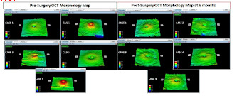
Macular Abnormality after Successful Surgery for Idiopathic Macular Hole Assessed Using Optical Coherence Tomography | Crimson Publishers
Macular Abnormality after Successful Surgery for Idiopathic Macular Hole Assessed Using Optical Coherence Tomography by Nazimul Hussain in Medical & Surgical Ophthalmology Research
Purpose: Macular changes following Internal Limiting Membrane peeling after successful Macular hole closure using optical coherence tomography.
Methods: 5 eyes of 5 patients were included in the study. The inclusion criteria were idiopathic full thickness macular hole and completed at least 6 months follow up. All patients underwent optical coherence tomography assessment before and after surgery until 6 months. Macular thickness and morphology map was analyzed before and after surgery.
Results: There were 3 males and 2 females. The age range from 45 to 59 years. Conspicuous irregular surface of the inner retina on Optical Coherence Tomography surface topography was evident in all eyes at 6 months. This was also associated with downward slope of the temporal macula seen on surface topography. There was trend towards decrease in subfield thickness especially in the T1, S1, I1 and N1 6 months after surgery. The average decrease was -60.4 microns in T1, -20.4 microns in S1, -13.0 microns in I1 and -23.4 microns in N1.
Conclusion: In this small case series brilliant blue assisted ILM peeling after successful macular whole closure showed evidence of decrease in macular thickness in all first subfield quadrant and downward slope of the temporal macula as well as conspicuous irregularity of the inner retina.
https://crimsonpublishers.com/msor/fulltext/MSOR.000517.php
For more open access journals in Crimson Publishers please click on link: https://crimsonpublishers.com
For More Articles on Medical Research Please click on: https://crimsonpublishers.com/msor/
#Crimson Publishers#Crimson Publishers Journals#Crimson Publishers LLC#Journal of Ophthalmology and Ophthalmic Surgery#Ophthalmology open access journals
18 notes
·
View notes
Text
Lupine Publishers|To Compare the Retinal and Choroidal Thickness in Fuchs Uveitis Syndrome Patients with Healthy Controls Using Optical Coherence Tomography Seen at Tertiary Care Center
To Compare the Retinal and Choroidal Thickness in Fuchs Uveitis Syndrome Patients with Healthy Controls Using Optical Coherence Tomography Seen at Tertiary Care Center

Abstract
Purpose: To compare the retinal and choroidal thickness in Fuchs Uveitis Syndrome patients with healthy controls using optical coherence tomography seen at tertiary care center.
Method: 16 patients with unilateral Fuchs Uveitis Syndrome (FUS) and 20 healthy control subjects were enrolled in this retrospective study. Spectral domain optical coherence tomography was used to measure the central foveal thickness (CFT), retinal nerve fiber layer (RNFL) thickness and sub foveal choroidal thickness (SFCT) of the eyes with FUS and compared with the unaffected fellow eye and healthy controls.
Results: The mean age of the patients was 35.2 ± 8 years. Seven patients (60%) were male, and nine (40%) were female. Diffuse stellate keratic precipitates (KPs) were seen in all patients. Mild anterior chamber reaction was noted 62.5%. Heterochromia was observed in 87.5% of the eyes, cataract in 43.8% of the eyes, and vitreous cells and debris in 37.5% of the eyes. The mean CFT was (249.7 ± 27.4𝜇m) in affected eyes, (251.5± 23.1 𝜇m) in unaffected eyes of FUS and (253.3 ± 29.2 𝜇m) in healthy control which were statistically insignificant on comparison (𝑝 value > 0.05). The mean SFCT was significantly thinner in eyes with FUS (272.47 ± 13.12 μm) than in the fellow eyes without FUS (316.37 ± 16.13 μm; p = 0.04). and control (320.27 ± 18.15). The average RNFL thickness was found to be (106 ± 14.2 μm) in eyes with FUS, (108 ± 16.5μm) in eyes without FUS and (112 ±13.8μm) in healthy control which were statistically not significant (p value > 0.05).
Conclusion: We conclude that affected eyes in patients with FUS tend to have thinner choroids as compared to unaffected fellow eyes which is consistent with previous studies. It might be associated with chronic inflammation induced choroidal is chaemic changes. Future studies using swept source OCT will further improve precision to get choroid findings more accurate.
Keywords:Choroid; Fuchs Uveitis Syndrome; SD-OCT; Retinal Nerve Fiber Layer
Introduction
Fuchs’ uveitis syndrome (FUS) is an intraocular inflammatory condition that involves anterior segment, lens, vitreous humor and optic disc. It accounts for 2-11% of all uveitis cases [1]. Although many genetic, immunological, vascular and sympathetic theories have been proposed, recent studies show evidence of rubella or herpes simplex virus, toxoplasma association in some cases of FUS [2]. It is a frequently unilateral and typical age at presentation is 30-40 years [3,4]. The diagnostic criteria include stellate keratic precipitates (KPs) scattered diffusely all over cornea, chronic lowgrade anterior chamber reactions, iris atrophy with or without heterochromia, absence of posterior synechiae, vitreous opacities and macular edema [5]. It affects both genders equally and the prognosis is usually good [6]. Iris atrophy and heterochromia are due to atrophy and depigmentation of all iris layers, although it is not pathognomonic. Recently, fluorescein angiography (FA) had provided better information about the posterior segment involvement in FUS [7-9]. Optical Coherent Tomography (OCT) is a routinely employed noninvasive tool in detecting most posterior pole retinal abnormalities. The Enhanced Depth Imaging (EDI) in spectral domain (SD) OCT and swept source (SS) OCT allow visualizing the choroid more accurately [10,11]. Some studies documented the changes in choroidal thickness using EDI-OCT in uveitic conditions, such as Vogt–Koyanagi–Harada (VKH) disease, Behçet disease (BD), ocular sarcoidosis providing remarkable information about disease activity [12,13]. The natural course of the disease is slow to progress, without substantial reduction of visual acuity until significant vitreous opacities or cataract develop. FUS is often misdiagnosed or diagnosed delay leading to posterior subcapsular opacity and elevated IOP mainly due to long-term corticosteroid therapy. There are very few reports in the literature comparing macular, RNFL and choroidal thickness in FUS patients. The purpose of the current study was to measure central foveal thickness (CFT), subfoveal choroidal thickness (SFCT) and retinal nerve fiber layer (RNFL) thickness, in patients with FUS and to compare these with the unaffected fellow eyes and the age, sex and refractive equivalent-matched healthy controls. To the best of our knowledge, it is the first comparative study to evaluate SFCT and other parameters in FUS using spectral domain SD-OCT on FUS from central India.
Material and Methods
This retrospective study was conducted at a tertiary care center from central India. The medical records of 16 patients with FUS and 20 healthy controls between september 2018 and august 2021 were reviewed. Data including age, sex, ocular and medical history, ophthalmic examination, laboratory work-up and OCT parameters were retrieved from patient record. Aii patients had best-corrected visual acuity (BCVA), slit-lamp biomicroscopy, Goldmann applanation tonometry and dilated fundoscopy. The inclusion criteria were diagnosed unilateral FUS based on clinical findings and fundus sufficient for its visualization on clinical examination. Bilateral FUS, history of previous ocular surgery, eyes with dense cataract or media opacity obscuring the visualization of choroid, presence of a coexisting ocular or systemic disease and use of any topical or systemic medications. were excluded from study. The control group consisted of age-, sex-, and refractive equivalent-matched healthy controls who visited ophthalmology clinic for routine examination. The laboratory investigations included complete blood counts, angiotensin converting enzyme, mantoux test and venereal disease research laboratory tests etc. To exclude other causes of anterior or intermediate uveitis. The CFT and RNFL thickness of eyes with FUS were evaluated with SD-OCT and compared with the unaffected fellow eyes and healthy controls. The EDI mode was used to evaluate SFCT. SFCT was defined as the vertical distance from the outermost hyperreflective line of the retinal pigment epithelium (RPE) to choroid-sclera junction under the center of the fovea and was measured using the calipers available within the software. The RNFL thickness (from the inner margin of the internal limiting membrane to the outer margin of the RNFL layer) was automatically segmented using software version. Average RNFL was used for analysis. Statistical analyses were performed with R version 4.0.5. Data were recorded as the mean ± standard deviation (SD). Quantitative data was analyzed using one way analysis of variance (ANOVA) test. An independent 𝑡-test and Chi-square test were used to compare variables between patients with FUS and healthy control subjects. A value of 𝑝< 0.05 was considered to be statistically significant for all analyses.
Results
16 patients with FUS (9 females and 7 males) and 20 healthy control subjects (11 females and 9 males) were included for analysis in this study. The mean age of FUS patients was 35.2±8 years, while it was 38.4± 6years for controls, which was statistically insignificant (𝑝>0.05) (Table 1). Gender differences in both groups were also statistically insignificant (𝑝>0.05). Blurred vision and floaters were the most frequent presenting symptoms {5 patients, (31.3%) and 4 patients (25%) respectively}. Other symptoms included red eye (1 patient, 6.3%), photophobia (1 patient, 6.3%). Three patients were asymptomatic (18.75%), and the diagnosed during a routine eye examination (Table 2). Iris atrophy was present in 5 patients. Heterochromia was present in 14 patients. Cataract was present in 7 patients. Stellate keratic precipitates were noted in all patients. Mild anterior chamber reactions were seen in 10 patients. Vitreous cells or debris were observed in 6 patients (Table 3). The cause of visual impairment was cataract in all cases. The central foveal thickness was 249.7±27.4𝜇m in the affected eyes, 251.5±23.1 𝜇m in the unaffected eyes of FUS patients, and 253.3±29.2 𝜇m in control subjects. There were no significant differences in central foveal thickness between the affected and unaffected eyes of FUS patients (𝑝 = 0.93) or between the affected eyes of FUS patients and the eyes of control subjects (𝑝 = 0.9). The mean SFCT was found to be 272.47 ± 13.12μm in eyes with FUS and 316.37± 16.13μm in eyes without FUS (p = 0.04) and 320.27 ± 18.15𝜇m in the eyes of healthy control subjects. There was choroidal thinning at fovea in the affected eyes of FUS patients compared with the unaffected eyes or control subjects, Average RNFL thickness was 106 ±14.2𝜇m in the affected eyes of FUS patients, 108±16.5𝜇min the unaffected eyes of FUS patients, and 112 ± 13.8𝜇m in the eyes of healthy control subjects (Table 4). No significant differences in RNFL thickness were observed between the affected and unaffected eyes of FUS patients (𝑝 = 0.92) or between the affected eyes of FUS patients and the eyes of control subjects (𝑝 = 0.76)
Table 1: Demographic analysis.
Table 2: Presenting symptoms.
Table 3: Characteristic of FUS patients.
Table 4: Thickness of fovea, sub foveal choroid, and RNFL in FUS & control.
Discussion
To our knowledge, very few comparative studies on FUS patients have been done till yet. In the present study, we compared central foveal thickness, retinal nerve fiber layer and subfoveal choroidal thickness between the affected eyes, the unaffected eyes of FUS patients and healthy controls from central India. Although we found choroidal thinning at fovea in the affected eyes of FUS patients compared with the unaffected eyes of FUS patients or control subjects, there was no statistically significant difference in RNFL and central foveal thickness. FUS was first described in 1906, recent studies have focused on various viral agents and the etiology of FUS remains controversial. In FUS, persistence of chronic lowgrade inflammation for years leads to various degrees of atrophy of the iris and ciliary body. As the choroid being more vulnerable to the effects of the inflammatory and vascular diseases than other tissues, imaging of choroid is important for understanding the pathophysiology of various diseases. EDI mode using SD-OCT devices enables cross-sectional, high resolution better visualization of the choroid. Many studies have documented the effect of various acute and chronic ocular inflammatory conditions on choroidal abnormalities. Nakayama et al. showed that choroidal thickness as a marker for the degree of choroidal inflammation in acute Vogt- Koyanagi-Harada disease using EDI-OCT [14]. Kim et al. found an increase in subfoveal choroidal thickness in the acute phase of Behcet’s posterior uveitis [15]. Multiple studies suggest that increased blood flow and choroidal effusion is the mechanism responsible for choroidal thickening in acute inflammation [16,17]. However, Coskun et al. observed the thinning of subfoveal choroid in chronic Behcet’sassociated posterior uveitis probably due to chronic inflammation induced fibrosis [18]. Very few studies from Turkey and Italy like Balci and Ozsutsus, Kardes et al. and Carquaglia et al. showed the comparison between the affected and fellow eye in FUS patients [19-21] (Table 5). The study by Kardes et al. had findings consistent with the study of Balci and Ozsutsus. We propose that chronic anterior chamber and vitreous inflammation in eyes with FUS may affect choroidal perfusion, which may result in ischaemic changes leading to atrophy and fibrosis of the choroid tissue, thereby reducing choroidal thickness. Limitations of our study is being retrospective nature and small sample size. We have used SD OCT in this study and with advent of new imaging methods designed specifically for the choroid, SS OCT which allows examination of the choriocapillaris, and larger choroidal vessels more clearly can further improve precision to determine the inner and outer boundaries of the choroid findings in the future. To conclude, we found subfoveal thinner choroid in FUS as compared to uninvolved fellow eyes, but we could not find a statistically significant comparison of foveal and RNFL thickness in FUS versus unaffected eye. Our findings are also consistent with those of previous studies. As this is the first analysis of its kind from central India, longitudinal studies with large sample size would help in more understanding of the effect of chronic inflammation on the choroid in FUS in future.
Table 5: Comparison between different studies of mean sub foveal choroidal thickness (SFCT).
For more information about Trends in Ophthalmology Open Access Journal archive page click on below link
https://lupinepublishers.com/ophthalmology-journal/archive.php
For more information about lupine publishers page click on below link
https://lupinepublishers.com/index.php
#lupine publishers#lupine publishers group#lupine publishers LLC#trends in ophthalmology open access journal#tooaj
2 notes
·
View notes
Text
Clinical Image Journal

Clinical Image Journal: Journal of Clinical Images accepting articles in the form of images, image case, clinical images journal, image of journal, clinical image illustrations journal, clinical research image journal, clinical research imaging journal etc. Journal paves a great platform to access the recent developments of the clinical and medical world and is employed for publishing the varied case reports & clinical images that pertains to many clinical and medical conditions. Clinical Image Journal: Journal of Clinical Images is an international peer reviewed open access journal focused on publishing the most complete, reliable source of research information, current developments, and clinically interesting, trainees and researchers in all surgical subspecialties, as well as clinicians in related fields. Clinical Image Journal: Journal of Clinical Images is internationally peer reviewed and provides major understanding of diagnosis of many diseases, their management and their therapeutic strategies that aims in improving health outcomes globally. Case reports and clinical images are required altogether areas of medicine and involves research using the human volunteers who are intended to contribute to the clinical and medical knowledge.
Journal Homepage: https://www.literaturepublishers.org/
Manuscript Submission
Authors are requested to submit their manuscript by using Online Manuscript Submission Portal:
(or) also invited to submit through the Journal E-mail Id: [email protected]
The mission of Clinical Image Journal: Journal of Clinical Images is to publish, in a timely manner, the very best clinical research around the world with special attention to the impact of medical imaging on patient care. Clinical Image Journal: Journal of Clinical Images publications cover all imaging modalities, radiology issues related to patients, policy and practice improvements, and clinically-oriented imaging physics and informatics. Clinical Image Journal: Journal of Clinical Images is a valuable resource for practicing radiologists, radiologists-in-training and other clinicians with an interest in imaging.
Papers are carefully peer-reviewed and selected by our experienced subject editors who are leading experts spanning the range of imaging sub-specialties, which include: Body Imaging- Breast Imaging- Cardiothoracic Imaging- Imaging Physics and Informatics- Molecular Imaging and Nuclear Medicine- Musculoskeletal and Emergency Imaging- Neuroradiology- Practice, Policy & Education- Pediatric Imaging- Vascular and Interventional Radiology.
Clinical Image Journal: Journal of Clinical Images Scope
Case reports / Clinical Images can be prospective or retrospective and examine the effects of an intervention in more than one patient. All case reports and clinical Images submitted need to comply with the relevant reporting criteria. It is dedicated to publishing Medical Case Reports, Clinical Images, Case Series and Clinical Videos. The following classifications and topics related to it will be considered for publication in the Journal but not limited to the following fields.
Neurology Image Journal, Oncology Image Journal, Dentistry Image Journal, Surgery Image Journal, Cardiology Image Journal, Nutrition and Dietetics Image Journal, Ophthalmology Image Journal, Gastroenterology Image Journal, Internal Medicine Image Journal, Nephrology Image Journal, Palliative Care Image Journal, Physiotherapy Image Journal, Radiation Oncology Image Journal, Sleep Disorders Image Journal & Sleep Studies Image Journal, Anesthesiology Image Journal, Emergency Medicine Image Journal and Critical, Forensic Image Journal and Legal Medicine Image Journal, Infectious Disease Image Journal, Infertility Case reports, Neurological Surgery Image Journal, Obstetrics Image Journal and Gynecology Image Journal, Otolaryngology Image Journal, Pharmacology Image Journal and Therapeutics Image Journal, Physical Medicine Image Journal & Rehabilitation Image Journal, Radiology Image Journal, Dermatology Image Journal, Endocrinology Image Journal, Diabetes Image Journal & Metabolism Image Journal, Orthopedics Image Journal & Rheumatology Image Journal, Pathology- Anatomic & Clinical Image Journal, Pulmonary Image Journal Disease, Preventive Medicine Image Journal, Respiratory Medicine Image Journal, Urology Image Journal, Oral Medicine Image Journal, ENT Image Journal, Geriatric Medicine Image Journal, Maxillofacial Surgery Image Journal, Neonatology Image Journal, Nuclear Medicine Image Journal, Pain Management Image Journal, Pediatrics Image Journal, Psychiatry Image Journal, Sexual Health Image Journal, Vascular Medicine Image Journal, Family Medicine Image Journal and Public Health Image Journal, Allergy Image Journal & Immunology Image Journal, Diabetology Image Journal, Hematology Image Journal.
1 note
·
View note
Text
Journal of Clinical Images

Clinical Image Journal: Journal of Clinical Images accepting articles in the form of images, image case, clinical images journal, image of journal, clinical image illustrations journal, clinical research image journal, clinical research imaging journal etc. Journal paves a great platform to access the recent developments of the clinical and medical world and is employed for publishing the varied case reports & clinical images that pertains to many clinical and medical conditions. Clinical Image Journal: Journal of Clinical Images is an international peer reviewed open access journal focused on publishing the most complete, reliable source of research information, current developments, and clinically interesting, trainees and researchers in all surgical subspecialties, as well as clinicians in related fields. Clinical Image Journal: Journal of Clinical Images is internationally peer reviewed and provides major understanding of diagnosis of many diseases, their management and their therapeutic strategies that aims in improving health outcomes globally. Case reports and clinical images are required altogether areas of medicine and involves research using the human volunteers who are intended to contribute to the clinical and medical knowledge.
Journal Homepage: https://www.literaturepublishers.org/
Manuscript Submission
Authors are requested to submit their manuscript by using Online Manuscript Submission Portal:
https://www.literaturepublishers.org/submit.html
(or) also invited to submit through the Journal E-mail Id: [email protected]
The mission of Clinical Image Journal: Journal of Clinical Images is to publish, in a timely manner, the very best clinical research around the world with special attention to the impact of medical imaging on patient care. Clinical Image Journal: Journal of Clinical Images publications cover all imaging modalities, radiology issues related to patients, policy and practice improvements, and clinically-oriented imaging physics and informatics. Clinical Image Journal: Journal of Clinical Images is a valuable resource for practicing radiologists, radiologists-in-training and other clinicians with an interest in imaging.
Papers are carefully peer-reviewed and selected by our experienced subject editors who are leading experts spanning the range of imaging sub-specialties, which include: Body Imaging- Breast Imaging- Cardiothoracic Imaging- Imaging Physics and Informatics- Molecular Imaging and Nuclear Medicine- Musculoskeletal and Emergency Imaging- Neuroradiology- Practice, Policy & Education- Pediatric Imaging- Vascular and Interventional Radiology.
Clinical Image Journal: Journal of Clinical Images Scope
Case reports / Clinical Images can be prospective or retrospective and examine the effects of an intervention in more than one patient. All case reports and clinical Images submitted need to comply with the relevant reporting criteria. It is dedicated to publishing Medical Case Reports, Clinical Images, Case Series and Clinical Videos. The following classifications and topics related to it will be considered for publication in the Journal but not limited to the following fields.
Neurology Image Journal, Oncology Image Journal, Dentistry Image Journal, Surgery Image Journal, Cardiology Image Journal, Nutrition and Dietetics Image Journal, Ophthalmology Image Journal, Gastroenterology Image Journal, Internal Medicine Image Journal, Nephrology Image Journal, Palliative Care Image Journal, Physiotherapy Image Journal, Radiation Oncology Image Journal, Sleep Disorders Image Journal & Sleep Studies Image Journal, Anesthesiology Image Journal, Emergency Medicine Image Journal and Critical, Forensic Image Journal and Legal Medicine Image Journal, Infectious Disease Image Journal, Infertility Case reports, Neurological Surgery Image Journal, Obstetrics Image Journal and Gynecology Image Journal, Otolaryngology Image Journal, Pharmacology Image Journal and Therapeutics Image Journal, Physical Medicine Image Journal & Rehabilitation Image Journal, Radiology Image Journal, Dermatology Image Journal, Endocrinology Image Journal, Diabetes Image Journal & Metabolism Image Journal, Orthopedics Image Journal & Rheumatology Image Journal, Pathology- Anatomic & Clinical Image Journal, Pulmonary Image Journal Disease, Preventive Medicine Image Journal, Respiratory Medicine Image Journal, Urology Image Journal, Oral Medicine Image Journal, ENT Image Journal, Geriatric Medicine Image Journal, Maxillofacial Surgery Image Journal, Neonatology Image Journal, Nuclear Medicine Image Journal, Pain Management Image Journal, Pediatrics Image Journal, Psychiatry Image Journal, Sexual Health Image Journal, Vascular Medicine Image Journal, Family Medicine Image Journal and Public Health Image Journal, Allergy Image Journal & Immunology Image Journal, Diabetology Image Journal, Hematology Image Journal.
0 notes
Text
Prevalence and Comparison of Visual Acuity Charts in School Going Children with Low Vision in Garhwal Himalayan Region-Juniper Publishers

Abstract
Aim: To compare the effectiveness of visual acuity charts used in school going children with Low Vision.
Materials and Methods: Two groups of 40 each of the children age less than 15 years of age, 40 each in study and control group attending the out patients department were selected after fulfilling the inclusion criteria. All the children were tested with Snellen’s and Lea’s symbol chart. The response of each child with both type charts was recorded. Testing of both groups was done with and without correction.
Results: In groups, female preponderance, 24(60%) in study group, 22(55%) were female children in control group. Majority of childrenin study group had refractive error (40%) and amblyopia (37.5%) while in control group 60% of children were found to have myopia. In control group, before correction, visual acuity was more than 6/18 in 13 (32.5%) children in Snellen’s chart and 22 (55%) children in Lea’s chart while 27 (67.5%) children were less than 6/18 in Snellen’s chart, 18 (45%) children in Lea’s chart. In study group, before correction visual acuity was less than 6/18 in 37 (93.2%) in Snellen‘s chart and 32 (79.4%) children in Lea’s chart while after correction the visual acuity was more than 6/18 in (17) 41.3% of cases in Snellen‘s chart and (30) 75.8% of cases in Lea’s chart.
Conclusion: Visual performance depends not only on size and distance, but also on contrast, illumination, and arrangement of targets. Study also concluded myopia and astigmatism is the commonest refractive error among school going children.
Introduction
Vision or visual perception is the complex integration of light sense, form sense, contrast sense and colour sense. Visual acuity measurement involves the determination of a threshold level of vision. Visual acuity is represented as the reciprocal of the minimal angle of resolution (the smallest letters resolved) at a given distance at high contrast. Visual acuity testing especially in low vision depends on targets, distance, lighting, background, contrast, ocular condition, concentration and intellectual level of the patients. Selection of a test is dependent on ability to respond, developmental age and time of testing. Type of tests used, therefore, will modify responses elicited. This is true in children especially in those with low vision [1]. Different types of visual acuity charts which are using now a days to Snellens acuity chart, Lea symbols test, Bailey lowie chart, ferris Log MAR charts, Wateloo charts, HOTV tests, Landolts test types and Lighthouse flash card test.
Aim
To compare the effectiveness of visual acuity charts used in school going children with Low Vision.
Materials and Methods
This is the prospective and comparative non randomized study done in outpatient department of a tertiary eye care centre over a period of 1 year. Two groups of 40 each of the children attending the Pediatric outpatients department were selected. 40 were included as control group of the children with low vision while 40 were included in the study group. All the children in both the groups were tested with Snellen’s chart and Lea’s symbol chart. Chart used was according to their developmental age. Those in the low vision group were tested at different distances also. The response of each child with both type charts was recorded. Testing of both groups was done by the same person and both groups were tested with and without correction.
Inclusion criteria
Both sexes
Children with age less than 15 years
All normal and refractive error children
Children with low vision
Exclusion criteria
Children above 16 years
Children with multi handicap
Infants.
Basic Protocol
A standard protocol was used to collect and document all the details regarding the cases included in this study. Detailed information about the history and complaints of the patients were taken. This included the type of visual problems, duration of symptoms and any history of predisposing factors like refractive errors, family history, systemic disease etc.,
Data collection
Data includes demographic details of children (age, sex,), type of refractive error, and duration of symptoms, prior history of using glasses, surgery and visual acuity at presentation were collected. Complete ocular examination details of each patient such as visual acuity for distant vision (checked with Snellen’s acuity chart and Lea’s symbol chart), Slit lamp examination, fundus examination (done with +90D lens) and binocular indirect ophthalmoscopy, were recorded. Data regarding subjective correction were also noted. Visual acuity was measured before and after correction with Snellen’s chart and Lea’s symbol chart was also noted.
Procedure of testing in Snellen’s chart
For testing distant visual acuity, the patient was seated at a distance of 6 m from the Snellen’s chart, so that the rays of light are practically parallel and the patient exerts minimal accommodation. The charts were properly illuminated not less than 20 foot-candles. The patient was asked to read the chart with each eye separately and the visual acuity was recorded as a frication, the numerator being the distance of the patient from the letters and the denominator being the smallest letters accurately read. When the patient was able to read up to 6 m the visual acuity was recorded as 6/6, which is normal. Similarly, depending upon the smallest line that the patient can read from the distance of 6m, his or her vision was recorded as 6/9, 6/12, 6/18, 6/24, 6/36, 6/60. If one cannot see the top line from 6m, he or she was asked to slowly walk towards the chart till one can read the top line. Depending upon the distance at which one can read the top line; the vision was record as 5/60, 4/60, 3/60, 2/60, 1/60.
If the patient was unable to read the top line even from 1m, he or she was asked to count fingers of the examiner. His or her vision was recorded as CF-3, CF-2, CF-1, or CF close to face, depending upon the distance at which the patient was able to count fingers. When the patient failed to count fingers, the examiner moved his or her hand close to the patient’s face. If one can appreciate the hand movements, the examiner noted whether the patient can perceive light or not, if yes, vision was recorded as projection of If the patient was unable to read the top line even from 1m, he or she was asked to count fingers of the examiner. His or her vision was recorded as CF-3, CF-2, CF-1, or CF close to face, depending upon the distance at which the patient was able to count fingers. When the patient failed to count fingers, the examiner moved his or her hand close to the patient’s face. If one can appreciate the hand movements, the examiner noted whether the patient can perceive light or not, if yes, vision was recorded as projection of
Procedure of testing in Lea’s chart
The child was allowed to stand or sit at a table with the response card in front, eyes at a 10‑foot distance from the chart. The child must be conditioned to match symbols by pointing to the same symbol on the response card as was being shown with a flash card or pointed to on the chart. Begin screening with one person holding an occluder over the child’s left eye, another person pointed to the symbols on the Lea wall chart. The screener pointed to the symbols must be careful not to cover the box around the symbols as this could affect the results of the test. The child should point to the corresponding symbol on the response card. Start with the top line of the chart and continue downward showing one letter per line. If the child reaches the bottom line, show the remaining three symbols. If the child misses any of the symbols, go to the line above and show four different symbols in that line. If the child matches them correctly, proceed downward. To receive credit for a line, the child must correctly match each of the four different symbols on the line. The number recorded as the visual acuity is the smallest line the child can read correctly. The procedure is repeated for the left eye.
Results
This prospective and comparative study was conducted at the tertiary eye care hospital over a period of one year between May 2012 to April 2013. 160 eyes of 80 patients with refractive errors were included in this study.
Age distribution
In control group (normal children), 6 of 40 (15%) were 0-5 age group, and 9 (22.5%) children were 6-10 age group and 25 (62.5%) children were 11-15 age group (Table 1).
In study group (low vision children), 4 of 40, (10%) children were 0-5 age group and 9 (22.5%) children were 6-10 age group and 27 (67.5%) children were 11-15 age group (Table 1).
Sex distribution
In control group, 16 of 40 (40%) were male children and 24(60%) were female children (Table 2).
In study group, 18 of 40 (45%) were male children and 22(55%) were female children (Table 2).
Diagnosis of low vision
In study group, 16 of 40 (40%) had refractive errors and 15 (37.5%) children had amblyopia and 5 (12.5%) children had corneal disorders and 2 (5%) children had glaucoma (Table 3).
Type of refractive errors
In control group, of the 40 children, 24 were found to be myopia (60%), hypermetropia in 2 (5%) and astigmatism in 14 (35%) children.
In study group, of the 40 children, it was found that myopia in 24 (60%), hypermetropia in 10 (25%) and astigmatism in 6 (15%) children. (Table 4).
Visual acuity
Control group:
a) Before correction: In the control group, of 40 children, visual acuity was more than 6/18 in 13 (32.5%) children in Snellen’s chart and 22 (55%) children in Lea’s chart. 27 (67.5%) children were less than 6/18 in Snellen’s chart, 18 (45%) children in Lea’s chart (Table 5).
b) After correction: In the control group of 40 children had visual acuity was more than 6/18 in 40 (100%) children in both Snellen’s and Lea’s chart (Table 5). There was different in visual acuity with different charts in 22.5% of cases before correction. Visual acuity did not differ between charts, in all cases after correction.
Study group:
a) Before correction: In the study group, out of 40 cases, the visual acuity was more than 6/18 in 3 (6.8%) children in Snellen’s chart, 8 (20.6%) children in Lea’s chart. Visual acuity was less than 6/18 in 37 (93.2%) children in Snellen‘s chart and 32 (79.4%) children in Lea’s chart (Table 6).
b) After correction: In the study group, out of 40 children, the visual acuity was more than 6/18 in (17) 41.3% of cases in Snellen‘s chart and (30) 75.8% of cases in Lea’s chart. Visual acuity was less than 6/18 in 23 (58.7%) children in Snellen’s chart and 10 (24.2%) children in Lea’s chart (Table 6).
Discussion
This prospective and comparative study was conducted in the tertiary eye care hospital from May 2014 to February 2015. 160 eyes of 80 patients with refractive errors were included in this study. Niroula et al [2] reported that distribution of myopia was found to be higher (4.05%) than the hyperopia (1.24%) and astigmatism (1.14%). Niroula et al [2] in the present study, myopia was in 24 (60%), hypermetropia in 2 (5%) and astigmatism in 14 (35%) children. In study group out of 40 children we found to be myopia in 24 (60%), hypermetropia in 10 (25%) and astigmatism in 6 (15%) children. Tong et al reported bearing in mind that the visual acuity measurements were performed by two different groups of professionals, visual acuity screening using the ETDRS method appears to be more accurate than the simplified charts for the detection of myopia or any refractive errors in children. Tong et al [3] In present study Lea’s symbol chart was found to be better than Snellen’s chart by measuring the accurate visual acuity among the normal as well as the low vision children. Dobson et al [4] reported correlation between visual acuity results obtained with the two charts was high. There was no difference in absolute inter-eye acuity difference measured with the two acuity charts. However, on average, Lea’s Symbols acuity scores were one log MAR line better than Bailey-Lovie acuity scores, and this difference increased with worse visual acuity. Cyert L et al [5], Lueder G [6] In present study also there was no much difference in normal children comparatively than low vision children.
Tong et al [7] studied modified ETDRS visual acuity chart can be used to predict refractive errors in school children in Singapore in a sensitive and specific manner using a referral criterion of worse than or equal to 0.28 logarithm of the minimum angle of resolution. Dobson et al [8] reported in the comparitive visual acuity study of young children, in whom the primary source of reduced visual acuity was astigmatism-related amblyopia, the Lea’s symbols chart produced visual acuity scores that were about 0.5 lines better than visual acuity scores obtained with ETDRS charts. Dobson et al [8] Similarly, in the present study also Lea’s symbol visual chart produced visual acuity scores better than the Snellen’s chart especially in low vision children. Uzma et al [9] found out that myopia was present in almost 51% of the study population, she stressed on provision of health education, periodic visual screening programs, and primary eye care by trained health care personnel in the elementary schools will prevent the prevalence of refractive errors and common ocular diseases in school children. Gupta et al [10] found out that refractive errors were the most common ocular disorders among school children of age 6-16 years.
Dandona et al [11] in his study stated that the prevalence of uncorrected refractive error, especially myopia, was higher in urban children. He further emphasized on the eye screening of school children. However, the approach used may be different for urban and rural school children [11,12].
Conclusion
Visual performance depends not only on size and distance, but also on contrast, illumination, and arrangement of targets. These factors, affected in low vision, may modify responses during testing, thus giving high false negatives. Use of appropriate charts is, therefore, mandatory while assessing children with low vision. This study also concluded myopia and astigmatism is the commonest refractive error among school going children. Lea’s chart is easier to measure visual acuity in low vision school going children than the Snellen’s chart. It was observed that the visual acuity differed with different charts, in low vision cases, before and after correction. The difference is dependent on the individual’s absolute level of visual acuity. Visual acuity is easy to measure in children by Lea’s chart.
Acknowledgement
The authors are thankful to Dr. Amjad Salman, Dr. Preeti Pant and Divyam Pandey for their timely help.
To know more about JOJ Ophthalmology Please click on : https://juniperpublishers.com/jojo/index.php
To know more about Open access Journals Publishers Please click on : Juniper Publishers
0 notes
Text
Journal of Clinical and Medical Case Reports

Journal of Clinical and Medical Case Reports publishes clinical case reports, medical case series, medical case studies, medical case reports and clinical images for publication that fall under the scope of all clinical and medical studies. Journal of Clinical and Medical Case Reports mainly focuses on symptoms, signs, diagnosis, treatment, and follow-up of patient disease in different areas of the journal in diagnostic case report and treatment.
Journal Homepage: https://www.literaturepublishers.org/
Journal of Clinical and Medical Case Reports is a peer-reviewed open access high impact factor indexed Journal that publishes highly cited research work conducted as case reports in the medical field on various types of diseases, covering their respective clinical journal case reports, medical journal case report, clinical reports, medical case reports, clinical images, clinical case reports, journal of medical case reports and diagnosis issues.
Scope and Keywords: Journal of Clinical and Medical Case Reports, Open Journal of Clinical and Medical Case Reports, Journal of Medical Case Reports, Clinical and Medical Case Reports, Journal of Clinical Images and Medical Case Reports, Journal of Clinical Studies & Medical Case Reports, Journal of Clinical and Medical Case Studies, International Journal of Clinical and Medical Cases, Journal of Clinical Medicine, Clinical Case Reports, International Medical Case Reports Journal, Archives of Clinical and Medical Case Reports, Case Reports - A journal for medical case reports, International Journal of Clinical Case Reports and Reviews, Japanese Journal of Clinical and Medical Case Reports etc.
Journal of Clinical and Medical Case Reports
Journal publishes only high quality articles from all over the world. Journal of Clinical and Medical Case Reports follows double blinded peer review process. All Editors are active and Editorial Board Members belonging to reputed institutions from abroad. They are senior faculty members, doctors, scientist and research fellows etc. Journal regularly releasing issues with good number of articles in the form of clinical images and case reports.
Scope of Clinical and Medical Case Reports Journal
Authors can also find this journal in their scope on the basis these keywords: medical case reports journals, journal of medical case reports, clinical image, cardiology case reports, case reports cardiology, case reports in cardiology, case reports pediatrics, pediatrics case reports, ent journal, case reports hematology, hematology case reports, journal of otolaryngology head and neck surgery, orthopaedics & traumatology, case reports gastroenterology, case reports in gastroenterology, gastroenterology case report, clinical case report journal, International journal of surgery case reports, case images, dermatology case reports, case report in ophthalmology, case report ophthalmology, case reports in surgery, clinical image journal, journal of surgical case reports, ophthalmology case report, journal of clinical imaging science, literature publishers, cardiology case report journals, journal of traumatology, case reports in nephrology, case reports nephrology, nephrology case reports, clinical images in medicine, journal of pediatric surgery case reports, medical image analysis journals, journal of medical case reports impact factor, otolaryngology case reports, clinical case reports impact factor, case reports otolaryngology, surgical case reports, journal of orthopedic case reports, case reports in neurological medicine, best case report journals, journal of otology and laryngology and, clinical imaging impact factor etc.
Medical Case Report Journal scope also includes medical advancements with an aim towards special techniques that are implementing in all aspects of the human anatomy journal. The body image journal is running with a strong desire to provide knowledge on recent scientific research and advances in the field of Clinical and Medical Studies. The aim of the clinical imaging journal is to collect an article in the Journal of Clinical and Medical Case Reports across all clinical imaging science, medical imaging science and clinical fields, thereby integrating international medical case reports and clinical knowledge.
We feel honored to associate with and invite scientists and researchers to submit their original research/ medical case report journal/ body imaging journal/ clinical imaging journal/ clinical imaging science in International journal of clinical and medical images and case reports work for publication in literature publishers: journal of clinical and medical case studies and reports. This journal considers articles in the form of a research article, review article, short communication, opinion, Image, Case reports and commentary.
Journal of Clinical and Medical Case Reports covers all the areas of Medical Science Journal that includes: case reports in oncology, oncology case reports journal, case reports in cardiology, journal of cardiology case reports, international journal of surgery case reports, case reports in surgery, journal of surgery case reports, general surgery case report, surgical case reports journal, surgery case reports journal, journal of dermatological case reports, case reports in dermatology, case reports in endocrinology, case reports endocrinology, case reports in gastroenterology, gastroenterology case report journals, case reports in hematology, case reports in nephrology, orthopedic surgery case reports, journal of orthopedic case reports, case reports in pediatrics, journal of pediatric surgery case reports, case reports in microbiology, clinical microbiology case reports, case reports in genetics, case reports in toxicity, case reports in neuroscience, case reports in ophthalmology, case reports in andrology and gynecology, case reports in dentistry, case reports in odontology, case reports in otolaryngology, case reports in ENT, case report in head and neck surgery etc.
Journal of Clinical and Medical Case Reports encourages authors and scientists all over the world to submit their work related to various diseases, clinical trials, radiology, surgery, basic research, epidemiology, and palliative care. At a time when the research on drug delivery is taking place at a tremendous phase.
Manuscript Submission
Authors are requested to submit their manuscript by using Online Manuscript Submission Portal:
https://www.literaturepublishers.org/submit.html
(or) also invited to submit through the Journal E-mail Id: [email protected]
#literaturepublishers#Journal of Clinical and Medical Case Reports#clinical images#medicalcasereportsjournal
0 notes
Last Seen Blogs

oops-prow-did-it-again
New year, new tries.

bareshellestates
Bareshell Estates

vitamin-bara
Vita's tasteful pin-ups

mkautoindia
MK Auto India | 2W & 3W Clutch manufacturer in India

let-da-music
What doesn't kill you